The ferrule effect: a literature review - OSTEOCOM
The ferrule effect: a literature review - OSTEOCOM
The ferrule effect: a literature review - OSTEOCOM
Create successful ePaper yourself
Turn your PDF publications into a flip-book with our unique Google optimized e-Paper software.
RE VIE W<br />
<strong>The</strong> <strong>ferrule</strong> <strong>effect</strong>: a <strong>literature</strong> <strong>review</strong><br />
N. R. Stankiewicz 1 & P. R. Wilson 2<br />
1 General Dental Practice, Bath, UK; 2 School of Dental Science,<strong>The</strong> University of Melbourne, Melbourne, Australia<br />
Abstract<br />
Stankiewicz NR, Wilson PR. <strong>The</strong> <strong>ferrule</strong> <strong>effect</strong>: a <strong>literature</strong><br />
<strong>review</strong>. International Endodontic Journal, 35, 575^581, 2002.<br />
Literature <strong>review</strong> A <strong>ferrule</strong> is a metal ring or cap<br />
used to strengthen the end of a stick or tube. It has<br />
been proposed that the use of a <strong>ferrule</strong> as part of the<br />
core or arti¢cial crown may be of bene¢t in<br />
reinforcing root-¢lled teeth. A <strong>review</strong> of the <strong>literature</strong><br />
investigating this e¡ect is presented. <strong>The</strong> <strong>literature</strong><br />
Introduction<br />
Successful restoration of root-¢lled teeth requires an<br />
e¡ective coronal seal, protection of the remaining tooth,<br />
restored function and acceptable aesthetics. A postretained<br />
crown may be indicated to ful¢l these requirements.<br />
However, one mode of failure of the post-restored<br />
tooth is root fracture.<strong>The</strong>refore, the crown and post preparation<br />
design features that reduce the chance of root<br />
fracture would be advantageous.<br />
A <strong>ferrule</strong> is a metal ring or cap intended for strengthening.<br />
<strong>The</strong> word probably originates from combining<br />
the Latin for iron (ferrum) and bracelets (viriola) (Brown<br />
1993). Adental <strong>ferrule</strong> is an encircling band of cast metal<br />
around the coronal surface of the tooth. It has been proposed<br />
that the use of a <strong>ferrule</strong> as part of the core or arti-<br />
¢cial crown may be of bene¢t in reinforcing root-¢lled<br />
teeth. A protective, or ‘<strong>ferrule</strong> e¡ect’could occur owing<br />
to the <strong>ferrule</strong> resisting stresses such as functional lever<br />
forces, the wedging e¡ect of tapered posts and the lateral<br />
forces exerted during the post insertion (Sorensen &<br />
Engelman 1990).<br />
Correspondence: Associate Professor Peter R.Wilson, School of Dental<br />
Science, 711 Elizabeth Street, Melbourne, Vic 3000, Australia<br />
(Tel.: þ613 93410275; fax: þ613 93410339; e-mail: prwilson@unimelb.<br />
edu.au).<br />
demonstrates that a <strong>ferrule</strong> e¡ect occurs owing to<br />
the arti¢cial crown bracing against the dentine<br />
extending coronal to the crown margin. Overall, it<br />
can be concluded that a <strong>ferrule</strong> is desirable, but should<br />
not be provided at the expense of the remaining<br />
tooth/root structure.<br />
Keywords: dental prosthesis design, <strong>ferrule</strong>, post<br />
and core, tooth.<br />
Received 30 November 2001; accepted1March 2002<br />
A <strong>literature</strong> search was conducted using the Medline<br />
database to ¢nd papers that have examined the <strong>ferrule</strong><br />
e¡ect or made reference to it. Papers were found by<br />
searching for the word‘<strong>ferrule</strong>’.Those pertaining to dentistry<br />
were then obtained and read to see whether they<br />
contributed in examining the <strong>ferrule</strong> e¡ect. Some of<br />
the references used inthese papers provided further articles<br />
of interest.<br />
Laboratory-based investigation of the<br />
<strong>ferrule</strong> <strong>effect</strong><br />
Most research investigating the <strong>ferrule</strong> e¡ect has been<br />
conducted in the laboratory. <strong>The</strong> complexity of the oral<br />
environment prevents clear extrapolation owing to the<br />
simplicity of the experiments.<br />
Studies without use of artificial crowns<br />
<strong>The</strong> concept of an extracoronal ‘brace’ has been<br />
proposed (Rosen 1961) and de¢ned as a‘‘. . .subgingival<br />
collar or apron of gold which extends as far as<br />
possible beyond the gingival seat of the core and<br />
completely surrounds the perimeter of the cervical part<br />
of the tooth. It is an extension of the restored crown<br />
which, by its hugging action, prevents shattering of the<br />
root.’’<br />
ß 2002 Blackwell Science Ltd International Endodontic Journal, 35, 575^581, 2002 575
<strong>The</strong> <strong>ferrule</strong> <strong>effect</strong> Stankiewicz & Wilson<br />
Rosen & Partida-Rivera (1996) tested this concept<br />
using 76 extracted maxillary lateral incisors that had<br />
the crown sectioned to a level 1 mm coronal to the<br />
cementoenamel junction. Half of the teeth were further<br />
prepared with a bevelled shoulder 2 mm high and<br />
0.25 mm wide at the base, having an angle of convergence<br />
of 68. A gold casting, which represented the collar<br />
portion of a crown, was then cemented onto these teeth.<br />
Screw posts were then inserted and tightened with<br />
incremental torque until the root or post fracture<br />
occurred. <strong>The</strong> collar signi¢cantly reduced the incidence<br />
of root fracture. However, the rotational application of<br />
force in a continuous manner would be rarely present<br />
in the mouth, and implies independent movement of<br />
the post and collar.<br />
Buccal dentine thickness<br />
<strong>The</strong> in£uence of dentine thickness (buccal to the<br />
post space) on the resistance to root fracture has been<br />
investigated (Tjan & Whang 1985). This study used 40<br />
extracted maxillary central incisors divided into<br />
four groups. <strong>The</strong> control group had 1 mm of remaining<br />
buccal dentine. One of the test groups also had 1 mm<br />
of remaining buccal dentine and a 608 bevel. <strong>The</strong><br />
other two test groups had 2 and 3 mm of remaining<br />
buccal dentine, and no bevel. Cast post and cores<br />
were cemented into the test teeth, but no crowns were<br />
placed.<br />
<strong>The</strong> teeth then underwent compressive loading until<br />
they failed. <strong>The</strong> incorporation of a bevel produced a core<br />
that provided a metal collar.<strong>The</strong> authors concluded from<br />
their study that the incorporation of the metal collar<br />
did not increase resistance to root fracture. No signi¢cant<br />
di¡erences were noted betweenthe varying dentine<br />
wall thickness, although both the groups with only<br />
1 mm of dentine all failed owing to fracture rather than<br />
cement failure. This is of particular interest as di¡erent<br />
modes of failure may be easier to manage, i.e. a loose post<br />
versus a fractured root.<br />
Modified collar<br />
<strong>The</strong> e¡ect of a cervical metal collar was re-examined<br />
(Barkhordar et al.1989). This study was based on that of<br />
Tjan & Whang (1985) but used a modi¢ed collar design.<br />
Twenty extracted maxillary central incisors were<br />
divided into two groups; those with and those without<br />
a collar. Both the groups had 1 mm of buccal dentine,<br />
but the test group had a 2-mm collar preparation with<br />
approximately 38 of wall taper, and a total convergence<br />
of 68. Cast post and cores were then cemented but no<br />
crowns were used. <strong>The</strong> teeth then underwent compressive<br />
loading until root fracture. Barkhordar et al. (1989)<br />
found that a metal collar signi¢cantly increased resistance<br />
to root fracture. <strong>The</strong>y also observed di¡erent fracture<br />
patterns in the collared teeth compared to those<br />
without collars. <strong>The</strong> collared group predominantly<br />
underwent patterns of horizontal fracture whereas the<br />
teeth without collars mainly exhibited patterns of vertical<br />
fracture (splitting).<br />
Modified collar and buccal dentine<br />
thickness<br />
<strong>The</strong> e¡ectiveness of a cervical collar with a thicker buccal<br />
dentine wall was investigated (Joseph & Ramachandran<br />
1990). Forty extracted maxillary central incisors<br />
were divided into four groups;1and 2 mm of buccal dentine,<br />
with and without 608 bevels. <strong>The</strong> subsequent testing<br />
was similar to that used by Tjan & Whang (1985).<br />
<strong>The</strong> authors concluded that the use of a 2-mm collar<br />
increased the resistance of the tooth to root fracture.<br />
<strong>The</strong> authors noted there was no signi¢cant di¡erence<br />
between the mean failure load of the two groups with<br />
cervical collars, regardless of the amount of remaining<br />
buccal dentine.<br />
Collar with resin teeth<br />
<strong>The</strong> e¡ect of a cervical collar was investigated using<br />
photoelastic stress analysis on resin teeth resembling<br />
canines (Loney et al.1990). Resin was necessary to conduct<br />
the photoelastic analysis but it must be emphasized<br />
that resin has a sti¡ness betweena sixth to a quarter that<br />
of dentine. Eight teeth were duplicated from a mould of<br />
a master tooth. <strong>The</strong> master tooth was prepared with 2mm<br />
incisal reduction, 1.5-mm buccal reduction and an<br />
angled shoulder, and a 0.5-mm lingual chamfer. <strong>The</strong><br />
master tooth was then reduced another 4 mm to form<br />
a £at plane for the core. Four of the resin teeth replicated<br />
this preparation; the other four had a 1.5-mm bevel<br />
around the £at plane. Cast post and cores were cemented<br />
in all the teeth. Crowns were not placed. Each tooth<br />
was then placed under a 400-g load at 1528 to its long<br />
axis, sectioned and then examined at ¢ve points. <strong>The</strong><br />
shear stresses were less varied in the collared group,<br />
however, the teeth without collars exhibited signi¢cantly<br />
lower shear stresses at the three of the ¢ve points<br />
examined. From their ¢ndings, Loney et al. (1990) concluded<br />
that the used of a core collar did not produce a<br />
<strong>ferrule</strong> e¡ect.<br />
576 International Endodontic Journal, 35, 575^581, 2002 ß 2002 Blackwell Science Ltd
Figure 1 <strong>The</strong> provision of a cervical<br />
collar as part of the core without an<br />
arti¢cial crown. Although a bevelled<br />
collar/<strong>ferrule</strong> has been depicted, a<br />
butt ¢nish or chamfer could also be<br />
used.<br />
Bonded posts and broken down teeth<br />
<strong>The</strong> in£uence of a metal collar on the structurally compromised<br />
teeth, with and without resin reinforcement<br />
has been examined (Saupe et al. 1996). Forty extracted<br />
maxillary central incisors were divided into two categories:<br />
resin reinforced and non-reinforced. <strong>The</strong>se were<br />
further divided into roots with and without collars. <strong>The</strong><br />
collar design was similar to that used by Barkhordar<br />
et al. (1989), having a 2-mm collar with 38 of taper on<br />
the root wall. All the teeth were prepared to simulate<br />
structurally compromised roots, with only 0.5^<br />
0.75 mm of dentine at the cementoenamel junction.<br />
<strong>The</strong> resin-reinforced roots were prepared using a visible<br />
light-cured resin composite bonded to the internal root<br />
surface. Cast post and cores were cemented in all teeth<br />
using a resin cement. All the other in vitro studies (without<br />
crowns) <strong>review</strong>ed used zinc phosphate as a luting<br />
cement. Prior to loading the teeth, the roots were coated<br />
with rubber to simulate the periodontal ligament, then<br />
embedded in resin. <strong>The</strong> teeth were loaded until failure,<br />
which was detected by a sudden release of the load on<br />
the test tooth. Resin reinforcement signi¢cantly<br />
increased the resistance to failure, but the use of the collar<br />
in the resin reinforced group was found to be of no<br />
bene¢t.<br />
Torsion<br />
<strong>The</strong> use of a cervical collar on a post was found to be<br />
of particular bene¢t in increasing the resistance of<br />
the post and core to torsional forces (Hemmings et al.<br />
1991). Where a very tapered bevel of 458 was used, an<br />
increase in resistance to torsional forces 13 times that<br />
of the control group was seen. <strong>The</strong> study did not use<br />
crowns over the cores. <strong>The</strong> authors made the important<br />
point that a metal cervical collar may be aesthetically<br />
unacceptable where the metal is visible at the gingival<br />
margin.<br />
Summary of studies without use of<br />
artificial crowns<br />
<strong>The</strong>se studies give mixed results as to the e¡ectiveness of<br />
a cervical collar inproducinga <strong>ferrule</strong> e¡ect (Fig. 1).Even<br />
so, the results of these studies should be interpreted with<br />
caution, as none of them used arti¢cial crowns on the<br />
cores.This was noted by Loney et al. (1990), who felt that<br />
such tests were still valid, as they helped to determine<br />
an optimal post and core form. <strong>The</strong>y believed this would<br />
be of importance in crowns that had lost cement or<br />
become loose, resulting in the post and core being placed<br />
under increased forces.<br />
Despite the confusion over the usefulness of a cervical<br />
collar, cores that are made witha <strong>ferrule</strong> create technical<br />
problems. <strong>The</strong> design requires increased casting expansion<br />
at the <strong>ferrule</strong>d part of the core, and a decrease in<br />
casting expansion inthe post. Ina comparison of casting<br />
techniques, it was demonstrated that this could not be<br />
achieved to a clinically acceptable level, despite the<br />
authors trying ¢ve di¡erent casting techniques (Campagni<br />
et al. 1993). <strong>The</strong>ir results raise concern about the<br />
¢ndings inthe studies which did employa core witha <strong>ferrule</strong>,<br />
as the technical problems encountered with casting<br />
may have in£uenced the outcomes of testing.<br />
Studies with artificial crowns<br />
Stankiewicz & Wilson <strong>The</strong> <strong>ferrule</strong> <strong>effect</strong><br />
Sixty extracted maxillary incisors were used to investigate<br />
the e¡ect of six di¡erent tooth preparation designs<br />
on the resistance to failure (Sorensen & Engelman<br />
1990). All the teeth had post and cores cemented, over<br />
which a crown was then cemented. Each tooth was<br />
loaded at1308 to its long axis until it failed which encompassed<br />
either displacement of the crown or post, or fracture<br />
of the root or post.<br />
One designwas a1308 sloped shoulder from the base of<br />
the core to the margin.Whereas this design has a <strong>ferrule</strong><br />
between the crown and margins and also between the<br />
ß 2002 Blackwell Science Ltd International Endodontic Journal, 35, 575^581, 2002 577
<strong>The</strong> <strong>ferrule</strong> <strong>effect</strong> Stankiewicz & Wilson<br />
core and tooth, it did not increase resistance to failure or<br />
fracture.<br />
Two groups had a 908 shoulder without any coronal<br />
dentinal extension, and one of these groups also had a<br />
1-mm-wide 608bevel ¢nish line.<strong>The</strong> placement of abevel<br />
at the crown margin in these groups did not increase<br />
fracture resistance.This was inaccordancewiththe¢ndings<br />
of Tjan & Whang (1985).<br />
Two groups had a signi¢cantly di¡erent mean failure<br />
load compared with the other four groups. <strong>The</strong>se two<br />
designs had a 908 shoulder and a 1-mm-wide 608 bevel<br />
¢nish line. One of the groups had a 1-mm coronal dentinal<br />
extension.<strong>The</strong> otherhad a 2-mm-wide coronal dentinal<br />
extension and a 1-mm-wide 608 contrabevel at the<br />
tooth^core junction. As failure threshold was not significantly<br />
di¡erent between these two groups, it may be<br />
inferred that the contrabevel is of no bene¢t. Furthermore,<br />
and most importantly, the coronal extension of<br />
dentine above the shoulder is the design feature which<br />
increases the resistance to failure, and thus imparts a<strong>ferrule</strong><br />
e¡ect.<br />
Sorensen & Engelman (1990) advised that as much<br />
coronal tooth as possible should be preserved, and a<br />
butt-joint margin between the core and tooth be used,<br />
i.e. minimal taper. <strong>The</strong>y went on to suggest that the <strong>ferrule</strong><br />
e¡ect be de¢ned as ‘‘...a 3608 metal collar of the<br />
crown surrounding the parallel walls of the dentine<br />
extending coronal to the shoulder of the preparation.<br />
<strong>The</strong> result is an elevation in resistance form of the crown<br />
from the extension of dentinal tooth structure.’’<br />
An investigation of root fracture related to the post<br />
selectionand crowndesignalso considered thein£uence<br />
of the <strong>ferrule</strong> e¡ect (Milot & Stein1992). Forty-eight resin<br />
maxillary central incisors were divided into three<br />
groups. One of the groups had a cast post and core, the<br />
other two groups used a direct post systemwith a cermet<br />
cement core. Half the teeth in each group had a 1-mm<br />
concave bevel apical to the margin. Although the dimensions<br />
of the tooth preparations were not disclosed, the<br />
illustrations indicated that in all teeth there was dentine<br />
extending coronal to the margin. <strong>The</strong>refore, the <strong>ferrule</strong><br />
e¡ect would have been expected in both the control<br />
and test groups in this experiment.<br />
Crowns were cemented on all the teeth, which were<br />
then loaded with a compressive force until they fractured.<br />
<strong>The</strong> force was applied at 1208 to the long axis of<br />
each tooth. <strong>The</strong> teeth with the bevel had an increased<br />
resistance to root fracture. Milot & Stein (1992) proposed<br />
that the bevel produced a <strong>ferrule</strong> e¡ect and the gingival<br />
extension of the metal collar provided support at the<br />
point of leverage. <strong>The</strong> use of a cermet/glass-ionomer<br />
cement as a core material, as in this study, is not recommended<br />
owing to its lack of strength (McLean 1998).<br />
<strong>The</strong> authors noted some cracking and crazing of the<br />
cement cores prior to crown placement, but did not<br />
report any signi¢cant di¡erences in fracture resistance<br />
between the teeth with cast and cermet cement cores.<br />
Ferrule length<br />
Based on the e¡ective <strong>ferrule</strong> demonstrated by Sorensen<br />
& Engelman (1990), the in£uence of <strong>ferrule</strong> length on<br />
resistance to preliminary failure was investigated (Libman<br />
& Nicholls 1995). <strong>The</strong> authors de¢ned preliminary<br />
failure as the propagation of a crack in or around the luting<br />
cement of the crown. Twenty-¢ve extracted maxillary<br />
central incisors were split into ¢ve groups; a<br />
control group and four test groups. <strong>The</strong> test groups had<br />
<strong>ferrule</strong> lengths of 0.5,1,1.5 and 2 mm.<strong>The</strong> teethwereprepared<br />
with1-mm-wide shoulders.<strong>The</strong> test teeth had cast<br />
post and cores cemented and the control group did not.<br />
All the teeth were restored with cast crowns. <strong>The</strong> teeth<br />
were subjected to cyclic loading until preliminary failure<br />
was detected, using a strain gauge. <strong>The</strong> control group<br />
and the teeth with 1.5- and 2-mm <strong>ferrule</strong>s were found<br />
to be signi¢cantly better than the teeth with 0.5- and 1mm<br />
<strong>ferrule</strong>s in resistance to preliminary failure. <strong>The</strong><br />
authors concluded that1.5 mm should be the minimum<br />
<strong>ferrule</strong> length when restoring a root-¢lled maxillary<br />
central incisor with a post- and core-retained crown.<br />
Libman & Nicholls (1995) paid particular attention to<br />
the shortcomings of some methods of in vitro testing.<br />
<strong>The</strong> use of cyclic loading in their study was based upon<br />
the rationale that failure within the dental complex<br />
was associated with repeated fatigue loads rather than<br />
a single fracture-inducing load. <strong>The</strong> authors also<br />
acknowledged that their study did not duplicate the<br />
deformability of the periodontal ligament. Furthermore,<br />
the <strong>ferrule</strong> height in the study was constant around<br />
the circumference of the tooth, which may di¡er from<br />
the clinical situation where the ¢nish line follows the<br />
morphology of the interproximal gingivae.<br />
<strong>The</strong> conclusions of Libman & Nicholls (1995) were<br />
based on rigid parameters (Gegau¡ 2000). <strong>The</strong>ir study<br />
used short (8-mm long) and narrow (1.25-mm diameter)<br />
posts and there was no simulation of periodontal support.<strong>The</strong>ir<br />
¢ndingof a1.5-mm minimume¡ective<strong>ferrule</strong><br />
may not be the case clinically.<br />
Cyclic loading was adopted in an investigation of the<br />
in£uence of post and <strong>ferrule</strong> length on resistance to failure<br />
(Isidor et al. 1999). Using 90 extracted bovine teeth,<br />
they studied three post lengths (5, 7.5 and 10 mm) and<br />
578 International Endodontic Journal, 35, 575^581, 2002 ß 2002 Blackwell Science Ltd
two <strong>ferrule</strong> lengths (1.25 and 2.5 mm). Bovine teethwere<br />
selected in an attempt to reduce variability. <strong>The</strong> teeth<br />
were restored with prefabricated titanium posts and<br />
resin composite cores. <strong>The</strong> teeth were all crowned, then<br />
subjected to cyclic loading until the crown or post dislodged<br />
or the post or root fractured. <strong>The</strong> fracture resistance<br />
of the specimens increased with the <strong>ferrule</strong><br />
length, but was not enhanced by increased post length.<br />
<strong>The</strong> authors note that this is of particular signi¢cance<br />
in re-evaluating studies which have investigated post<br />
length and design without a crown in their experiment<br />
methodology. Furthermore, it would suggest that an<br />
increase in post length, to achieve an increase in retention<br />
does not decrease resistance to failure.<br />
Type of post and core<br />
Most of the studies investigating the <strong>ferrule</strong> e¡ect have<br />
used cast post and cores. Milot & Stein (1992) used direct<br />
posts with cermet cores, but found no signi¢cant di¡erence<br />
in fracture resistance compared with the teeth they<br />
restored with cast post and cores. <strong>The</strong> posts were all<br />
cemented with zinc phosphate cement. Al-Hazaimeh &<br />
Gutteridge (2001) investigated the e¡ect of a <strong>ferrule</strong> on<br />
central incisors when a direct post and composite resin<br />
core was used. <strong>The</strong> prefabricated posts and composite<br />
resin cores were placed in 20 central incisor teeth. <strong>The</strong><br />
posts and crowns were luted with a resin cement. Ten<br />
of the teeth had a 2-mm <strong>ferrule</strong>, the others had no <strong>ferrule</strong>.<br />
Analysis following compressive loading until failure<br />
demonstrated no signi¢cant di¡erence between the<br />
two groups. <strong>The</strong> authors proposed that the strength<br />
a¡orded by the resin may have masked any bene¢t provided<br />
by the <strong>ferrule</strong>.<br />
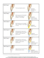
Figure 2 Coronal extension of dentine<br />
above the shoulder provides an<br />
e¡ective <strong>ferrule</strong>.<br />
<strong>The</strong> authors observed a high mean failure load for<br />
both groups, which they attributed to the resin luting<br />
cement. Although there was no di¡erence between the<br />
two groups’resistance to failure, the modes of failure differed.<br />
<strong>The</strong> group with a <strong>ferrule</strong> underwent oblique fracture,<br />
whereas the group without a <strong>ferrule</strong> largely<br />
underwent vertical root fracture.<br />
Summary of studies with artificial crowns<br />
<strong>The</strong>se studies showed that coronal extension of dentine<br />
above the shoulder increases the resistance to failure<br />
(Fig. 2).This <strong>ferrule</strong> e¡ect may be signi¢cantly enhanced<br />
when the coronal extension of dentine is at least<br />
1.5 mm. This e¡ect has been demonstrated in teeth<br />
restored with cast post and cores. A <strong>ferrule</strong> in teeth<br />
restored with direct posts and resin cores may not o¡er<br />
any additional bene¢t.<br />
In all the in vitro studies, both with and without<br />
crowns, only single rooted teeth have been investigated.<br />
<strong>The</strong> in£uence of the <strong>ferrule</strong> e¡ect on multi-rooted teeth<br />
remains an area for further research.<br />
<strong>The</strong> <strong>ferrule</strong> <strong>effect</strong> in vivo<br />
Stankiewicz & Wilson <strong>The</strong> <strong>ferrule</strong> <strong>effect</strong><br />
<strong>The</strong>re appear to be no reports of prospective clinical<br />
investigations of the <strong>ferrule</strong> e¡ect. A retrospective study<br />
of the survival rate of two post designs was conducted<br />
byTorbjo« rneret al. (1995).<strong>The</strong>ir study involved <strong>review</strong>ing<br />
the records of 638 patients, with a total of 788 posts;<br />
the patients were not examined. Seventy-two of the posts<br />
failed. Most (46 cases) failed owing to the loss of retention.<br />
It was observed that all the post fractures (six cases)<br />
occurred in teeth with a ‘‘. . . lack of a <strong>ferrule</strong> e¡ect of<br />
ß 2002 Blackwell Science Ltd International Endodontic Journal, 35, 575^581, 2002 579
<strong>The</strong> <strong>ferrule</strong> <strong>effect</strong> Stankiewicz & Wilson<br />
the metal collar at the crown margin area . . .’’. <strong>The</strong><br />
remainder failed owing to root fracture. <strong>The</strong>ir study did<br />
not state how manycrowns surveyed had a <strong>ferrule</strong>.From<br />
the work of Sorensen & Engelman (1990) and Libman<br />
& Nicholls (1995) it has been shown that the position<br />
and length of the <strong>ferrule</strong> is of signi¢cance. Torbjo« rner<br />
et al. (1995) did not indicate whether these design features<br />
were recorded in the patients’ ¢les. Furthermore,<br />
radiographs do not allow assessment of the amount of<br />
tissue remaining under a crown with a metal substructure.<br />
<strong>The</strong>refore, it would be prudent for future studies<br />
tomake some recordof the <strong>ferrule</strong>, orevenconsiderkeeping<br />
dies.<br />
A classi¢cation of the single-rooted pulpless teeth<br />
based upon the amount of remaining supragingival<br />
tooth structure has been recommended (Kurer 1991).<br />
Five classes of pulpless tooth were described; class I has<br />
su⁄cient coronal tissue for a crown preparation, II has<br />
coronal tissue but requires a core, and III has no coronal<br />
tissue. <strong>The</strong> classes IVand V refer to the complications of<br />
intraosseous fracture and periodontal disease. However,<br />
this classi¢cation does not account for a minimum e¡ective<br />
<strong>ferrule</strong> length. It would perhaps be of value to add<br />
a further subclassi¢cation of <strong>ferrule</strong> length to the Class<br />
II type of pulpless tooth. A suitable distinction would<br />
be less than or at least 2 mm-<strong>ferrule</strong> length.<br />
Preoperative tooth assessment<br />
Inassessing whetheratooth should be restored, the clinician<br />
must consider the amount of remaining supragingival<br />
tissue. Libman & Nicholls (1995) demonstrated<br />
in vitro that the minimum e¡ective <strong>ferrule</strong> should be<br />
1.5 mm of coronal dentine extending beyond the preparation<br />
margin. Below this length, there is a signi¢cant<br />
decrease in resistance to preliminary failure. Without<br />
supportive in vivo research the clinician is left to question<br />
whether satisfactory treatment can still be provided<br />
where a <strong>ferrule</strong> is absent or that is shorter than that<br />
advised in this in vitro study.<br />
A minimum <strong>ferrule</strong> length of 2 mm to compensate for<br />
the di⁄culties of intraoral tooth preparation has been<br />
recommended (McLean1998). CitingapaperbyFreeman<br />
et al. (1998), it was noted that even where a1-mm <strong>ferrule</strong><br />
was attempted extraorally, it was di⁄cult to achieve.<br />
Overcompensation is thus advisable to achieve an adequate<br />
length of parallel dentine, to produce an e¡ective<br />
<strong>ferrule</strong>.<br />
<strong>The</strong> <strong>ferrule</strong> length that may be obtained will be in£uenced<br />
by the ‘biologic width’. This is de¢ned as ‘‘. . . the<br />
dimension of the junctional epithelial and connective<br />
tissue attachment to the root above the alveolar crest’’<br />
(Sivers & Johnson 1985). If unpredictable bone loss and<br />
in£ammation is to be avoided, the crown margin should<br />
be at least 2 mm from the alveolar crest. It has been<br />
recommended that at least 3 mm should be left to avoid<br />
impingement onthe coronalattachment of theperiodontal<br />
connective tissue (Fugazzotto & Parma-Benfenait<br />
1984). <strong>The</strong>refore, at least 4.5 mm of supra-alveolar tooth<br />
structure may be required to provide an e¡ective <strong>ferrule</strong>.<br />
In those clinical situations where there is insu⁄cient<br />
<strong>ferrule</strong> length, even where margins are placed subgingivally,<br />
the clinician may consider surgical crown<br />
lengthening or orthodontic extrusion. This allows the<br />
distance between the crown margin and alveolar crest<br />
tobe widened, and increases the potential <strong>ferrule</strong>length.<br />
Methods to increase <strong>ferrule</strong> lengthwill reduce the root<br />
length and result in more tooth loss, possibly making<br />
the crown to root ratio unfavourable. Furthermore, both<br />
procedures will add to the cost of restoring the tooth,<br />
prolong treatment time and cause discomfort to the<br />
patient. <strong>The</strong> study by Al-Hazaimeh & Gutteridge (2001)<br />
suggests that the use of a resin-bonded direct post and<br />
resin core may be a preferable alternative where a <strong>ferrule</strong><br />
can not easily be obtained.<br />
<strong>The</strong> e¡ect of crown lengthening to establish a <strong>ferrule</strong><br />
on static load failure was investigated (Gegau¡ 2000).<br />
Using restorative composite resin to make root analogues,<br />
twogroups of10 teethwere tested. One group simulated<br />
teeth that had received crown lengthening and a<br />
<strong>ferrule</strong> in the preparation; the other group were not<br />
crown lengthened and were without a <strong>ferrule</strong> and no<br />
remaining clinical crown. All the teeth had cast post<br />
retained cores and crowns cemented prior to testing.<br />
Both the alveolar bone and periodontal ligament were<br />
simulated in the study.<strong>The</strong> teeth were subjected to a load<br />
by a universal testing machine until failure (peak load).<br />
<strong>The</strong> group that had crown lengthening and a <strong>ferrule</strong><br />
demonstrated a signi¢cantly lower failure load.<br />
Gegau¡ (2000) noted that the apical relocation of the<br />
¢nish line following crown lengthening resulted in a<br />
decrease in the cross section of the preparation. Subsequently,<br />
this reduction in tissue combined with an<br />
altered crown to root ratio may result in weakening of<br />
the tooth. He alsopointed out that orthodontic extrusion<br />
may be preferable to crown lengthening as it results in<br />
a smaller change in the crown to root ratio. <strong>The</strong> results<br />
of that study suggested that the overall importance of a<br />
<strong>ferrule</strong> at the expense of remaining tooth structure<br />
was unclear.<br />
Abalance betweenthe <strong>ferrule</strong>lengthobtainedand the<br />
remaining root is thereforeneeded.<strong>The</strong>se considerations<br />
580 International Endodontic Journal, 35, 575^581, 2002 ß 2002 Blackwell Science Ltd
are obviouslybest made prior to root canaltreatment. If a<br />
suitable <strong>ferrule</strong> length cannot be obtained, the patient<br />
should be informed of the potential compromise.<br />
Conclusions<br />
Laboratory evidence shows in some circumstances that<br />
a <strong>ferrule</strong> e¡ect occurs owing to the crown bracing<br />
against the dentine extending coronal to the crown margin.<br />
Furthermore, a signi¢cant increase in resistance<br />
to failure in single rooted teeth is observed where this<br />
dentine extends at least1.5 mm. However, the cost of getting<br />
this support in teeth with no coronal dentine is loss<br />
of tooth tissue.Whenassessing atooth prior to root treatment<br />
and subsequent restoration with a crown (if<br />
needed), a <strong>ferrule</strong> would be desirable but not at the<br />
expense of the remaining tooth/root structure.<br />
Acknowledgements<br />
<strong>The</strong> authors wish to thank Mr Lucas Sproson for producing<br />
the ¢gures.<br />
References<br />
Al-Hazaimeh N, Gutteridge DL (2001) An in vitro study into the<br />
e¡ect of the <strong>ferrule</strong> preparation on the fracture resistance of<br />
crowned teeth incorporating prefabricated post and composite<br />
core restorations. International Endodontic Journal 34,<br />
40^6.<br />
Barkhordar RA, Radke R, Abbasi J (1989) E¡ect of metal collars<br />
on resistance of endodontically treated teeth to root fracture.<br />
Journal of Prosthetic Dentistry 61,676^8.<br />
Brown L, ed. (1993) <strong>The</strong> New Shorter Oxford Dictionary onHistorical<br />
Principles. Oxford, UK: Clarendon Press. 223.<br />
CampagniWV, Reisbick MH, Jugan M (1993) Acomparison of an<br />
accelerated technique for casting post-and-core restorations<br />
with conventional techniques. Journal of Prosthodontics 2,<br />
159^66.<br />
Freeman MA, NichollsJL, KyddWL, HarringtonGW (1998)Leakage<br />
associated with load fatigue-induced preliminary failure<br />
of full crowns placed over three di¡erent post and core<br />
systems. Journal of Endodontics 24, 26^32.<br />
Fugazzotto PA, Parma-Benfenait S (1984) Pre-prosthetic periodontal<br />
considerations. Crown length and biologic width. Quintessence<br />
International 12,1247^56.<br />
Stankiewicz & Wilson <strong>The</strong> <strong>ferrule</strong> <strong>effect</strong><br />
Gegau¡ AG (2000) E¡ect of crown lengthening and <strong>ferrule</strong> placement<br />
on static load failure of cemented cast post-cores<br />
and crowns. Journal of Prosthetic Dentistry 84,169^79.<br />
Hemmings KW, King PA, Setchell DJ (1991) Resistance to torsional<br />
forces of various post and core designs. Journal of<br />
Prosthetic Dentistry 66, 325^9.<br />
Isidor F, Brondum K, Ravnholt G (1999) <strong>The</strong> in£uence of post<br />
length and crown <strong>ferrule</strong> length on the resistance to cyclic<br />
loading of bovine teeth with prefabricated titanium posts.<br />
InternationalJournal of Prosthodontics 12,78^82.<br />
Joseph J, Ramachandran G (1990) Fracture resistance of dowel<br />
channel preparation with various dentin thickness. Journal<br />
of FODI 1990,32^5.<br />
Kurer HG (1991) <strong>The</strong> classi¢cation of single-rooted, pulpless<br />
teeth. Quintessence International 22,939^43.<br />
LibmanWJ, NichollsJI (1995) Load fatigue of teeth restored with<br />
cast posts and cores and complete crowns. International<br />
Journal of Prosthodontics 8,155^61.<br />
Loney RW, Kotowicz WE, McDowell GC (1990) Three-dimensional<br />
photoelastic stress analysis of the <strong>ferrule</strong> e¡ect in cast<br />
post and cores. Journal of Prosthetic Dentistry 63,506^12.<br />
McLean A (1998) Predictably restoring endodontically treated<br />
teeth. Journal of the Canadian Dental Association 64,782^7.<br />
Milot P, Stein RS (1992) Root fracture in endodontically treated<br />
teeth related to post selection and crown design. Journal of<br />
Prosthetic Dentistry 68, 428^35.<br />
Rosen H (1961) Operative procedures on mutilated endodontically<br />
treated teeth. Journal of Prosthetic Dentistry11,973^86.<br />
Rosen H, Partida-Rivera M (1996) Iatrogenic fracture of roots<br />
reinforced with a cervical collar. Operative Dentistry 11,<br />
46^50.<br />
SaupeWA, Gluskin AH, Radke RAJr (1996) Acomparative study<br />
of fracture resistance between morphologic dowel and cores<br />
and a resin-reinforced dowel system in the intraradicular<br />
restoration of structurally compromised roots. Quintessence<br />
International 27, 483^91.<br />
Sivers JE, Johnson GK (1985) Periodontal and restorative considerations<br />
for crown lengthening. Quintessence International<br />
12,833^6.<br />
Sorensen JA, Engelman MJ (1990) Ferrule design and fracture<br />
resistance of endodontically treated teeth. Journal of<br />
Prosthetic Dentistry 63,529^36.<br />
Tjan AHL,Whang SB (1985) Resistance to root fracture of dowel<br />
channels with various thickness of buccal dentin walls.<br />
Journal of Prosthetic Dentistry 53, 496^500.<br />
Torbjo« rnerA, Kerlsson S, O« dman PA (1995) Survival rateandfailure<br />
characteristics for two post designs. Journal of Prosthetic<br />
Dentistry 73, 439^44.<br />
ß 2002 Blackwell Science Ltd International Endodontic Journal, 35, 575^581, 2002 581


