Open vs Closed Centripetal Build-up Technique - Osteocom.net
Open vs Closed Centripetal Build-up Technique - Osteocom.net
Open vs Closed Centripetal Build-up Technique - Osteocom.net

You also want an ePaper? Increase the reach of your titles
YUMPU automatically turns print PDFs into web optimized ePapers that Google loves.
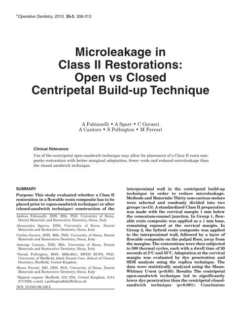
© Operative Dentistry, 2010, 35-3, 308-313<br />
Microleakage in<br />
Class II Restorations:<br />
<strong>Open</strong> <strong>vs</strong> <strong>Closed</strong><br />
<strong>Centripetal</strong> <strong>Build</strong>-<strong>up</strong> <strong>Technique</strong><br />
A Fabianelli • A Sgarr • C Goracci<br />
A Cantoro • S Pollington • M Ferrari<br />
Clinical Relevance<br />
Use of the centripetal open-sandwich technique may allow for placement of a Class II resin composite<br />
restoration with better marginal adaptation, fewer voids and reduced microleakage than<br />
the closed sandwich technique.<br />
SUMMARY<br />
Purpose: This study evaluated whether a Class II<br />
restoration in a flowable resin composite has to be<br />
placed prior to (open-sandwich technique) or after<br />
(closed-sandwich technique) construction of the<br />
Andrea Fabianelli, DDS, MSc, PhD, University of Siena,<br />
Dental Materials and Restorative Dentistry, Siena, Italy<br />
Alessandra Sgarra, DDS, University of Siena, Dental<br />
Materials and Restorative Dentistry, Siena, Italy<br />
Cecilia Goracci, DDS, MSc, PhD, University of Siena, Dental<br />
Materials and Restorative Dentistry, Siena, Italy<br />
Amerigo Cantoro, DDS, MSc, University of Siena, Dental<br />
Materials and Restorative Dentistry, Siena, Italy<br />
*Sarah Pollington, BDS, MMedSci, MFDS RCPS, PhD,<br />
University of Sheffield, Adult Dental Care, School of Clinical<br />
Dentistry, Sheffield, United Kingdom<br />
Marco Ferrari, MD, DDS, PhD, University of Siena, Dental<br />
Materials and Restorative Dentistry, Siena, Italy<br />
*Reprint request: Sheffield, S10 2TA, United Kingdom, 0114<br />
2717928; e-mail: s.pollington@sheffield.ac.uk<br />
DOI: 10.2341/09-128-L<br />
interproximal wall in the centripetal build-<strong>up</strong><br />
technique in order to reduce microleakage.<br />
Methods and Materials: Thirty non-carious molars<br />
were selected and randomly divided into two<br />
gro<strong>up</strong>s (n=15). A standardized Class II preparation<br />
was made with the cervical margin 1 mm below<br />
the cementum-enamel junction. In Gro<strong>up</strong> 1, flowable<br />
resin composite was applied as a 1 mm base,<br />
remaining exposed at the cervical margin. In<br />
Gro<strong>up</strong> 2, the hybrid resin composite was applied<br />
to the interproximal wall, followed by a layer of<br />
flowable composite on the pulpal floor, away from<br />
the margins. The restorations were then subjected<br />
to 500 thermal cycles, each with a dwell time of 20<br />
seconds at 5°C and 55°C. Adaptation at the cervical<br />
margin was evaluated by dye pe<strong>net</strong>ration and<br />
SEM analysis using the replica technique. The<br />
data were statistically analyzed using the Mann-<br />
Whitney U-test (p
Fabianelli & Others: Microleakage and <strong>Centripetal</strong> <strong>Build</strong>-<strong>up</strong> <strong>Technique</strong>s<br />
Flowable resin composite placed under hybrid<br />
resin composites in Gro<strong>up</strong> 1 provided better marginal<br />
adaptation and fewer voids. However, neither<br />
Gro<strong>up</strong> 1 nor Gro<strong>up</strong> 2 was able to completely<br />
prevent microleakage.<br />
INTRODUCTION<br />
With the increasing demand for esthetic treatment<br />
options in restorative dentistry, an interest in longevity<br />
and reliability of resin composite restorations has<br />
grown. Resin composites represent the material most<br />
commonly used as an alternative to amalgam for Class<br />
II restorations. Resin composites have been employed<br />
for many years. While their wear resistance has been<br />
satisfactorily improved in recent years, 1 difficulties in<br />
achieving an adequate interfacial seal and a valid<br />
interproximal contact point can still limit the clinical<br />
success of resin composite Class II restorations. 2 Both<br />
adapting resin composite to cervical walls and adjusting<br />
interproximal contact points are often considered<br />
critical steps. 2-3 Hassan and others4 and Bichacho5 have<br />
proposed the centripetal build-<strong>up</strong> technique for placing<br />
posterior resin composite restorations. This technique<br />
replaces lost tooth structure from the periphery<br />
towards the center of the cavity, thereby achieving better<br />
marginal adaptation to the pulpal floor. 5 The<br />
authors of the current study suggest incremental<br />
insertion in combination with centripetal resin composite<br />
build-<strong>up</strong>, thus transforming a Class II into a<br />
Class I restoration. The use of thin metal matrix bands<br />
and wooden wedges eliminates the need for transparent<br />
matrix bands, which may lead to poor contact<br />
areas and anatomical proximal contours. 6<br />
Also, sectional metal matrices can be utilized, along<br />
with ring retainers that exert pressure, thus allowing<br />
for proper modeling of the proximal contacts. In addition,<br />
the thin proximal layer of resin composite can<br />
expect to achieve complete curing and, thus, develop<br />
adequate mechanical properties. The use of enamel<br />
shades for the first interproximal layer, followed by<br />
dentin shades, leads to predictable and satisfactory<br />
esthetic results. The layer subsequently placed on the<br />
pulpal floor is believed to eventually fill any voids present<br />
at the cervical margin. The ability of the centripetal<br />
build-<strong>up</strong> technique to improve the marginal<br />
seal has been confirmed by recent laboratory-based<br />
studies. 7-8<br />
A relevant factor for the clinical failure of posterior<br />
resin composite restorations is the stress generated at<br />
the tooth-restoration interface due to competition<br />
between the rigid bond and polymerization shrinkage. 9<br />
This may compromise the quality of the seal primarily<br />
at the pulpal margins located below the enamelcementum<br />
junction of Class II restorations. In the<br />
attempt to improve the marginal seal, many strategies<br />
have been proposed, such as applying a combination of<br />
309<br />
materials and using different curing regimes. 10 The use<br />
of a flexible lining of flowable resin composite has been<br />
advised. 11 Flowable composites are microhybrid resins<br />
with a 60%-70% by weight load of filler particles ranging<br />
in size from 0.7 to 1.0 microns. In vitro studies have<br />
shown that such resin composites exhibit a substantially<br />
lower modulus of elasticity, which enables<br />
increased elastic deformation to absorb polymerization<br />
shrinkage stresses, thus minimizing open margins,<br />
especially at the cervical level. 12<br />
This laboratory study evaluated whether the flowable<br />
resin composite in a Class II resin composite<br />
restoration should be placed before (open-sandwich<br />
technique) or after (closed-sandwich technique) construction<br />
of the interproximal wall in the centripetal<br />
build-<strong>up</strong> technique. The quality of the marginal seal<br />
was evaluated with microleakage. SEM observations<br />
were also undertaken to verify the presence of marginal<br />
gaps, as well as to visualize the morphological<br />
aspects of the tooth-restoration interface. The null<br />
hypothesis was that there is no difference between the<br />
open and closed centripetal build-<strong>up</strong> technique with<br />
regard to microleakage at the gingival margin of a<br />
Class II resin composite restoration placed below the<br />
cementum-enamel junction.<br />
METHODS AND MATERIALS<br />
Specimen Preparation<br />
Thirty caries-free, unrestored human molars were<br />
selected and stored in a 1% chloramine solution for <strong>up</strong><br />
to three months. A standardized adhesive Class II<br />
preparation was made in the mesial and occlusal surface.<br />
The cervical margin of the interproximal box was<br />
placed 1 mm below the cementum-enamel junction.<br />
Occlusally, the tooth was reduced by 2 mm and the cavity<br />
was 3 mm wide. The proximal box was 4 mm wide<br />
bucco-lingually; whereas, the pulpal and axial walls<br />
measured to be 2 mm deep. The dimensions of the prepared<br />
cavities were checked with a Boley gauge. A ±0.3<br />
mm tolerance in the measurements was considered<br />
acceptable for including the specimen in the trial. No<br />
bevels were added to any margin of the preparations.<br />
The teeth were randomly divided into two gro<strong>up</strong>s of<br />
15 specimens each. All the specimens were restored<br />
with the adhesive Bond Force (Tokuyama, Tokyo,<br />
Japan), the flowable resin composite Palfique Estelite<br />
LV (Tokuyama) and the hybrid all-purpose resin composite<br />
Estelite Sigma (Tokuyama). Chemical composition<br />
and batch numbers of the materials are summarized<br />
in Table 1. A Brenner metal matrix was used to<br />
create the interproximal wall.<br />
In Gro<strong>up</strong> 1 (open-sandwich technique), the cavity<br />
was air-dried and the bonding agent was rubbed in for<br />
20 seconds, air-dried and light-cured for 10 seconds.<br />
The flowable resin composite was applied as a 1-mm
310 Operative Dentistry<br />
Table 1: Adhesive and Resin Composites Tested in This Study<br />
Material Type Composition<br />
Bond Force All-in-one self-etch adhesive Phosphoric acid monomer, Bisphenol A<br />
(Tokuyama, Tokyo, Japan) di(2-hydroxy propoxy) dimethacrylate<br />
Batch #4T10787 (Bis-GMA),<br />
Triethylene glycol dimethacrylate,<br />
2-Hydroxyethyl methacrylate (HEMA),<br />
Camphorquinone, alcohol and purified<br />
water.<br />
Estelite Sigma<br />
(Tokuyama, Tokyo, Japan)<br />
Microhybrid resin composite 0.2 µm SiO2-ZrO2 (spherical)<br />
composite filler (82wt%),<br />
LOT W805 methacrylate monomers<br />
Bis-GMA/TEGDMA<br />
Camphorquinone<br />
Palfique Estelite LV<br />
(Tokuyama, Tokyo, Japan)<br />
Flowable resin composite 0.2 µm SiO2-ZrO2(spherical) composite filler (42wt%)<br />
LOT 312 methacrylate monomers<br />
Bis-GMA/TEGDMA<br />
Camphorquinone<br />
thick base at the cervical margin according to the centripetal<br />
open-sandwich technique (Figure 1) and lightcured<br />
(LCU, 3M ESPE, Seefeld, Germany) for 20 seconds.<br />
Then, a 2-mm increment of hybrid resin composite,<br />
shade C2, was applied on the gingival wall of the<br />
proximal box and packed interproximally towards the<br />
metal matrix, causing the resin to climb <strong>up</strong>ward strictly<br />
in contact with the inner surface of the matrix band.<br />
This increment was adapted and light-cured. Subsequent<br />
2-mm thick layers were placed in horizontal<br />
increments toward the occlusal margin of the cavity.<br />
In Gro<strong>up</strong> 2, wherein the centripetal closed-sandwich<br />
technique was followed (Figure 2), after the application<br />
of the same bonding agent, the interproximal wall was<br />
created using 1-mm thick increments of the hybrid resin<br />
Figure 1: Specimen restored with the centripetal open-sandwich technique:<br />
the flowable resin composite is demonstrated by the red area.<br />
Figure 2: Specimen restored with the centripetal closed-sandwich technique:<br />
the flowable resin composite is demonstrated by the red area.<br />
composite singularly<br />
light-cured for 20<br />
seconds. After creating<br />
the interproximal<br />
wall, a 1 mm<br />
layer of flowable<br />
composite was placed<br />
on the pulpal floor<br />
and light-cured.<br />
Then, subsequent 2mm<br />
thick increments<br />
of hybrid<br />
resin composite were<br />
placed and lightcured.<br />
The same<br />
number of increments<br />
was used for<br />
the two techniques.<br />
The restorations<br />
were then subjected to 500 thermal cycles, each with a dwell<br />
time of 20 seconds at 5C° and 55C°.<br />
Impressions of the entire marginal area and interproximal<br />
walls were taken with a polyether impression<br />
material (Impregum, 3M ESPE) and epoxy resin replicas<br />
were cast (Epoxy Cure Resin, Buehler, IL, USA) for<br />
SEM marginal analysis. The specimens were mounted<br />
on aluminum stubs, coated with a colloid silver paint<br />
and sputtered with gold palladium (Edwards S105B<br />
Sputter Coater, London, England). The specimens<br />
were then observed under SEM (JEOL JSM-6060LV,<br />
Tokyo, Japan) to evaluate adaptation of the resin composites<br />
at the gingival margin with both techniques.<br />
Dye Pe<strong>net</strong>ration Test<br />
Nail varnish was applied to coat the foramina and the<br />
entire specimen surface, leaving a 1-mm window<br />
around the cavity margins. The teeth were then<br />
immersed in a 2% methylene blue solution for six<br />
hours. After rinsing the methylene blue solution off<br />
with distilled water, the specimens were embedded in<br />
acrylic resin and longitudinally sectioned with a diamond<br />
saw (Isomet, Buehler, IL, USA) at three different<br />
levels in the mesio-distal direction. The first cut was<br />
positioned in the center of the restorations, while the<br />
two remaining sections were cut along the lingual and<br />
buccal walls, approximately at the interface between<br />
the restoration and the cavity wall.<br />
The extent of dye pe<strong>net</strong>ration at the cervical margin<br />
was assessed under an optical microscope (Nikon<br />
SMZ645, Nikon, Japan) at 25x magnification and<br />
scored as: 0 = no pe<strong>net</strong>ration; 1 = pe<strong>net</strong>ration not<br />
exceeding the middle of the cervical wall; 2 = pe<strong>net</strong>ration<br />
past the middle of the cervical wall; 3 = pe<strong>net</strong>ration<br />
along the axial wall.<br />
Microleakage scores were independently assigned by<br />
two examiners and, in case of disagreement between
Fabianelli & Others: Microleakage and <strong>Centripetal</strong> <strong>Build</strong>-<strong>up</strong> <strong>Technique</strong>s<br />
their evaluations, the worse score was considered for<br />
statistical analysis.<br />
Statistical Analysis<br />
The Mann-Whitney U-test was applied to assess statistical<br />
significance of the difference in microleakage<br />
scores between the two experimental gro<strong>up</strong>s. The level<br />
of statistical significance was set at p
312 Operative Dentistry<br />
can be realized as a part of an in vitro screening of new<br />
adhesive restorative materials, preliminarily to clinical<br />
testing. It should be pointed out that laboratory<br />
data provides less reliable evidence than in vivo trials.<br />
As a matter of fact, the contribution of microleakage to<br />
restoration failure remains controversial and the clinical<br />
relevance of interfacial dye pe<strong>net</strong>ration is still the<br />
object of discussion. 27 No operative technique or adhesive<br />
system has been proven to completely prevent<br />
microleakage and no correlation between gap width<br />
and tracer pe<strong>net</strong>ration was reported in a recent laboratory<br />
study. 28 Also, in the current study, in none of the<br />
specimens obtained with either restorative procedure<br />
was dye pe<strong>net</strong>ration completely impeded. Moreover,<br />
the methods of microleakage testing have not yet been<br />
standardized. A systematic review of microleakage<br />
tests for restorative materials concluded that a comparison<br />
of study results was impossible, due to the<br />
variability of the employed methodologies. 29<br />
As an example, different dye tracers are available for<br />
use in microleakage studies. Recently, Heitze and others<br />
reported that there is no significant difference in<br />
tracer pe<strong>net</strong>ration between fuchsin, silver nitrate and<br />
methylene blue. 30 Methylene blue is one of the most<br />
common tracers and can be used in different concentrations,<br />
from 0.5% <strong>up</strong> to 5%. 31 It was pointed out that,<br />
because of the small surface area of the particles<br />
(approximately 0.52 nm2 ), methylene blue may lead to<br />
an overestimation of leakage at the tooth-restoration<br />
interface, particularly with self-etch adhesives in relation<br />
to their increased hydrophilicity. 32<br />
It is also disputed how many sections per tooth<br />
should be evaluated in dye pe<strong>net</strong>ration scoring. The<br />
evaluation of dye pe<strong>net</strong>ration scores is performed on<br />
one or more cuts of the specimen, and this method may<br />
be less sensitive than a three-dimensional evaluation. 33<br />
However, it is believed that the use of three cuts of one<br />
specimen may avoid under-estimation of in vitro<br />
microleakage. 34<br />
Still another controversial issue is the dwelling time<br />
in the dye tracer. It has been reported that storage<br />
time in the tracer is not a relevant factor. 35 Conversely,<br />
another study documented that longer dwelling periods<br />
can lead to over-diffusion of the tracer and higher<br />
microleakage scores. 23 There is also no standardized<br />
protocol for thermocycling, as several different regimens<br />
have been proposed to simulate clinical function.<br />
36<br />
In the current study, a new all-in-one self-etch dental<br />
adhesive that allows for simplification of the bonding<br />
procedure was used. Although etch-and-rinse threestep<br />
formulations are still regarded as the gold standard<br />
of adhesive systems, self-etch adhesives of the<br />
latest generation have given promising results both in<br />
laboratory and clinical studies. 37<br />
CONCLUSIONS<br />
According to the methodology proposed and within the<br />
limitations of an in vitro study, the following conclusions<br />
can be drawn:<br />
1. The centripetal open-sandwich technique produced<br />
a significantly more effective seal at the<br />
cervical margin of Class II resin composite<br />
restorations than the centripetal closed-sandwich<br />
technique, with better marginal adaptation<br />
and less voids.<br />
2. Neither restorative procedure was able to fully<br />
prevent dye pe<strong>net</strong>ration.<br />
(Received 27 April 2009)<br />
References<br />
1. Hickel R, Manhart J & García-Godoy F (2000) Clinical<br />
results and new developments of direct posterior restorations<br />
American Journal of Dentistry 13(Spec No) 41D-54D.<br />
2. Wilson NH, Dunne SM & Gainsford ID (1997) Current<br />
materials and techniques for direct restorations in posterior<br />
teeth. Part 2: Resin composite systems International<br />
Dental Journal 47(4) 185-193.<br />
3. Franchi C, Lo Guercio AD, Reis A & Carrilho MRO (2002) A<br />
novel filling technique for packable composite resin in Class<br />
II restorations Journal of Esthetic and Restorative<br />
Dentistry 14(3) 149-157.<br />
4. Hassan K, Mante F, List G & Dhuru V (1987) A modified<br />
incremental filling technique for Class II composite restorations<br />
The Journal of Prosthetic Dentistry 58(2) 153-156.<br />
5. Bichacho N (1994) The centripetal build-<strong>up</strong> for composite<br />
resin posterior restorations Practical Periodontics &<br />
Aesthetic Dentistry 6(3) 17-23.<br />
6. Christensen GJ (1992) Don’t underestimate the Class II<br />
resin Journal of the American Dental Association 123(3)<br />
103-104.<br />
7. Szep S, Frank H, Kenzel B, Gerhardt T & Heidemann D<br />
(2001) Comparative study of composite resin placement:<br />
<strong>Centripetal</strong> build-<strong>up</strong> versus incremental technique<br />
Practical Proceedings & Aesthetic Dentistry 13(3) 243-250.<br />
8. Ghavamnasiri M, Moosavi H & Tahvildarnejad N (2007)<br />
Effect of centripetal and incremental methods in Class II<br />
composite resin restorations on gingival microleakage<br />
Journal of Contemporary Dental Practice 8(2) 113-120.<br />
9. Feilzer AJ, de Gee AJ & Davidson CL (1987) Setting stress<br />
in composite resin I relation to configuration of restoration<br />
Journal of Dental Research 66(11) 1636-1639.<br />
10. Fabianelli A, Pollington S, Davidson CL, Cagidiaco MC &<br />
Goracci C (2007) The relevance of microleakage studies<br />
International Dentistry SA 9(3) 64-74.<br />
11. Kemp-Sholte CM & Davidson CL (1990) Marginal integrity<br />
related to bond strength and strain capacity of composite<br />
resin restorative systems The Journal of Prosthetic<br />
Dentistry 64(6) 658-664.
Fabianelli & Others: Microleakage and <strong>Centripetal</strong> <strong>Build</strong>-<strong>up</strong> <strong>Technique</strong>s<br />
12. Unterbrink GL & Liebenberg WH (1999) Flowable resin<br />
composites as “filled adhesives”: Literature review and clinical<br />
recommendations Quintessence International 30(4)<br />
249-257.<br />
13. Korkmaz Y, Ozel E & Attar N (2007) Effect of flowable composite<br />
lining on microleakage and internal voids in Class II<br />
composite restorations Journal of Adhesive Dentistry 9(2)<br />
189-194.<br />
14. Attar N, Tam LE & McComb D (2003) Flow, strength, stiffness<br />
and radiopacity of flowable resin composites Journal<br />
of the Canadian Dental Association 69(8) 516-521.<br />
15. Chuang SF, Liu JK & Jin YT (2001) Microleakage and<br />
internal voids in Class II composite restorations with flowable<br />
composite linings Operative Dentistry 26(2) 193-200.<br />
16. Ferrari M, Cagidiaco MC & Mason PM (1994)<br />
Micromorphologic relationship between resin and dentin in<br />
Class II restorations: An in vivo and in vitro investigation<br />
by scanning electron microscopy Quintessence International<br />
25(12) 861-866.<br />
17. Carvalho RM, Pereira JC, Yoshiyama M & Pashley DH<br />
(1996) A review of polymerisation contraction: The influence<br />
of stress development versus stress relief Operative<br />
Dentistry 21(1) 17-24.<br />
18. Lucena-Martin C, Gonzales-Rodriguez MP, Ferrer-Luque<br />
CM, Robles-Gijon V & Navajas JM (2001) Influence of time<br />
and thermocycling on marginal sealing of several dentin<br />
adhesive systems Operative Dentistry 26(6) 550-555.<br />
19. Cara RR, Fleming GJ, Palin WM, Walmsley AD & Burke FJ<br />
(2007) Cuspal deflection and microleakage in premolar<br />
teeth restored with resin-based composites with and without<br />
an intermediary flowable layer Journal of Dentistry<br />
35(6) 482-489.<br />
20. Gueders AM, Charpentier JF, Albert AI & Geerts SO (2006)<br />
Microleakage after thermocycling of 4 etch and rinse and 3<br />
self-etch adhesives with and without a flowable composite<br />
lining Operative Dentistry 31(4) 450-455.<br />
21. Ziskind D, Adell I, Teperovich E & Peretz B (2005) The<br />
effect of an intermediate layer of flowable composite on<br />
microleakage in packable composite restorations<br />
International Journal of Paediatric Dentistry 15(5) 349-354.<br />
22. Efes BG, Dorter C, Gomec Y & Koray F (2006) Two-year<br />
clinical evaluation of ormocer and nanofill composite with<br />
and without a flowable liner Journal of Adhesive Dentistry<br />
8(2) 119-126.<br />
23. Chersoni S, S<strong>up</strong>pa P, Grandini S, Goracci C, Monticelli F,<br />
Yiu C, Huang C, Prati C, Breschi L, Ferrari M, Pashley DH<br />
& Tay FR (2004) In vivo and in vitro permeability of one<br />
step self-etch adhesives Journal of Dental Research 83(6)<br />
459-464.<br />
24. Alomari QD, Reinhardt JW & Boyer DB (2001) Effect of liners<br />
on cusp deflection and gap formation in composite<br />
restoration Operative Dentistry 26(4) 406-411.<br />
25. Bayne SC, Thompson JY, Swift EJ, Stamatiades P &<br />
Wilkerson M (1998) A characterization of first generation<br />
flowable composites Journal of the American Dental<br />
Association 129(5) 567-577.<br />
313<br />
26. Kidd EAM (1976) Microleakage: A review Journal of<br />
Dentistry 4(5) 199-206.<br />
27. Heintze SD (2007) Systematic reviews: I. The correlation<br />
between laboratory tests on marginal quality and bond<br />
strength. II. The correlation between marginal quality and<br />
clinical outcome The Journal of Adhesive Dentistry (9<br />
S<strong>up</strong>plement 1) 77-106.<br />
28. Idriss S, Abduljabbar T, Habib C & Omar R (2007) Factors<br />
associated with microleakage in Class II resin composite<br />
restorations Operative Dentistry 32(1) 60-66.<br />
29. Raskin A, D’Hoore W, Gonthier S, Degrange M & Dejou J<br />
(2001) Reliability of in vitro microleakage test: A literature<br />
review The Journal of Adhesive Dentistry 3(4) 295-308.<br />
30. Heintze S, Forjanic M & Cavalleri A (2008) Microleakage of<br />
Class II restorations with different tracers—Comparison<br />
with SEM quantitative analysis Journal of Adhesive<br />
Dentistry 10(4) 259-267.<br />
31. Owens BM, Lim DY & Arheart KL (2003) The effect of<br />
antimicrobial pre-treatments on the performance of resin<br />
composite restorations Operative Dentistry 28(6) 716-722.<br />
32. Ernst CP, Galler P, Willershausen B & Haller B (2008)<br />
Marginal integrity of Class V restorations: SEM versus dye<br />
pe<strong>net</strong>ration Dental Materials 24(3) 319-327.<br />
33. Gale MS, Darvell BW & Cheung GSP (1994) Three-dimensional<br />
reconstruction of microleakage pattern using a<br />
sequential grinding technique Journal of Dentistry 22(6)<br />
370-375.<br />
34. Raskin A, Tassery H, D’Hoore W, Gonthier S, Vreven J,<br />
Degrange M & Déjou J (2003) Influence of the number of<br />
section on reliability of in vitro microleakage evaluations<br />
American Journal of Dentistry 16(3) 207-210.<br />
35. Hilton TJ (2002) Can modern restorative procedures and<br />
materials reliably seal cavities? In vitro investigations.<br />
Part 2 American Journal of Dentistry 15(4) 279-289.<br />
36. Ebert J, Loffler C, Roggendorf MJ, Petschelt A &<br />
Frankenberger F (2009) Clinical adhesive sealing of the<br />
pulp chamber following endodontic treatment: Influence of<br />
thermomechanical loading on microleakage Journal of<br />
Adhesive Dentistry 11(4) 311-317.<br />
37. Owens BM & Johnson WW (2007) Effect of single step<br />
adhesives on the marginal permeability of Class V resin<br />
composites Operative Dentistry 32(1) 67-72.<br />
38. Van Meerbeek B, De Munck J, Yoshida Y, Inoue S, Vargas<br />
M, Vijay P, Van Landuyt K, Lambrechts P & Vanherle G<br />
(2003) Buonocore Memorial Lecture. Adhesion to enamel<br />
and dentin: Current status and future challenges Operative<br />
Dentistry 28(3) 215-235.<br />
39. Silveira de Araùjo C, Incerti da Silva T, Ogliari FA,<br />
Meireles SS, Piva E & Demarco FF (2006) Microleakage of<br />
seven adhesives systems in enamel and dentin The Journal<br />
of Contemporary Dental Practice 7(5) 26-33.