ACE Class I ACE Class II ACE Class III ACE Class IV ... - OSTEOCOM
ACE Class I ACE Class II ACE Class III ACE Class IV ... - OSTEOCOM
ACE Class I ACE Class II ACE Class III ACE Class IV ... - OSTEOCOM

Create successful ePaper yourself
Turn your PDF publications into a flip-book with our unique Google optimized e-Paper software.
<strong>ACE</strong> <strong>Class</strong> I<br />
Thinning of palatal enamel<br />
Treatment:<br />
No restorative<br />
treatment<br />
<strong>ACE</strong> <strong>Class</strong> <strong>II</strong><br />
Dentin exposure on the palatal<br />
aspect (contact areas), no<br />
damage to incisal edges<br />
Treatment:<br />
Direct or indirect<br />
palatal composites<br />
<strong>ACE</strong> <strong>Class</strong> <strong>II</strong>I<br />
2 mm<br />
Dentin exposure on the palatal<br />
aspect, damage to incisal edges<br />
(< 2 mm)<br />
Treatment:<br />
Palatal veneers<br />
<strong>ACE</strong> <strong>Class</strong> <strong>IV</strong><br />
2 mm<br />
Extended dentin exposure on<br />
the palatal aspect, loss of tooth<br />
length (> 2 mm), preserved<br />
facial enamel<br />
Treatment:<br />
Sandwich approach<br />
<strong>ACE</strong> <strong>Class</strong> V<br />
Extended dentin exposure on<br />
the palatal aspect, loss of tooth<br />
length (> 2 mm), loss of<br />
facial enamel<br />
Treatment:<br />
Sandwich approach<br />
(experimental)<br />
<strong>ACE</strong> <strong>Class</strong> VI<br />
Advanced loss of tooth structure<br />
leading to pulp necrosis<br />
Treatment:<br />
Sandwich approach<br />
(highly experimental)<br />
The International Journal of Periodontics & Restorative Dentistry<br />
© 2010 BY QUINTESSENCE PUBLISHING CO, INC. PRINTING OF THIS DOCUMENT IS RESTRICTED TO PERSONAL USE ONLY.<br />
NO PART OF THIS ARTICLE MAY BE REPRODUCED OR TRANSMITTED IN ANY FORM WITHOUT WRITTEN PERMISSION FROM THE PUBLISHER.
559<br />
<strong>Class</strong>ification and Treatment of the<br />
Anterior Maxillary Dentition Affected by<br />
Dental Erosion: The <strong>ACE</strong> <strong>Class</strong>ification<br />
Francesca Vailati, MD, DMD, MSc*<br />
Urs Christoph Belser, DMD, Prof Dr Med Dent**<br />
Erosive tooth wear is a serious problem with very costly consequences.<br />
Intercepting patients at the initial stages of the disease is critical to avoid significant<br />
irreversible damages to their dentition and to benefit from still favorable<br />
conditions when it comes to clinical performance of the restorative measures<br />
proposed. In this article, a new classification is proposed to quantify the severity<br />
of the dental destruction and to guide clinicians and patients in the therapeutic<br />
decision-making process. The classification is based on several parameters<br />
relevant for both the selection of treatment and the assessment of the prognosis,<br />
such as dentin exposure in the palatal tooth contact areas, alterations at the level<br />
of the incisal edges, and ultimately, loss of pulp vitality. (Int J Periodontics<br />
Restorative Dent 2010;30:559–571.)<br />
*Senior Lecturer, Department of Fixed Prosthodontics and Occlusion, School of Dental<br />
Medicine, University of Geneva, Geneva, Switzerland; Private Practice, Geneva,<br />
Switzerland.<br />
**Chairman, Department of Fixed Prosthodontics and Occlusion, School of Dental<br />
Medicine, University of Geneva, Geneva, Switzerland.<br />
Correspondence to: Dr Francesca Vailati, rue Barthélemy-Menn 19, Geneva, Switzerland<br />
1205; email: francesca.vailati@unige.ch.<br />
In modern society, dental erosion has<br />
become one of the major causes of<br />
the loss of mineralized tooth structure.<br />
Several surveys have pointed<br />
out a high and still increasing prevalence,<br />
especially among young individuals<br />
(eg, 37% of 14-year-olds in<br />
the United Kingdom present signs of<br />
palatal enamel erosion). 1–12 Signs of<br />
dental erosion that may be easily evident<br />
at an early stage include:<br />
“glossy” (smooth, glazed) enamel,<br />
yellowing of the teeth from the underlying<br />
dentin, increased incisal translucency,<br />
and cupping of the occlusal<br />
surfaces. While the presence of dental<br />
caries normally leads clinicians to<br />
take action immediately, in the case<br />
of dental erosion, many clinicians prefer<br />
to postpone any dental treatment<br />
until the patient is older, even though<br />
literature confirms that direct clinical<br />
observation is an unreliable method<br />
for monitoring the rates of tooth<br />
wear. 13,14 To play down this problem<br />
is frequently the preferred approach,<br />
which is understandable since many<br />
clinicians do not feel comfortable<br />
proposing an extensive dental rehabilitation<br />
to young individuals who<br />
are still asymptomatic and unaware of<br />
Volume 30, Number 6, 2010<br />
© 2010 BY QUINTESSENCE PUBLISHING CO, INC. PRINTING OF THIS DOCUMENT IS RESTRICTED TO PERSONAL USE ONLY.<br />
NO PART OF THIS ARTICLE MAY BE REPRODUCED OR TRANSMITTED IN ANY FORM WITHOUT WRITTEN PERMISSION FROM THE PUBLISHER.
560<br />
the problem. What such clinicians are<br />
not considering, however, is that<br />
these patients sooner or later will<br />
need to restore their jeopardized<br />
dentition anyway. The debate over<br />
whether it is preferable to start earlier<br />
with a lighter, less invasive rehabilitation<br />
or later with a highly<br />
aggressive but eventually more resistant<br />
one is still open.<br />
The aim of this article is to convince<br />
clinicians that in the specific case<br />
of dental erosion, hesitation in undertaking<br />
the adequate treatment will<br />
inevitably lead to further degradation<br />
of the patient’s dentition. To persuade<br />
the patients and to obtain informed<br />
consent for treatment, it is necessary<br />
to quantify the dental destruction and<br />
to make a prognosis on the future<br />
progression of the disease if no treatment<br />
would be undertaken. It would<br />
appear that the existing erosion<br />
assessment indices and classifications<br />
have not led to a broad respective<br />
awareness among dental care<br />
providers to date. This may be<br />
because these tools are rather complex<br />
and difficult to use in a daily practice<br />
set-up, since they have been<br />
primarily designed for scientific purposes.<br />
Furthermore, practical experience<br />
indicates that all relevant signs<br />
linked to the various progression<br />
stages of generalized dental erosion<br />
could be assessed clinically by mainly<br />
examining the anterior dentition.<br />
This finding may help to simplify the<br />
diagnostic process significantly.<br />
Consequently, a new classification,<br />
the anterior clinical erosive classification<br />
(<strong>ACE</strong>), has been proposed<br />
to provide clinicians with a practical<br />
tool to grade the dental status of<br />
each patient and to subsequently<br />
relate it to the appropriate treatment.<br />
Maxillary anterior teeth and<br />
dental erosion<br />
Disease progression<br />
In the case of dental erosion, the<br />
palatal aspect of the maxillary anterior<br />
teeth usually appears to be the<br />
most affected portion of the dentition,<br />
particularly in patients with an<br />
intrinsic etiology (eg, gastric reflux,<br />
psychiatric diseases). At an early stage,<br />
acid-caused destruction can be very<br />
subtle and thus difficult to discover<br />
because of the somewhat hidden<br />
location of the palatal tooth surfaces,<br />
especially if the disease progresses<br />
slowly. Patients frequently do not present<br />
signs of tooth sensitivity, even in<br />
the presence of dentin exposure.<br />
Often, the erosive wear will manifest<br />
too late, when irreversible damage<br />
has already taken place and costly<br />
restorative treatments are required.<br />
At the initial stage, only an attentive<br />
and trained eye can detect the<br />
more yellowish color resulting from<br />
the thinning of the enamel in the central<br />
palatal portion of the clinical<br />
crown. The cingula appear flatter and<br />
their surfaces are very shiny.<br />
The next step of erosive wear<br />
leads to a weakening of the incisal<br />
edges, which is first noticeable by an<br />
increase in translucency. Furthermore,<br />
the presence of caries or <strong>Class</strong> <strong>II</strong>I<br />
restorations may contribute additionally<br />
to the weakening of the facial<br />
aspect of the tooth. In extreme<br />
instances, a complete loss of the<br />
incisal edge may result, which<br />
depends strongly on the original overbite<br />
and overjet configuration and on<br />
the location of the occlusal contact<br />
area. For example, in a patient with a<br />
slight vertical overlap (overbite), the<br />
risk of incisal fracture is very high<br />
because of the destructive combination<br />
of erosion and the focal attrition<br />
of the antagonist teeth. In fact, at an<br />
early stage of enamel erosion, chipping<br />
is frequently visible in the form of<br />
irregularities at the incisal edges. On<br />
the other hand, in patients with a<br />
deep bite interarch configuration, the<br />
maxillary anterior teeth may present a<br />
pronounced concave morphology on<br />
their palatal aspect before any effect<br />
on the length of the clinical crown<br />
manifests. In extreme situations, the<br />
loss of the tooth structure may<br />
become so extensive that the pulp<br />
chamber (or its original extent) can be<br />
identified on the palatal aspect.<br />
Surprisingly, such teeth frequently<br />
keep their vitality; however, they may<br />
respond less quickly to the vitality test.<br />
In advanced stages, when the<br />
labial tooth structure has been undermined<br />
too much, the facial surfaces<br />
fracture and the clinical crowns suddenly<br />
appear reduced in length.<br />
Finally, especially in deep bite<br />
patients, the vertical overlap may be<br />
aggravated by the supraeruption of<br />
the anterior segments.<br />
Traditional reconstructive versus<br />
adhesive therapy<br />
Following the guidelines for conventional<br />
oral rehabilitation concepts,<br />
structurally compromised teeth<br />
The International Journal of Periodontics & Restorative Dentistry<br />
© 2010 BY QUINTESSENCE PUBLISHING CO, INC. PRINTING OF THIS DOCUMENT IS RESTRICTED TO PERSONAL USE ONLY.<br />
NO PART OF THIS ARTICLE MAY BE REPRODUCED OR TRANSMITTED IN ANY FORM WITHOUT WRITTEN PERMISSION FROM THE PUBLISHER.
561<br />
should receive complete crown coverage.<br />
To place the associated<br />
restoration margins at the gingival<br />
level, a significant amount of the<br />
remaining volume of the clinical crown<br />
has to be removed during tooth<br />
preparation to provide the required<br />
vertical path of insertion for the crown.<br />
In other terms, preparing such teeth<br />
for crowns will substantially aggravate<br />
the destruction of mineralized tissue<br />
that was initiated by the erosive<br />
process. Not infrequently, elective<br />
endodontic treatment will be necessary,<br />
mostly accompanied by the use<br />
of posts, to assure intraradicular retention<br />
of the crowns to be cemented.<br />
To avoid these types of invasive<br />
treatment modalities and to keep the<br />
teeth vital, an experimental approach<br />
to restoring the maxillary anterior<br />
teeth of patients affected by severe<br />
dental erosion is currently under<br />
investigation at the University of<br />
Geneva School of Dental Medicine<br />
(Geneva Erosion study) by the authors<br />
of this research. A minimally invasive<br />
treatment concept that consists of<br />
reconstructing the palatal aspect with<br />
composite restorations, followed by<br />
the restoration of the facial aspect<br />
with ceramic veneers, is promoted.<br />
The treatment objective is reached<br />
by the most conservative approach<br />
possible, since the remaining tooth<br />
structure is preserved and located in<br />
the center between two different<br />
restorations (the sandwich approach)<br />
and performed at two different time<br />
points. When it comes to the preservation<br />
of mineralized tooth structure,<br />
such an ultraconservative approach<br />
cannot be matched by any type of<br />
complete crown coverage.<br />
The type of restoration best indicated<br />
to restore the palatal aspect of<br />
the eroded maxillary anterior teeth<br />
(direct or indirect composite restorations)<br />
is selected according to the<br />
amount of the anterior interocclusal<br />
space obtained after an increase in<br />
the vertical dimension of occlusion. If<br />
the space is limited (< 1 mm), the<br />
composite restorations can be fabricated<br />
free-hand, saving time and<br />
money (there is no laboratory fee for<br />
the palatal onlays and only one clinical<br />
appointment is required). If the<br />
interocclusal distance between the<br />
anterior teeth is significant, however,<br />
free-hand resin composites could<br />
prove to be rather challenging.<br />
When the teeth present a combination<br />
of compromised palatal,<br />
incisal, and facial aspects, it is difficult<br />
to visualize the optimal final morphology<br />
of the teeth, particularly<br />
while restoring only the palatal<br />
aspect with rubber dam in place.<br />
Thus, the results may be unpredictable<br />
and highly time consuming.<br />
Under such conditions, fabricating<br />
palatal onlays in a laboratory clearly<br />
presents some advantages, including<br />
superior wear resistance and higher<br />
precision during fabrication of the<br />
definitive form. A series of articles<br />
on full-mouth adhesive rehabilitation<br />
address this in detail. 15–17 One of the<br />
criticisms to the sandwich approach<br />
is the work and cost associated with<br />
the fabrication of two separate<br />
restorations for each tooth. However,<br />
only with two independent restorations<br />
are two different paths of insertion<br />
possible, and the tooth<br />
preparation can therefore be kept<br />
minimal. Even the most conservative<br />
preparation for all-ceramic crowns<br />
could not achieve this level of tooth<br />
preservation.<br />
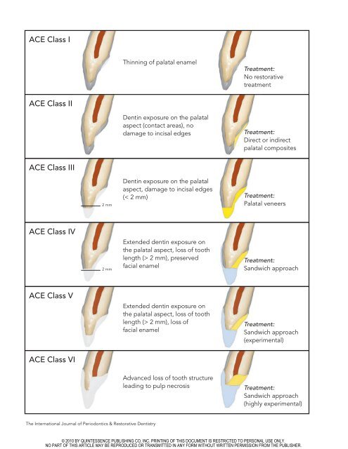
<strong>ACE</strong> classification<br />
Assessment of the severity of dental<br />
erosion is complicated because of the<br />
subjectivity of the methods of evaluation<br />
and the possible presence of<br />
wear cofactors (parafunctional habits,<br />
hyposalivation, wear resulting from<br />
tooth malposition, aging, coarse diet,<br />
inappropriate tooth-brushing techniques,<br />
abrasive toothpastes, etc). In<br />
addition, the rating scales selected<br />
by investigators may be somewhat<br />
complicated to translate in a clinical<br />
environment, and early alterations are<br />
difficult to locate, even with the support<br />
of photography, study casts, and<br />
attentive clinical examination. 18–26<br />
Several authors have proposed<br />
classifications and indices addressing<br />
either tooth wear in general 25 or<br />
including diagnostic criteria for erosive<br />
tooth wear specifically. 26 Most recently,<br />
Bartlett et al 18 published a new<br />
scoring system, termed basic erosive<br />
wear examination (BEWE), designed<br />
for both scientific and clinical purposes.<br />
It was the authors’ twofold objective<br />
to provide a simple tool for use in<br />
general practice and to permit more<br />
scientifically oriented comparisons<br />
with already existing indices.<br />
Furthermore, the BEWE aimed to<br />
augment the awareness of tooth erosion<br />
among general practitioners and<br />
to provide a respective guide for treatment<br />
when indicated. Finally, the<br />
BEWE was intended to stop the continued<br />
proliferation of new indices, as<br />
Volume 30, Number 6, 2010<br />
© 2010 BY QUINTESSENCE PUBLISHING CO, INC. PRINTING OF THIS DOCUMENT IS RESTRICTED TO PERSONAL USE ONLY.<br />
NO PART OF THIS ARTICLE MAY BE REPRODUCED OR TRANSMITTED IN ANY FORM WITHOUT WRITTEN PERMISSION FROM THE PUBLISHER.
562<br />
Table 1<br />
The <strong>ACE</strong> classification<br />
Palatal Palatal Incisal edge Facial Pulp Suggested<br />
enamel dentin length enamel vitality therapy<br />
<strong>Class</strong> I Reduced Not exposed Preserved Preserved Preserved No restorative treatment<br />
<strong>Class</strong> <strong>II</strong> Lost in contact Minimally exposed Preserved Preserved Preserved Palatal composites<br />
areas<br />
<strong>Class</strong> <strong>II</strong>I Lost Distinctly exposed Lost ≤ 2 mm Preserved Preserved Palatal onlays<br />
<strong>Class</strong> <strong>IV</strong> Lost Extensively exposed Lost > 2 mm Preserved Preserved Sandwich approach<br />
<strong>Class</strong> V Lost Extensively exposed Lost > 2 mm Distinctively Preserved Sandwich approach<br />
reduced/lost<br />
(experimental)<br />
<strong>Class</strong> VI Lost Extensively exposed Lost > 2 mm Lost Lost Sandwich approach<br />
(highly experimental)<br />
Fig 1 <strong>ACE</strong> <strong>Class</strong> I: (left) Frontal and (right)<br />
occlusal views. Very early detection of the<br />
erosive problem. All the cingula lost their<br />
microanatomical details. The enamel<br />
appears very shiny. Even though there is not<br />
yet dentin exposure, small chipping of the<br />
enamel at the incisal edge is visible (minimal<br />
vertical overlap). Considering the patient’s<br />
age (25 years) and etiology (bulimia), this<br />
patient has a high risk of deteriorating<br />
toward a more severe stage in a short period<br />
of time.<br />
it was hoped to represent a consensus<br />
within the specialized scientific community.<br />
Nevertheless, there is still an<br />
undisputable need for a classification<br />
that directly and specifically focuses<br />
on the anterior maxillary dentition,<br />
where loss of mineralized tissue<br />
because of erosion, as minute as it<br />
may be at an early stage of the disease,<br />
can be assessed easily.<br />
Clinicians not involved in epidemiologic<br />
surveys clearly need the<br />
least complicated approach to classify<br />
each patient rapidly and to<br />
decide on the most appropriate<br />
treatment plan. Thus, the prerequisite<br />
for a precise and rapid assessment<br />
is a diagnostic instrument that<br />
is based on a limited number of key<br />
parameters and that guides the clinician<br />
in a logical and systematic way.<br />
As a consequence, these two fundamental<br />
paradigms have been instrumental<br />
in the development and<br />
finalization of the proposed <strong>ACE</strong> classification<br />
(Table 1).<br />
The <strong>ACE</strong> classification is strictly<br />
related to the clinical observation of<br />
the status of the anterior maxillary<br />
teeth, which are generally the most<br />
damaged. Patients are grouped into<br />
six classes, and for each class, a dental<br />
treatment plan is suggested. The<br />
classification is based on five parameters<br />
relevant for the selection of<br />
the treatment and the assessment of<br />
the prognosis: the dentin exposure in<br />
the contact areas, the preservation of<br />
the incisal edges, the length of the<br />
remaining clinical crown, the presence<br />
of enamel on the vestibular surfaces,<br />
and the pulp vitality.<br />
<strong>ACE</strong> <strong>Class</strong> I: Flattened cingula<br />
without dentin exposure<br />
Suggested therapy:<br />
No restorative treatment<br />
This is the earliest stage of dental<br />
erosion. The enamel is present but<br />
thinner. The palatal aspect of the<br />
teeth may appear more yellowish in<br />
the central portion of the underlying<br />
dentin and more white at the periphery<br />
with the presence of thicker enamel<br />
(Fig 1).<br />
The International Journal of Periodontics & Restorative Dentistry<br />
© 2010 BY QUINTESSENCE PUBLISHING CO, INC. PRINTING OF THIS DOCUMENT IS RESTRICTED TO PERSONAL USE ONLY.<br />
NO PART OF THIS ARTICLE MAY BE REPRODUCED OR TRANSMITTED IN ANY FORM WITHOUT WRITTEN PERMISSION FROM THE PUBLISHER.
Fig 2 <strong>ACE</strong> <strong>Class</strong> <strong>II</strong>: Pretreatment (left) frontal and (center) occlusal views and (right) posttreatment occlusal view. In this patient, the palatal<br />
aspects present areas of dentin exposure at the level of the contact points. The incisal edges were still intact. An early conservative rehabilitation<br />
was planned, and all maxillary anterior teeth were restored using an indirect approach (palatal veneers), while the posterior teeth<br />
received direct composite restorations.<br />
For patients in this category, no<br />
restorative treatment is recommended.<br />
However, preventive measures<br />
(eg, occlusal guard, fluoride gel) are<br />
mandatory. Most of all, the etiology<br />
should be investigated and the cause<br />
of the dental erosion eliminated.<br />
Since the enamel layer is still intact,<br />
100% recovery is possible at this<br />
stage if the patient is capable of preventing<br />
further tissue loss.<br />
<strong>ACE</strong> <strong>Class</strong> <strong>II</strong>: Dentin exposure<br />
on the palatal aspect (contact<br />
areas), no damage to the incisal<br />
edges<br />
Suggested therapy: Direct or<br />
indirect palatal onlays<br />
In this group of patients, the enamel<br />
at the level of the palatal aspect of the<br />
maxillary teeth is more compromised<br />
and small areas of dentin are<br />
exposed, generally related to the<br />
contact points of the opposing dentition<br />
(Fig 2). Since the mandibular<br />
anterior teeth are rarely affected by<br />
erosion, their incisal edges, composed<br />
of enamel, typically remain<br />
563<br />
intact and act like chisels, damaging<br />
the maxillary anterior teeth in a very<br />
aggressive manner (focal attrition).<br />
Since the occlusal contacts are now<br />
composed of softer dentin, it is reasonable<br />
to anticipate that the loss of<br />
tooth structure will worsen at a faster<br />
rate, especially if the cause of the erosion<br />
is not under control. This is the<br />
reason why the dental status of<br />
patients affected by dental erosion<br />
may deteriorate quickly after an initial<br />
slow start (Fig 3). Nobody can predict<br />
exactly how each patient will evolve;<br />
nevertheless, parameters such as age<br />
and etiology of the dental erosion<br />
can guide the clinician to predict the<br />
steepness of the curve of the disease<br />
progression and to justify early intervention.<br />
A bulimic patient in his or her<br />
early 20s who already presents<br />
exposed areas of dentin (<strong>Class</strong> <strong>II</strong>) is at<br />
a higher risk of deteriorating the dentition<br />
compared to a patient in his or<br />
her 50s who suffers from gastric reflux<br />
that is kept under medical control.<br />
The first patient should be treated<br />
immediately, even though several<br />
authors recommend controlling the<br />
disease first. 27–29<br />
Volume 30, Number 6, 2010<br />
© 2010 BY QUINTESSENCE PUBLISHING CO, INC. PRINTING OF THIS DOCUMENT IS RESTRICTED TO PERSONAL USE ONLY.<br />
NO PART OF THIS ARTICLE MAY BE REPRODUCED OR TRANSMITTED IN ANY FORM WITHOUT WRITTEN PERMISSION FROM THE PUBLISHER.
564<br />
Tooth structure<br />
Enamel<br />
Coronal<br />
dentin<br />
Radicular<br />
dentin<br />
Adhesive therapy, <strong>ACE</strong> <strong>Class</strong> <strong>II</strong> patient<br />
Adhesive therapy, <strong>ACE</strong> <strong>Class</strong> <strong>II</strong>I patient<br />
Adhesive therapy, <strong>ACE</strong> <strong>Class</strong> <strong>IV</strong> patient<br />
Loss of tooth vitality<br />
No restorative treatment<br />
Conventional therapy<br />
Fig 3 Correlation between loss of tooth<br />
structure and the patient’s age in cases of<br />
dental erosion. The change in the steepness<br />
of the curve is related to the loss of enamel<br />
and the consequent dentin exposure in the<br />
contact areas. Several factors can add to<br />
the aggravation of the steepness of the<br />
curve (parafunctional habits, hyposalivation,<br />
lack of erosion control, acidic diet, etc).<br />
10 20 30 40 50 60 70 80 90 100<br />
Age (y)<br />
Since a psychologic problem is<br />
not often resolved quickly, protecting<br />
the remaining enamel and the<br />
exposed dentin from further damage<br />
is recommended, even though<br />
the restorations may have a less<br />
favorable prognosis under these specific<br />
conditions. 30–32 In the opinion<br />
of the authors of this paper, the<br />
palatal aspect of <strong>Class</strong> <strong>II</strong> patients<br />
should be restored as soon as possible,<br />
either by means of direct or indirect<br />
composite restorations (early not<br />
invasive rehabilitation).<br />
If the palatal wear has not yet<br />
affected the strength of the incisal<br />
edges and the length of the facial surfaces<br />
of the teeth is still intact, restoration<br />
of the palatal aspect of the<br />
maxillary anterior teeth could be the<br />
only treatment required. To obtain the<br />
necessary interocclusal space, adjunctive<br />
orthodontic treatment could be<br />
advocated, which allows the posterior<br />
teeth to be excluded from the treatment.<br />
However, not every patient<br />
accepts this possibility. A second<br />
option to obtain the anterior space<br />
needed consists of increasing the<br />
patient’s vertical dimension of occlusion.<br />
In this case, all the posterior<br />
teeth, at least in one arch, are restored<br />
with direct composite restorations<br />
without any tooth preparation. Since<br />
the dental destruction is intercepted<br />
at an early stage, there is not enough<br />
space for thicker, indirect posterior<br />
restorations; removing tooth structure<br />
to create the space for thicker restorations<br />
goes against the principles of<br />
minimal invasiveness. This early and<br />
extensive rehabilitation based on<br />
direct composites is not well accepted<br />
among clinicians, who think that<br />
restoring so many teeth with so-called<br />
”weak” restorations is an overtreatment<br />
for which a sufficient longevity<br />
would not be guaranteed. As a consequence,<br />
many clinicians prefer to<br />
wait until further damage has taken<br />
place to justify a full-mouth rehabilitation<br />
based on stronger restorations<br />
(onlays or crowns). Unfortunately,<br />
there are no clinical studies available<br />
to date showing which choice may be<br />
the most beneficial in the long term to<br />
<strong>ACE</strong> <strong>Class</strong> <strong>II</strong> patients: an immediate<br />
rehabilitation with weaker direct composites<br />
and no tooth preparation, or<br />
a later treatment with more resistant<br />
restorations but a more compromised<br />
dentition and more aggressive tooth<br />
preparation. Thus, further clinical<br />
research is needed.<br />
In the current investigation<br />
being undertaken by the authors of<br />
The International Journal of Periodontics & Restorative Dentistry<br />
© 2010 BY QUINTESSENCE PUBLISHING CO, INC. PRINTING OF THIS DOCUMENT IS RESTRICTED TO PERSONAL USE ONLY.<br />
NO PART OF THIS ARTICLE MAY BE REPRODUCED OR TRANSMITTED IN ANY FORM WITHOUT WRITTEN PERMISSION FROM THE PUBLISHER.
565<br />
Fig 4 <strong>ACE</strong> <strong>Class</strong> <strong>II</strong>I: (left) Pretreatment<br />
and (right) posttreatment views. In this deep<br />
bite patient, a severe loss of tooth structure<br />
at the level of the palatal aspect weakened<br />
the vestibular surfaces (note the high<br />
translucency), but the facial surface was<br />
almost intact (shortening of the clinical<br />
crown less than 2 mm). This patient<br />
required only palatal onlays. No further<br />
treatment was necessary to restore the<br />
maxillary anterior teeth. Note that all teeth<br />
were vital and maintained vitality after<br />
treatment.<br />
this research in Geneva, all patients<br />
(<strong>ACE</strong> <strong>Class</strong> <strong>II</strong>) involved were treated<br />
as early as possible. Since this<br />
prospective clinical study does not<br />
have a control group of patients who<br />
were left untreated and restored<br />
later with conventional therapy, comparison<br />
between the two different<br />
treatment plans is not possible. On<br />
the other hand, this clinical study will<br />
provide the first set of data helping<br />
to confirm (or reject) the clinical<br />
validity of this ultraconservative<br />
adhesive approach.<br />
<strong>ACE</strong> <strong>Class</strong> <strong>II</strong>I: Distinct dentin<br />
exposure on the palatal aspect,<br />
damage of the incisal edge<br />
length (≤ 2 mm)<br />
Suggested therapy: Palatal veneers<br />
If patients are left untreated, erosion<br />
and focal attrition will eventually lead<br />
to a weakening of the thickness of<br />
the incisal edges of the maxillary<br />
anterior teeth, especially if the vertical<br />
overlap (overbite) is not significant<br />
(Fig 4). When the incisal edges<br />
are affected, attentive patients start<br />
seeking help, driven mostly by esthetic<br />
concerns. Patients in this category<br />
are generally in their late 20s or early<br />
30s. Since not all of them are willing<br />
to receive orthodontic treatment to<br />
create interarch space in the anterior<br />
segments of their mouth, an increase<br />
of the vertical dimension of occlusion<br />
Volume 30, Number 6, 2010<br />
© 2010 BY QUINTESSENCE PUBLISHING CO, INC. PRINTING OF THIS DOCUMENT IS RESTRICTED TO PERSONAL USE ONLY.<br />
NO PART OF THIS ARTICLE MAY BE REPRODUCED OR TRANSMITTED IN ANY FORM WITHOUT WRITTEN PERMISSION FROM THE PUBLISHER.
566<br />
Fig 5 When the enamel frame is still present<br />
(mesial, distal, cervical, and vestibular<br />
aspects), the tooth presents a higher resistance<br />
to tensile forces. Adhesive restorations<br />
restoring the palatal aspect are<br />
subject to less bending forces, and their<br />
clinical performance is enhanced (tennis<br />
racket theory)<br />
is necessary and involves the reconstruction<br />
of the posterior teeth,<br />
which, at this stage, may present<br />
signs of erosion as well. The choice<br />
between indirect or direct composite<br />
restorations is based on the severity<br />
of the loss of tooth structure and<br />
sometimes on the financial status of<br />
the patient.<br />
The final restorative choice for the<br />
posterior quadrants (direct composite<br />
restorations or onlays) must always<br />
be driven by minimally invasive principles.<br />
Following the three-step<br />
technique to increase the vertical<br />
dimension of occlusion, the anterior<br />
maxillary teeth are restored with indirect<br />
restorations (composite palatal<br />
veneers), especially if the anterior<br />
space created with the increase in<br />
the vertical dimension of occlusion is<br />
more than 1 mm. Due to the minimal<br />
damage to the vestibular aspect of<br />
these anterior teeth, there is often no<br />
need for further treatment.<br />
If the vestibular surfaces of the<br />
maxillary anterior teeth are intact or<br />
only slightly damaged at the level of<br />
the incisal edges, facial veneers may<br />
be considered an overtreatment since<br />
the length could be reestablished by<br />
means of palatal veneers. An attempt<br />
should be made to match the color of<br />
the natural tooth with the palatal<br />
veneers, since the horizontal flat junction<br />
between the tooth and palatal<br />
veneers may be difficult in terms of<br />
color blending. Shade modification<br />
could always be attempted later if<br />
necessary. The clinician should have<br />
a discussion with each patient to<br />
determine if the patient could be satisfied<br />
esthetically without veneers.<br />
Even though no long-term followup<br />
data are available currently on the<br />
longevity of palatal veneers used to<br />
replace damaged incisal edges, these<br />
restorations have an acceptable prognosis<br />
for <strong>ACE</strong> <strong>Class</strong> <strong>II</strong>I patients. Often,<br />
all the margins of palatal veneers are<br />
bonded to enamel. Furthermore, the<br />
teeth involved still preserve their<br />
enamel frame. In fact, looking from<br />
the palatal aspect, this frame could be<br />
identified and comparable to the<br />
frame of a tennis racket (tennis racket<br />
theory, Fig 5).<br />
The mesial and distal walls of<br />
such erosion-affected teeth are generally<br />
still intact (unless <strong>Class</strong> <strong>II</strong>I<br />
restorations are present). The cervical<br />
palatal enamel is also mostly present<br />
as a band of 1 to 2 mm next to the<br />
gingival margin. Finally, the enamel at<br />
the vestibular aspect of the tooth is<br />
almost completely intact in this class<br />
of patients (less than a 2-mm loss of<br />
incisal edge length).<br />
The International Journal of Periodontics & Restorative Dentistry<br />
© 2010 BY QUINTESSENCE PUBLISHING CO, INC. PRINTING OF THIS DOCUMENT IS RESTRICTED TO PERSONAL USE ONLY.<br />
NO PART OF THIS ARTICLE MAY BE REPRODUCED OR TRANSMITTED IN ANY FORM WITHOUT WRITTEN PERMISSION FROM THE PUBLISHER.
567<br />
According to the tennis racket<br />
theory, compromised teeth with an<br />
almost intact enamel frame will show<br />
surprisingly high resistance to flexure<br />
during function (mastication or occlusion).<br />
As a consequence, palatal<br />
composite restorations, subject to<br />
less tensile forces, will last longer.<br />
Several studies have demonstrated<br />
the importance of the marginal<br />
ridges for posterior teeth.<br />
Restorations that extend to the mesial<br />
and distal aspect, such as mesial<br />
occlusal distal restorations, greatly<br />
affected the strength of the restored<br />
posterior teeth. 33–35 In the opinion of<br />
the current authors, the mesial and<br />
distal marginal ridges of the anterior<br />
teeth may have similar importance to<br />
that described for posterior teeth.<br />
Since their removal during palatal<br />
veneer preparation could dramatically<br />
compromise the flexure resistance<br />
of the tooth, the interproximal contact<br />
point should be removed minimally<br />
by means of an interproximal diamond<br />
strip or not be removed at all.<br />
<strong>ACE</strong> <strong>Class</strong> <strong>IV</strong>: Extended dentin<br />
exposure on the palatal aspect,<br />
loss of the incisal length of the<br />
tooth (> 2 mm), preserved facial<br />
enamel<br />
Suggested therapy:<br />
Sandwich approach<br />
Most patients in this category are<br />
aware of their dental problem since<br />
they have noticed the shortening of<br />
their clinical crowns and an increase in<br />
the translucency of the incisal edges,<br />
even though they might not realize<br />
the extent of the tooth destruction<br />
(Figs 6a to 6d). At this stage, the posterior<br />
teeth are often involved, especially<br />
the premolars. Since an increase<br />
in the vertical dimension of occlusion<br />
is mandatory to create the necessary<br />
interarch space for the restorative<br />
materials in the anterior and posterior<br />
segments, the three-step technique<br />
should be followed.<br />
To restore the anterior maxillary<br />
teeth, the sandwich approach is recommended.<br />
After the restoration of<br />
the palatal aspect with composite<br />
veneers, the treatment should be<br />
completed with ceramic facial<br />
veneers. The veneers are necessary<br />
not only because palatal veneers<br />
often do not match the color of the<br />
natural teeth, but also because there<br />
are no studies to document the longterm<br />
performance of such a large<br />
composite restoration in case the<br />
facial veneers are not placed.<br />
Some patients in the ongoing<br />
Geneva study have decided not to<br />
obtain facial veneers and are under<br />
strict monitoring. If the palatal<br />
veneers degrade at a quick rate,<br />
ceramic facial veneers could be fabricated<br />
at a later date. On the other<br />
hand, the remainder of <strong>ACE</strong> <strong>Class</strong> <strong>IV</strong><br />
patients all received the two anterior<br />
restorations, and the preliminary<br />
results (up to 4 years of follow-up<br />
without any clinical problems) are very<br />
encouraging (Figs 6e to 6h). While<br />
preparing these damaged teeth for<br />
facial veneers, attention should be<br />
given to not remove the facial enamel<br />
and transform these patients into<br />
<strong>ACE</strong> <strong>Class</strong> V cases. Additive techniques<br />
(tested by the diagnostic<br />
mock-up) or very thin veneers should<br />
be advocated. 36 For this second<br />
option, the technician should not be<br />
concerned with the final esthetic<br />
result (as for the crowns), since these<br />
teeth are generally still alive and their<br />
original color should not need heavy<br />
modification.<br />
<strong>ACE</strong> <strong>Class</strong> V: Extended dentin<br />
exposure on the palatal aspect,<br />
loss of the incisal length of the<br />
tooth (> 2 mm), distinct reduction/loss<br />
of the facial enamel<br />
Suggested therapy: Sandwich<br />
approach (experimental)<br />
Patients who are treated at this later<br />
stage, unfortunately, may not have a<br />
favorable long-term prognosis if their<br />
maxillary anterior teeth are restored<br />
using the sandwich approach (Fig 7).<br />
In addition to the reduced length of<br />
the remaining clinical crown, the lack<br />
of enamel on the facial aspect of the<br />
teeth compromises the quality of the<br />
bond of the definitive veneers and<br />
the flexure resistance.<br />
There are no long-term clinical<br />
studies reporting on the longevity of<br />
a sandwich approach in <strong>Class</strong> V<br />
patients. At the University of Geneva,<br />
patients in this category were treated<br />
following the adhesive technique<br />
since the alternative option (conventional<br />
therapy) would require devitalization<br />
of all compromised teeth.<br />
Preliminary data from the Geneva<br />
Erosion study show very promising<br />
results: the capacity of the sandwich<br />
approach to keep the vitality of all<br />
treated teeth, all rehabilitations<br />
achieved a very pleasing esthetic<br />
result, and tooth preservation was<br />
maximal. Nevertheless, patients<br />
Volume 30, Number 6, 2010<br />
© 2010 BY QUINTESSENCE PUBLISHING CO, INC. PRINTING OF THIS DOCUMENT IS RESTRICTED TO PERSONAL USE ONLY.<br />
NO PART OF THIS ARTICLE MAY BE REPRODUCED OR TRANSMITTED IN ANY FORM WITHOUT WRITTEN PERMISSION FROM THE PUBLISHER.
568<br />
Fig 6<br />
<strong>ACE</strong> <strong>Class</strong> <strong>IV</strong><br />
Figs 6a to 6d (left) Pretreatment and<br />
(right) postreatment views of an anterior<br />
maxillary restoration. This patient required a<br />
sandwich approach (composite palatal and<br />
ceramic facial veneers).<br />
Figs 6e to 6h (left) Pretreatment and<br />
(right) posttreatment views. In this patient,<br />
the combination of erosion and focal attrition<br />
led to a complete loss of the incisal<br />
edges (more than 2 mm). Composite<br />
veneers were used to restore the palatal<br />
aspect; even though ceramic facial veneers<br />
were planned to complete the treatment of<br />
these teeth, the patient decided to wait<br />
since the difference in shade was not visible<br />
at a normal communication distance (1-year<br />
follow-up). Note that all teeth kept their<br />
vitality after treatment.<br />
should be intercepted and treated<br />
whenever possible for an optimal<br />
clinical performance of their rehabilitation.<br />
<strong>ACE</strong> <strong>Class</strong> VI: Advanced loss of<br />
tooth structure leading to pulp<br />
necrosis<br />
Suggested therapy: Sandwich<br />
approach (highly experimental)<br />
Patients at this stage present a severely<br />
compromised dentition (Fig 8).<br />
Generally, even in the case of significant<br />
loss of palatal tooth structure,<br />
the pulp has time to withdraw and<br />
compromised teeth surprisingly preserve<br />
their vitality, a result of the slow<br />
progression of the erosive process.<br />
For a tooth to lose vitality because of<br />
dental erosion, a very severe and frequent<br />
acid attack (eg, bulimic or<br />
anorexic patients) is necessary, which<br />
overcomes the capacity of the pulp to<br />
protect itself, or simply an extreme<br />
destruction of its coronal dentin. In<br />
both cases, treatment prognosis may<br />
The International Journal of Periodontics & Restorative Dentistry<br />
© 2010 BY QUINTESSENCE PUBLISHING CO, INC. PRINTING OF THIS DOCUMENT IS RESTRICTED TO PERSONAL USE ONLY.<br />
NO PART OF THIS ARTICLE MAY BE REPRODUCED OR TRANSMITTED IN ANY FORM WITHOUT WRITTEN PERMISSION FROM THE PUBLISHER.
569<br />
Fig 7 <strong>ACE</strong> <strong>Class</strong> V: (left) Pretreatment and<br />
(right) posttreatment views (2-year followup).<br />
The dental destruction had involved<br />
almost two thirds of the crown length and<br />
the dentin was exposed on the facial<br />
aspect. The sandwich approach is considered<br />
experimental in these cases, since the<br />
ceramic facial veneers are bonded mainly to<br />
a reduced surface of dentin.<br />
Fig 8 <strong>ACE</strong> <strong>Class</strong> VI: (left) Pretreatment<br />
and (right) posttreatment views. The dental<br />
tissue destruction in this patient was so<br />
severe that two teeth were not vital at the<br />
time of the first consultation. Since the<br />
alternative was the extraction of the four<br />
maxillary incisors, the patient was treated<br />
following the sandwich approach. The 2-<br />
year clinical follow-up results are presented.<br />
Note that the palatal composite restorations<br />
were made directly in the mouth, and<br />
the veneers were fabricated by a laboratory<br />
technician selected by the patient for personal<br />
reasons (completed in collaboration<br />
with Dr H. Gheddaf Dam).<br />
be poor, especially if the erosion cannot<br />
be controlled.<br />
In the authors’ opinion, adhesive<br />
techniques should still be attempted,<br />
even though long-term results are<br />
lacking. The sandwich approach has<br />
the advantage of preserving the<br />
maximum tooth structure and, in<br />
most cases, the tooth vitality of the<br />
remaining teeth.<br />
.<br />
So far, in the Geneva Erosion<br />
study, patients in this category have<br />
maintained the vitality of all treated<br />
teeth. If loss of vitality occurs as a<br />
result of the severely affected pulp of<br />
these teeth, endodontic access will<br />
be made easier through the palatal<br />
veneer without damaging the facial<br />
veneer. This would be more difficult<br />
in cases of full coverage. Another<br />
advantage of the adhesive technique<br />
in cases of a later loss of vitality is that<br />
internal bleaching procedures could<br />
be done easily. On the contrary, in<br />
cases with conventional therapy, the<br />
option to change the shade of a discolored<br />
root visible after gingival<br />
recession is not available because of<br />
the presence of the post cemented<br />
in the root.<br />
Volume 30, Number 6, 2010<br />
© 2010 BY QUINTESSENCE PUBLISHING CO, INC. PRINTING OF THIS DOCUMENT IS RESTRICTED TO PERSONAL USE ONLY.<br />
NO PART OF THIS ARTICLE MAY BE REPRODUCED OR TRANSMITTED IN ANY FORM WITHOUT WRITTEN PERMISSION FROM THE PUBLISHER.
570<br />
Conclusion<br />
Dental erosion is a frequently underestimated<br />
pathology that affects an<br />
increasing number of young individuals.<br />
Intercepting patients at the initial<br />
stages of the disease is critical to<br />
avoid irreversible damage to their<br />
dentition and to guarantee a better<br />
clinical performance of the restorations<br />
selected. In this article, a new<br />
classification is proposed to quantify<br />
the severity of the dental destruction<br />
and to guide clinicians and patients in<br />
the decision-making process. The<br />
classification is based on several parameters<br />
relevant for both the selection<br />
of the treatment and the<br />
assessment of the prognosis, such as<br />
dentin exposure in the palatal tooth<br />
contact areas, alterations at the level<br />
of the incisal edges, and ultimately,<br />
loss of pulp vitality. Patients are<br />
grouped into six classes, and for each,<br />
a dental treatment plan is suggested.<br />
For patients in whom the severity<br />
varies depending on location, the<br />
most compromised anterior tooth is<br />
selected to decide which class the<br />
patient belongs to. Finally, with the<br />
exception of <strong>ACE</strong> <strong>Class</strong> <strong>II</strong>, where<br />
minor orthodontic tooth movement<br />
may be considered, treatment of the<br />
erosion requires a distinct augmentation<br />
of the existing vertical dimension<br />
of occlusion to create the<br />
necessary space to restore the maxillary<br />
anterior teeth. Consequently,<br />
direct or indirect restorations of the<br />
posterior quadrants must also be<br />
planned as an integral part of the<br />
definitive oral rehabilitation.<br />
Acknowledgment<br />
The authors would like to thank the following<br />
laboratory technicians and ceramists for their<br />
integral support in completing these complex<br />
cases: Alwin Schönenberger, Patrick Schnider,<br />
Pascal Müller, Serge Erpen, Sylvan Carciofo,<br />
and Sophie Zweiacker. Finally, the authors would<br />
like to acknowledge the collaboration of Dr<br />
Hamasat Gheddaf Dam, Dr Giovanna Vaglio, Dr<br />
Federico Prando, Dr Linda Grutter, Dr Tommaso<br />
Giovanni Rocca, and Dr Julian Luraschi.<br />
References<br />
1. Auad SM, Waterhouse PJ, Nunn JH,<br />
Moynihan PJ. Dental caries and its association<br />
with sociodemographics, erosion,<br />
and diet in schoolchildren from southeast<br />
Brazil. Pediatr Dent 2009;31:229–235.<br />
2. McGuire J, Szabo A, Jackson S, Bradley<br />
TG, Okunseri C. Erosive tooth wear among<br />
children in the United States: Relationship<br />
to race/ethnicity and obesity. Int J Paediatr<br />
Dent 2009;19:91–98 [erratum 2009;19:222].<br />
3. Van't Spijker A, Rodriguez JM, Kreulen<br />
CM, Bronkhorst EM, Bartlett DW, Creugers<br />
NH. Prevalence of tooth wear in adults. Int<br />
J Prosthodont 2009;22:35–42.<br />
4. El Aidi H, Bronkhorst EM, Truin GJ. A longitudinal<br />
study of tooth erosion in adolescents.<br />
J Dent Res 2008;87:731–735.<br />
5. Milosevic A, O'Sullivan E. Diagnosis, prevention<br />
and management of dental erosion:<br />
Summary of an updated national<br />
guideline. Prim Dent Care 2008;15:11–12.<br />
6. Milosevic A. Gastro-oesophageal reflux<br />
and dental erosion. Evid Based Dent<br />
2008;9:54.<br />
7. Shaughnessy BF, Feldman HA, Cleveland<br />
R, Sonis A, Brown JN, Gordon CM. Oral<br />
health and bone density in adolescents<br />
and young women with anorexia nervosa.<br />
J Clin Pediatr Dent 2008;33:87–92.<br />
8. Bartlett D. A new look at erosive tooth<br />
wear in elderly people. J Am Dent Assoc<br />
2007;138(suppl):21S–25S.<br />
9. Nunn JH. Prevalence of dental erosion and<br />
the implications for oral health. Eur J Oral<br />
Sci 1996;104:156–161.<br />
The International Journal of Periodontics & Restorative Dentistry<br />
© 2010 BY QUINTESSENCE PUBLISHING CO, INC. PRINTING OF THIS DOCUMENT IS RESTRICTED TO PERSONAL USE ONLY.<br />
NO PART OF THIS ARTICLE MAY BE REPRODUCED OR TRANSMITTED IN ANY FORM WITHOUT WRITTEN PERMISSION FROM THE PUBLISHER.
571<br />
10. Hinds K, Gregory JR. National Diet and<br />
Nutrition Survey: Children Aged 1? to 4?<br />
Years. Vol 2: Report of the Dental Survey.<br />
London: Office of Population Censuses<br />
and Surveys, 1995.<br />
11. O’Brien M. Children’s Dental Health in the<br />
United Kingdom 1993. London: Office of<br />
Population Censuses and Surveys, HMSO<br />
1994.<br />
12. Lussi A, Schaffner M, Hotz P, Suter P. Dental<br />
erosion in a population of Swiss adults.<br />
Community Dent Oral Epidemiol 1991;<br />
19:286–290.<br />
13. Taylor DF, Bayne SC, Sturdevant JR, Wilder<br />
AD. Comparison of direct and indirect<br />
methods for analyzing wear of posterior<br />
composite restorations. Dent Mater 1989;<br />
5:157–160.<br />
14. Leinfelder KF, Wilder AD Jr, Teixeira LC.<br />
Wear rates of posterior composite resins.<br />
J Am Dent Assoc 1986;112:829–833.<br />
15. Vailati F, Belser UC. Full-mouth adhesive<br />
rehabilitation of a severely eroded dentition:<br />
The three-step technique. Part 3. Eur<br />
J Esthet Dent 2008;3:236–257.<br />
16. Vailati F, Belser UC. Full-mouth adhesive<br />
rehabilitation of a severely eroded dentition:<br />
The three-step technique. Part 2. Eur<br />
J Esthet Dent 2008;3:128–146.<br />
17. Vailati F, Belser UC. Full-mouth adhesive<br />
rehabilitation of a severely eroded dentition:<br />
The three-step technique. Part 1. Eur<br />
J Esthet Dent 2008;3:30–44.<br />
18. Bartlett D, Ganss C, Lussi A. Basic Erosive<br />
Wear Examination (BEWE): A new scoring<br />
system for scientific and clinical needs.<br />
Clin Oral Investig 2008;12(suppl 1):S65–68.<br />
19. Young A, Amaechi BT, Dugmore C, et al.<br />
Current erosion indices—Flawed or valid?<br />
Clin Oral Investig 2008;12(suppl 1):S59–63.<br />
20. Holbrook WP, Ganss C. Is diagnosing<br />
exposed dentine a suitable tool for grading<br />
erosive loss? Clin Oral Investig 2008;<br />
12(suppl 1):S33–39.<br />
21. Ganss C. How valid are current diagnostic<br />
criteria for dental erosion? Clin Oral Investig<br />
2008;12(suppl 1):S41–49.<br />
22. Lussi A, Hellwig E, Zero D, Jaeggi T. Erosive<br />
tooth wear: Diagnosis, risk factors and prevention.<br />
Am J Dent 2006;19:319–325.<br />
23. Jaeggi T, Grüninger A, Lussi A. Restorative<br />
therapy of erosion. Monogr Oral Sci<br />
2006;20:200–214.<br />
24. Lussi A. Dental erosion clinical diagnosis<br />
and case history taking. Eur J Oral Sci 1996;<br />
104:191–198.<br />
25. Smith BG, Knight JK. An index for measuring<br />
the wear of teeth. Br Dent J 1984;<br />
156:435–438.<br />
26. Eccles JD. Dental erosion of nonindustrial<br />
origin. A clinical survey and classification.<br />
J Prosthet Dent 1979;42:649–653.<br />
27. Aranha AC, Eduardo Cde P, Cordás TA.<br />
Eating disorders. Part <strong>II</strong>: Clinical strategies<br />
for dental treatment. J Contemp Dent Pract<br />
2008;9:89–96.<br />
28. Aranha AC, Eduardo Cde P, Cordás TA.<br />
Eating disorders. Part I: Psychiatric diagnosis<br />
and dental implications. J Contemp<br />
Dent Pract 2008;9:73–81.<br />
29. Ali DA, Brown RS, Rodriguez LO, Moody<br />
EL, Nasr MF. Dental erosion caused by<br />
silent gastroesophageal reflux disease.<br />
J Am Dent Assoc 2002;133:734–737.<br />
30. Sundaram G, Wilson R, Watson TF, Bartlett<br />
D. Clinical measurement of palatal tooth<br />
wear following coating by a resin sealing<br />
system. Oper Dent 2007;32:539–543.<br />
31. Sundaram G, Bartlett D, Watson T. Bonding<br />
to and protecting worn palatal surfaces of<br />
teeth with dentine bonding agents. J Oral<br />
Rehabil 2004;31:505–509.<br />
32. Tay FR, Pashley DH. Resin bonding to cervical<br />
sclerotic dentin: A review. J Dent<br />
2004;32:173–196.<br />
33. Panitvisai P, Messer HH. Cuspal deflection<br />
in molars in relation to endodontic and<br />
restorative procedures. J Endod 1995;<br />
21:57–61.<br />
34. Reeh ES, Messer HH, Douglas WH.<br />
Reduction in tooth stiffness as a result of<br />
endodontic and restorative procedures.<br />
J Endod 1989;15:512–516.<br />
35. Reeh ES, Douglas WH, Messer HH.<br />
Stiffness of endodontically-treated teeth<br />
related to restoration technique. J Dent<br />
Res 1989;68:1540–1544.<br />
36. Magne P, Belser UC. Novel porcelain laminate<br />
preparation approach driven by a<br />
diagnostic mock-up. J Esthet Restor Dent<br />
2004;16:7–16.<br />
Volume 30, Number 6, 2010<br />
© 2010 BY QUINTESSENCE PUBLISHING CO, INC. PRINTING OF THIS DOCUMENT IS RESTRICTED TO PERSONAL USE ONLY.<br />
NO PART OF THIS ARTICLE MAY BE REPRODUCED OR TRANSMITTED IN ANY FORM WITHOUT WRITTEN PERMISSION FROM THE PUBLISHER.
572<br />
The International Journal of Periodontics & Restorative Dentistry<br />
© 2010 BY QUINTESSENCE PUBLISHING CO, INC. PRINTING OF THIS DOCUMENT IS RESTRICTED TO PERSONAL USE ONLY.<br />
NO PART OF THIS ARTICLE MAY BE REPRODUCED OR TRANSMITTED IN ANY FORM WITHOUT WRITTEN PERMISSION FROM THE PUBLISHER.