Texas Journal of Microscopy - Texas Society for Microscopy
Texas Journal of Microscopy - Texas Society for Microscopy
Texas Journal of Microscopy - Texas Society for Microscopy

You also want an ePaper? Increase the reach of your titles
YUMPU automatically turns print PDFs into web optimized ePapers that Google loves.
months [23] . The presence <strong>of</strong> As can disrupt the hepatic and<br />
renal enzymatic activity <strong>of</strong> catalase [44] , copper/zinc superoxide<br />
dismutase (Cu/Zn SOD) [72] , glutathione (GSH) [44] , glutathione<br />
reductase (GR) [44] , glucose-6-phosphate (G6P) [44] ,<br />
glutathione transferase (GST) [44,57] , glucose-6-phosphate<br />
dehydrogenase (G6Pdh), Na+/K+ ATPase [40,56] , nicotinamide<br />
adenine dinucleotide (NAD+) [6,16] , and pyruvate<br />
dehydrogenase (Pyr-dh) [54] . Other cellular effects include<br />
apoptosis, cell proliferation, cellular growth, chromosomal<br />
aberrations [7,39,43] , DNA demethylation [73] , DNA replication<br />
and DNA repair [7,43] , DNA strand breaks [7,13] , genotoxicity,<br />
mutagenesis, and phosphate substitution [2,7,20,21,24,26,33] .<br />
3. Pathology <strong>of</strong> Arsenic Toxicity in Humans<br />
Morphological and immunohistochemical effects <strong>of</strong> As<br />
have been documented in tissues at both the histological and<br />
ultrastructural levels. In general, As causes widespread endothelial<br />
cellular toxicity, resulting in capillary damage and<br />
tissue hypoxia [10] . Effects <strong>of</strong> arsenic damage to skin, gastrointestinal,<br />
cardiac, bone marrow, central nervous system,<br />
liver, pancreatic, gonadal, and renal tissues may be noticed<br />
at different stages <strong>of</strong> poisoning, particularly during chronic<br />
exposure [10].<br />
3.1 Liver Pathology<br />
In 1786, the tonic Fowler’s solution (KH 2AsO 4) was developed<br />
to treat psoriasis, an immune-mediated disease affecting<br />
the skin. The disease manifests as red scaly patches<br />
called plaques that are produced by excessive skin cell production<br />
and inflammation. It has been documented that in<br />
humans, prolonged use <strong>of</strong> Fowler’s solution in therapeutic<br />
doses caused hepatic ascites, non-cirrhotic portal hypertension,<br />
fibrosis, and cirrhosis [17] . Human liver fibrosis, cirrhosis<br />
and hepatoportal sclerosis due to As toxicity have been<br />
demonstrated to be linked to the disruption <strong>of</strong> the homeostasis<br />
<strong>of</strong> collagen, 4-hydroxyproline, phospholipids, cholesterol,<br />
and fatty acids [12] . Hepatic fibrosis is believed to develop<br />
from the oxidative stress induced by As [38] .<br />
Arsenical chemotherapeutic agents are used <strong>for</strong> the treatment<br />
<strong>of</strong> cancers such as chronic myeloid leukemia and<br />
Hodgkin’s disease [58,59] . Both therapies have been documented<br />
to produce mild hepatic sclerosis, perisinusoidal fibrosis,<br />
portal triad fibrosis and narrowing <strong>of</strong> portal venules [59] .<br />
Figure 1. Light micrograph <strong>of</strong> liver portal triad containing a<br />
bile duct (top arrow) and the hepatic artery (bottom arrow)<br />
but no distinguishable portal vein due to increased fibrosis.<br />
(With permission from <strong>Journal</strong> <strong>of</strong> Clinical Pathology [59] ).<br />
46 Tex. J. Micros. 37:2, 2006<br />
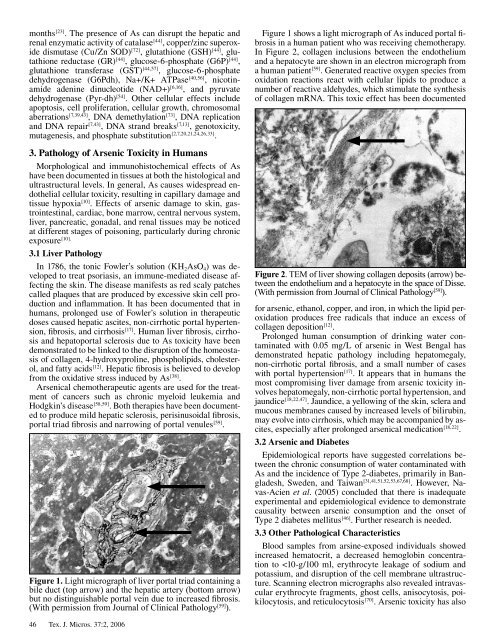
Figure 1 shows a light micrograph <strong>of</strong> As induced portal fibrosis<br />
in a human patient who was receiving chemotherapy.<br />
In Figure 2, collagen inclusions between the endothelium<br />
and a hepatocyte are shown in an electron micrograph from<br />
a human patient [59] . Generated reactive oxygen species from<br />
oxidation reactions react with cellular lipids to produce a<br />
number <strong>of</strong> reactive aldehydes, which stimulate the synthesis<br />
<strong>of</strong> collagen mRNA. This toxic effect has been documented<br />
Figure 2. TEM <strong>of</strong> liver showing collagen deposits (arrow) between<br />
the endothelium and a hepatocyte in the space <strong>of</strong> Disse.<br />
(With permission from <strong>Journal</strong> <strong>of</strong> Clinical Pathology [59] ).<br />
<strong>for</strong> arsenic, ethanol, copper, and iron, in which the lipid peroxidation<br />
produces free radicals that induce an excess <strong>of</strong><br />
collagen deposition [12] .<br />
Prolonged human consumption <strong>of</strong> drinking water contaminated<br />
with 0.05 mg/L <strong>of</strong> arsenic in West Bengal has<br />
demonstrated hepatic pathology including hepatomegaly,<br />
non-cirrhotic portal fibrosis, and a small number <strong>of</strong> cases<br />
with portal hypertension [17] . It appears that in humans the<br />
most compromising liver damage from arsenic toxicity involves<br />
hepatomegaly, non-cirrhotic portal hypertension, and<br />
jaundice [18,22,47] . Jaundice, a yellowing <strong>of</strong> the skin, sclera and<br />
mucous membranes caused by increased levels <strong>of</strong> bilirubin,<br />
may evolve into cirrhosis, which may be accompanied by ascites,<br />
especially after prolonged arsenical medication [18,22] .<br />
3.2 Arsenic and Diabetes<br />
Epidemiological reports have suggested correlations between<br />
the chronic consumption <strong>of</strong> water contaminated with<br />
As and the incidence <strong>of</strong> Type 2-diabetes, primarily in Bangladesh,<br />
Sweden, and Taiwan [31,41,51,52,53,67,68] . However, Navas-Acien<br />
et al. (2005) concluded that there is inadequate<br />
experimental and epidemiological evidence to demonstrate<br />
causality between arsenic consumption and the onset <strong>of</strong><br />
Type 2 diabetes mellitus [46] . Further research is needed.<br />
3.3 Other Pathological Characteristics<br />
Blood samples from arsine-exposed individuals showed<br />
increased hematocrit, a decreased hemoglobin concentration<br />
to