Texas Journal of Microscopy - Texas Society for Microscopy
Texas Journal of Microscopy - Texas Society for Microscopy
Texas Journal of Microscopy - Texas Society for Microscopy

You also want an ePaper? Increase the reach of your titles
YUMPU automatically turns print PDFs into web optimized ePapers that Google loves.
Figure 13. HJA watering yucca seedlings in the Botany Greenhouse on<br />
the ro<strong>of</strong> <strong>of</strong> the Life Science Building in 1957. Photo by Jean Arnott.<br />
<strong>of</strong> instant communication have greatly diminished that aspect<br />
<strong>of</strong> botany. However, now as then, one’s major pr<strong>of</strong>essor can still<br />
make or break any student—the choice <strong>of</strong> a pr<strong>of</strong>essor is not just<br />
important, it is critical!<br />
Much <strong>of</strong> my work on yucca was self-guided. I knew the general<br />
direction I wanted to go and Foster was not one to look over your<br />
shoulder. From time to time I would show Foster what I had been<br />
doing, especially if it was exceptional. Mostly, I went on my own<br />
way pushing the yucca research boundaries in various ways. I<br />
raised seedlings in the Botany Greenhouse on the ro<strong>of</strong> <strong>of</strong> Life<br />
Science Building (Fig. 13). During that time, however, I did some<br />
other research as the opportunity <strong>of</strong>fered itself. I will describe two<br />
cases to illustrate the point. I have already mentioned Cordyline<br />
observations.<br />
By my third year, I was working as Foster’s teaching Assistant<br />
in both Plant Anatomy and Plant Morphology. On occasion, Foster<br />
and I would go together to lunch at The True Blue Cafeteria, which<br />
was in “downtown” Berkeley. The journey involved walking down<br />
from the Life Science Building, through the grove <strong>of</strong> gigantic<br />
Eucalyptus trees, along Strawberry Creek and then out into the<br />
city and to the cafeteria; a total distance <strong>of</strong> ca. 0.3 miles. In those<br />
days, there was no urgency and we would walk along admiring<br />
what ever we came upon. Near the lower edge <strong>of</strong> the Eucalyptus<br />
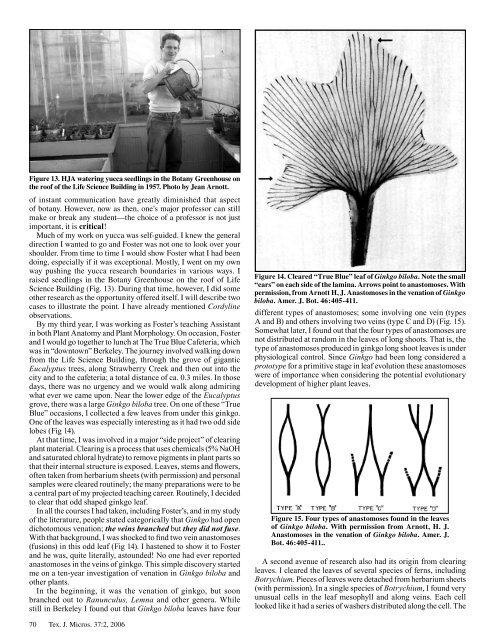
grove, there was a large Ginkgo biloba tree. On one <strong>of</strong> these “True<br />
Blue” occasions, I collected a few leaves from under this ginkgo.<br />
One <strong>of</strong> the leaves was especially interesting as it had two odd side<br />
lobes (Fig 14).<br />
At that time, I was involved in a major “side project” <strong>of</strong> clearing<br />
plant material. Clearing is a process that uses chemicals (5% NaOH<br />
and saturated chloral hydrate) to remove pigments in plant parts so<br />
that their internal structure is exposed. Leaves, stems and flowers,<br />
<strong>of</strong>ten taken from herbarium sheets (with permission) and personal<br />
samples were cleared routinely; the many preparations were to be<br />
a central part <strong>of</strong> my projected teaching career. Routinely, I decided<br />
to clear that odd shaped ginkgo leaf.<br />
In all the courses I had taken, including Foster’s, and in my study<br />
<strong>of</strong> the literature, people stated categorically that Ginkgo had open<br />
dichotomous venation; the veins branched but they did not fuse.<br />
With that background, I was shocked to find two vein anastomoses<br />
(fusions) in this odd leaf (Fig 14). I hastened to show it to Foster<br />
and he was, quite literally, astounded! No one had ever reported<br />
anastomoses in the veins <strong>of</strong> ginkgo. This simple discovery started<br />
me on a ten-year investigation <strong>of</strong> venation in Ginkgo biloba and<br />
other plants.<br />
In the beginning, it was the venation <strong>of</strong> ginkgo, but soon<br />
branched out to Ranunculus, Lemna and other genera. While<br />
still in Berkeley I found out that Ginkgo biloba leaves have four<br />
70 Tex. J. Micros. 37:2, 2006<br />
Figure 14. Cleared “True Blue” leaf <strong>of</strong> Ginkgo biloba. Note the small<br />
“ears” on each side <strong>of</strong> the lamina. Arrows point to anastomoses. With<br />
permission, from Arnott H, J. Anastomoses in the venation <strong>of</strong> Ginkgo<br />
biloba. Amer. J. Bot. 46:405-411.<br />
different types <strong>of</strong> anastomoses; some involving one vein (types<br />
A and B) and others involving two veins (type C and D) (Fig. 15).<br />
Somewhat later, I found out that the four types <strong>of</strong> anastomoses are<br />
not distributed at random in the leaves <strong>of</strong> long shoots. That is, the<br />
type <strong>of</strong> anastomoses produced in ginkgo long shoot leaves is under<br />
physiological control. Since Ginkgo had been long considered a<br />
prototype <strong>for</strong> a primitive stage in leaf evolution these anastomoses<br />
were <strong>of</strong> importance when considering the potential evolutionary<br />
development <strong>of</strong> higher plant leaves.<br />
Figure 15. Four types <strong>of</strong> anastomoses found in the leaves<br />
<strong>of</strong> Ginkgo biloba. With permission from Arnott, H. J.<br />
Anastomoses in the venation <strong>of</strong> Ginkgo biloba. Amer. J.<br />
Bot. 46:405-411..<br />
A second avenue <strong>of</strong> research also had its origin from clearing<br />
leaves. I cleared the leaves <strong>of</strong> several species <strong>of</strong> ferns, including<br />
Botrychium. Pieces <strong>of</strong> leaves were detached from herbarium sheets<br />
(with permission). In a single species <strong>of</strong> Botrychium, I found very<br />
unusual cells in the leaf mesophyll and along veins. Each cell<br />
looked like it had a series <strong>of</strong> washers distributed along the cell. The