Texas Journal of Microscopy - Texas Society for Microscopy
Texas Journal of Microscopy - Texas Society for Microscopy
Texas Journal of Microscopy - Texas Society for Microscopy

Create successful ePaper yourself
Turn your PDF publications into a flip-book with our unique Google optimized e-Paper software.
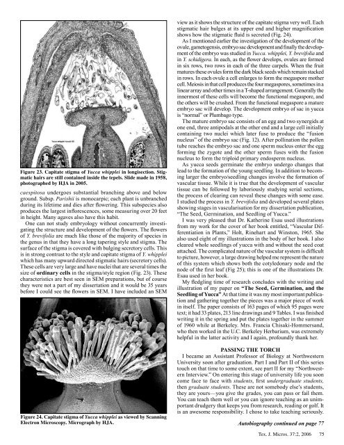
Figure 23. Capitate stigma <strong>of</strong> Yucca whipplei in longisection. Stigmatic<br />
hairs are still contained inside the tepels. Slide made in 1958,<br />
photographed by HJA in 2005.<br />
caespitosa undergoes substantial branching above and below<br />
ground. Subsp. Parishii is monocarpic; each plant is unbranched<br />
during its lifetime and dies after flowering. This subspecies also<br />
produces the largest inflorescences, some measuring over 20 feet<br />
in height. Many agaves also have this habit.<br />
One can not study embryology without concurrently investigating<br />
the structure and development <strong>of</strong> the flowers. The flowers<br />
<strong>of</strong> Y. brevifolia are much like those <strong>of</strong> the majority <strong>of</strong> species in<br />
the genus in that they have a long tapering style and stigma. The<br />
surface <strong>of</strong> the stigma is covered with bulging secretory cells. This<br />
is in strong contrast to the style and capitate stigma <strong>of</strong> Y. whipplei<br />
which has many upward directed stigmatic hairs (secretory cells).<br />
These cells are very large and have nuclei that are several times the<br />
size <strong>of</strong> ordinary cells in the stigma/style region (Fig. 23). These<br />
characteristics are best seen in SEM preparations, but <strong>of</strong> course<br />
they were not a part <strong>of</strong> my dissertation and it would be 35 years<br />
be<strong>for</strong>e I could see the flowers in SEM. I have included an SEM<br />
view as it shows the structure <strong>of</strong> the capitate stigma very well. Each<br />
stigmatic hair bulges at its upper end and higher magnification<br />
shows how the stigmatic fluid is secreted (Fig. 24).<br />
As I mentioned earlier the investigation <strong>of</strong> the development <strong>of</strong> the<br />
ovule, gametogensis, embryo sac development and finally the development<br />
<strong>of</strong> the embryo was studied in Yucca. whipplei, Y. brevifolia and<br />
in Y. schidigera. In each, as the flower develops, ovules are <strong>for</strong>med<br />
in six rows, two rows in each <strong>of</strong> the three carpels. When the fruit<br />
matures these ovules <strong>for</strong>m the dark black seeds which remain stacked<br />
in rows. In each ovule a cell enlarges to <strong>for</strong>m the megaspore mother<br />
cell. Meiosis in that cell produces the four megaspores, sometimes in a<br />
linear array and other times in a T-shaped arrangement. Generally the<br />
innermost <strong>of</strong> these cells will become the functional megaspore, and<br />
the others will be crushed. From the functional megaspore a mature<br />
embryo sac will develop. The development embryo <strong>of</strong> sac in yucca<br />
is “normal” or Plumbago type.<br />
The mature embryo sac consists <strong>of</strong> an egg and two synergids at<br />
one end, three antipodals at the other end and a large cell initially<br />
containing two nuclei which later fuse to produce the “fusion<br />
nucleus” <strong>of</strong> the embryo sac (Fig. 12). After pollination the pollen<br />
tube reaches the embryo sac and one sperm nucleus enter the egg<br />
<strong>for</strong>ming the zygote and the other sperm fuses with the fusion<br />
nucleus to <strong>for</strong>m the triploid primary endosperm nucleus.<br />
As yucca seeds germinate the embryo undergo changes that<br />
lead to the <strong>for</strong>mation <strong>of</strong> the young seedling. In addition to becoming<br />
larger the embryo/seedling changes involve the <strong>for</strong>mation <strong>of</strong><br />
vascular tissue. While it is true that the development <strong>of</strong> vascular<br />
tissue can be followed by laboriously studying serial sections,<br />
the process <strong>of</strong> clearing can reveal these changes with some ease.<br />
I studied the process in Y. brevifolia and developed several plates<br />
showing stages in vascularisation <strong>for</strong> my dissertation publication,<br />
“The Seed, Germination, and Seedling <strong>of</strong> Yucca.”<br />
I was very pleased that Dr. Katherine Esau used illustrations<br />
from my work <strong>for</strong> the cover <strong>of</strong> her book entitled, “Vascular Differentiation<br />
in Plants,” Holt, Rinehart and Winston, 1965. She<br />
also used eight <strong>of</strong> my illustrations in the body <strong>of</strong> her book. I also<br />
cleared whole seedlings <strong>of</strong> yucca with and without the seed coat<br />
attached. The complicated nature <strong>of</strong> the vascular system is difficult<br />
to picture, however, a large drawing helped me represent the nature<br />
<strong>of</strong> this system which shows both the cotyledonary node and the<br />
node <strong>of</strong> the first leaf (Fig 25); this is one <strong>of</strong> the illustrations Dr.<br />
Esau used in her book.<br />
My fledgling time <strong>of</strong> research concludes with the writing and<br />
illustration <strong>of</strong> my paper on “The Seed, Germination, and the<br />
Seedling <strong>of</strong> Yucca” At that time it was my most important publication<br />
and gathering together the pieces was a major piece <strong>of</strong> work<br />
in itself. The paper consists <strong>of</strong> 163 pages <strong>of</strong> which 95 pages were<br />
text; it had 33 plates, 213 line drawings and 9 Tables. I was finished<br />
writing it in the spring and put the plates together in the summer<br />
<strong>of</strong> 1960 while at Berkeley. Mrs. Francia Chisaki-Hommersand,<br />
who then worked in the U.C. Berkeley Herbarium, was extremely<br />
helpful in the latter activity and I again, pr<strong>of</strong>oundly thank her.<br />
PASSinG ThE Torch<br />
I became an Assistant Pr<strong>of</strong>essor <strong>of</strong> Biology at Northwestern<br />
University soon after graduation. Part I and Part II <strong>of</strong> this series<br />
touch on that time to some extent, see part II <strong>for</strong> my “Northwestern<br />
Interview.” On entering this stage <strong>of</strong> university life you soon<br />
come face to face with students, first undergraduate students,<br />
then graduate students. These are not somebody else’s students,<br />
they are yours—you give the grades, you can pass or fail them.<br />
You can teach them well or you can ignore teaching as an unimportant<br />
drudgery that keeps you from research, reading or golf. It<br />
is an awesome responsibility. I chose to take teaching seriously.<br />
Figure 24. Capitate stigma <strong>of</strong> Yucca whipplei as viewed by Scanning<br />
Electron <strong>Microscopy</strong>. Micrograph by HJA. Autobiography continued on page 77<br />
Tex. J. Micros. 37:2, 2006<br />
75