Texas Journal of Microscopy - Texas Society for Microscopy
Texas Journal of Microscopy - Texas Society for Microscopy
Texas Journal of Microscopy - Texas Society for Microscopy

Create successful ePaper yourself
Turn your PDF publications into a flip-book with our unique Google optimized e-Paper software.
X-ray<br />
76 Tex. J. Micros. 37:2, 2006 X-RAY<br />
<br />
ultra<strong>Microscopy</strong><br />
in the SEM<br />
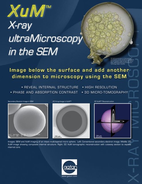
Secondary Electron Image in SEM 2D X-ray Image in XuM<br />
200 µm<br />
3D XuM Reconstruction<br />
Images: SEM and XuM imaging <strong>of</strong> an intact multi-layered micro sphere. Left: Conventional secondary electron image. Middle: 2D<br />
XuM image showing composite internal structure. Right: 3D XuM tomographic reconstruction with cutaway section to expose<br />
internal core.<br />
200 µm<br />
MICROSCOPY<br />
3D XuM reconstruction <strong>of</strong> a Ceramisphere .<br />
Ceramisphere sample provided by Ceramisphere<br />
Pty Ltd. Australia. Image courtesy <strong>of</strong> Dr. Sherry<br />
Mayo, CSIRO Manufacturing & Infrastructure<br />
Technology, Clayton, Australia.<br />
Image below the surface and add another<br />
dimension to microscopy using the SEM<br />
• REVEAL INTERNAL STRUCTURE • HIGH RESOLUTION<br />
• PHASE AND ABSORPTION CONTRAST • 3D MICRO-TOMOGRAPHY<br />
www.gatan.com