A Generic Revision and Phylogenetic Analysis of the Primnoidae
A Generic Revision and Phylogenetic Analysis of the Primnoidae
A Generic Revision and Phylogenetic Analysis of the Primnoidae

Create successful ePaper yourself
Turn your PDF publications into a flip-book with our unique Google optimized e-Paper software.
24 SMITHSONIAN CONTRIBUTIONS TO ZOOLOGY<br />
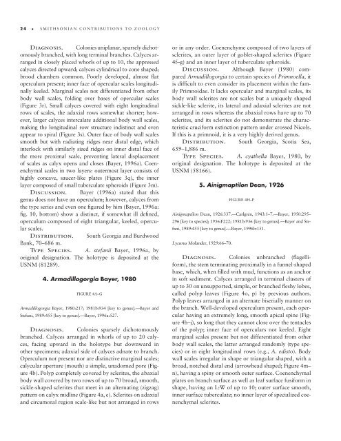
Diagnosis. Colonies uniplanar, sparsely dichotomously<br />
branched, with long terminal branches. Calyces arranged<br />
in closely placed whorls <strong>of</strong> up to 10, <strong>the</strong> appressed<br />
calyces directed upward; calyces cylindrical to cone shaped;<br />
brood chambers common. Poorly developed, almost fl at<br />
operculum present; inner face <strong>of</strong> opercular scales longitudinally<br />
keeled. Marginal scales not differentiated from o<strong>the</strong>r<br />
body wall scales, folding over bases <strong>of</strong> opercular scales<br />
(Figure 3r). Small calyces covered with eight longitudinal<br />
rows <strong>of</strong> scales, <strong>the</strong> adaxial rows somewhat shorter; however,<br />
larger calyces intercalate additional body wall scales,<br />
making <strong>the</strong> longitudinal row structure indistinct <strong>and</strong> even<br />
appear to spiral (Figure 3s). Outer face <strong>of</strong> body wall scales<br />
smooth but with radiating ridges near distal edge, which<br />
interlock with similarly sized ridges on inner distal face <strong>of</strong><br />
<strong>the</strong> more proximal scale, preventing lateral displacement<br />
<strong>of</strong> scales as calyx opens <strong>and</strong> closes (Bayer, 1996a). Coen -<br />
enchymal scales in two layers: outermost layer consists <strong>of</strong><br />
highly concave, saucer-like plates (Figure 3q), <strong>the</strong> inner<br />
layer composed <strong>of</strong> small tuberculate spheroids (Figure 3m).<br />
Discussion. Bayer (1996a) stated that this<br />
genus does not have an operculum; however, calyces from<br />
<strong>the</strong> type series <strong>and</strong> even one fi gured by him (Bayer, 1996a:<br />
fi g. 10, bottom) show a distinct, if somewhat ill defi ned,<br />
operculum composed <strong>of</strong> eight triangular, keeled, opercular<br />
scales.<br />
Distribution. South Georgia <strong>and</strong> Burdwood<br />
Bank, 70–686 m.<br />
Type Species. A. stefanii Bayer, 1996a, by<br />
original designation. The holotype is deposited at <strong>the</strong><br />
USNM (81289).<br />
4. Armadillogorgia Bayer, 1980<br />
FIGURE 4A–G<br />
Armadillogorgia Bayer, 1980:217; 1981b:934 [key to genus].—Bayer <strong>and</strong><br />
Stefani, 1989:455 [key to genus].—Bayer, 1996a:527.<br />
Diagnosis. Colonies sparsely dichotomously<br />
branched. Calyces arranged in whorls <strong>of</strong> up to 20 calyces,<br />
facing upward in <strong>the</strong> holotype but downward in<br />
o<strong>the</strong>r specimens; adaxial side <strong>of</strong> calyces adnate to branch.<br />
Operculum not present nor are distinctive marginal scales;<br />
calycular aperture (mouth) a simple, unadorned pore (Figure<br />
4b). Polyp completely covered by sclerites, <strong>the</strong> abaxial<br />
body wall covered by two rows <strong>of</strong> up to 70 broad, smooth,<br />
sickle-shaped sclerites that meet in an alternating (zigzag)<br />
pattern on calyx midline (Figure 4a, e). Sclerites on adaxial<br />
<strong>and</strong> circumoral region scale-like but not arranged in rows<br />
or in any order. Coenenchyme composed <strong>of</strong> two layers <strong>of</strong><br />
sclerites, an outer layer <strong>of</strong> goblet-shaped sclerites (Figure<br />
4f–g) <strong>and</strong> an inner layer <strong>of</strong> tuberculate spheroids.<br />
Discussion. Although Bayer (1980) compared<br />
Armadillogorgia to certain species <strong>of</strong> Primnoella, it<br />
is diffi cult to even consider its placement within <strong>the</strong> family<br />
<strong>Primnoidae</strong>. It lacks opercular <strong>and</strong> marginal scales, its<br />
body wall sclerites are not scales but a uniquely shaped<br />
sickle-like sclerite, its lateral <strong>and</strong> adaxial sclerites are not<br />
arranged in rows whereas <strong>the</strong> abaxial rows have up to 70<br />
sclerites, <strong>and</strong> its sclerites do not demonstrate <strong>the</strong> characteristic<br />
cruciform extinction pattern under crossed Nicols.<br />
If this is a primnoid, it is a very highly derived genus.<br />
Distribution. South Georgia, Scotia Sea,<br />
659–1,886 m.<br />
Type Species. A. cya<strong>the</strong>lla Bayer, 1980, by<br />
original designation. The holotype is deposited at <strong>the</strong><br />
USNM (58166).<br />
5. Ainigmaptilon Dean, 1926<br />
FIGURE 4H–P<br />
Ainigmaptilon Dean, 1926:337.—Carlgren, 1943:1–7.—Bayer, 1950:295–<br />
296 [key to species]; 1956:F222; 1981b:936 [key to genus].—Bayer <strong>and</strong> Stefani,<br />
1989:455 [key to genus].—Bayer, 1996b:151.<br />
Lycurus Mol<strong>and</strong>er, 1929:66–70.<br />
Diagnosis. Colonies unbranched (fl agelliform),<br />
<strong>the</strong> stem terminating proximally in a funnel-shaped<br />
base, which, when fi lled with mud, functions as an anchor<br />
in s<strong>of</strong>t sediment. Calyces arranged in terminal clusters <strong>of</strong><br />
up to 30 on unsupported, simple, or branched fl eshy lobes,<br />
called polyp leaves (Figure 4o, p) by previous authors.<br />
Polyp leaves arranged in an alternate biserially manner on<br />
<strong>the</strong> branch. Well-developed operculum present, each opercular<br />
having an extremely long, smooth apical spine (Figure<br />
4h–j), so long that <strong>the</strong>y cannot close over <strong>the</strong> tentacles<br />
<strong>of</strong> <strong>the</strong> polyp; inner face <strong>of</strong> operculars not keeled. Eight<br />
marginal scales present but not differentiated from o<strong>the</strong>r<br />
body wall scales, <strong>the</strong> latter arranged r<strong>and</strong>omly (type species)<br />
or in eight longitudinal rows (e.g., A. edisto). Body<br />
wall scales irregular in shape or triangular shaped, with a<br />
broad, notched distal end (arrowhead shaped; Figure 4m–<br />
n), having a spiny or smooth outer surface. Coenenchymal<br />
plates on branch surface as well as leaf surface fusiform in<br />
shape, having an L:W <strong>of</strong> up to 10; outer surface smooth,<br />
inner surface tuberculate; no inner layer <strong>of</strong> specialized coenenchymal<br />
sclerites.