A Generic Revision and Phylogenetic Analysis of the Primnoidae
A Generic Revision and Phylogenetic Analysis of the Primnoidae
A Generic Revision and Phylogenetic Analysis of the Primnoidae

You also want an ePaper? Increase the reach of your titles
YUMPU automatically turns print PDFs into web optimized ePapers that Google loves.
Smithsonian Institution<br />
Scholarly Press<br />
smithsonian contributions to zoology number 629<br />
A Chronology <strong>of</strong><br />
A <strong>Generic</strong> <strong>Revision</strong> <strong>and</strong><br />
Middle Missouri Plains<br />
<strong>Phylogenetic</strong> <strong>Analysis</strong><br />
Village Sites<br />
<strong>of</strong> <strong>the</strong> <strong>Primnoidae</strong><br />
(Cnidaria: Octocorallia)<br />
By Craig M. Johnson<br />
with contributions by<br />
Stephen D. Cairns <strong>and</strong> Frederick M. Bayer<br />
Stanley A. Ahler, Herbert Haas, <strong>and</strong> Georges Bonani
SERIES PUBLICATIONS OF THE SMITHSONIAN INSTITUTION<br />
Emphasis upon publication as a means <strong>of</strong> “diffusing knowledge” was<br />
expressed by <strong>the</strong> fi rst Secretary <strong>of</strong> <strong>the</strong> Smithsonian. In his formal plan<br />
for <strong>the</strong> Institution, Joseph Henry outlined a program that included<br />
<strong>the</strong> following statement: “It is proposed to publish a series <strong>of</strong> reports,<br />
giving an account <strong>of</strong> <strong>the</strong> new discoveries in science, <strong>and</strong> <strong>of</strong> <strong>the</strong> changes<br />
made from year to year in all branches <strong>of</strong> knowledge.” This <strong>the</strong>me <strong>of</strong><br />
basic research has been adhered to through <strong>the</strong> years by thous<strong>and</strong>s <strong>of</strong><br />
titles issued in series publications under <strong>the</strong> Smithsonian imprint, commencing<br />
with Smithsonian Contributions to Knowledge in 1848 <strong>and</strong><br />
continuing with <strong>the</strong> following active series:<br />
Smithsonian Contributions to Anthropology<br />
Smithsonian Contributions to Botany<br />
Smithsonian Contributions in History <strong>and</strong> Technology<br />
Smithsonian Contributions to <strong>the</strong> Marine Sciences<br />
Smithsonian Contributions to Museum Conservation<br />
Smithsonian Contributions to Paleobiology<br />
Smithsonian Contributions to Zoology<br />
In <strong>the</strong>se series, <strong>the</strong> Institution publishes small papers <strong>and</strong> full-scale<br />
monographs that report on <strong>the</strong> research <strong>and</strong> collections <strong>of</strong> its various<br />
museums <strong>and</strong> bureaus. The Smithsonian Contributions Series are<br />
distributed via mailing lists to libraries, universities, <strong>and</strong> similar institutions<br />
throughout <strong>the</strong> world.<br />
Manuscripts submitted for series publication are received by <strong>the</strong><br />
Smithsonian Institution Scholarly Press from authors with direct affi liation<br />
with <strong>the</strong> various Smithsonian museums or bureaus <strong>and</strong> are subject<br />
to peer review <strong>and</strong> review for compliance with manuscript preparation<br />
guidelines. General requirements for manuscript preparation are on <strong>the</strong><br />
inside back cover <strong>of</strong> printed volumes. For detailed submissions requirements<br />
<strong>and</strong> to review <strong>the</strong> “Manuscript Preparation <strong>and</strong> Style Guide for<br />
Authors,” visit <strong>the</strong> Submissions page at www.scholarlypress.si.edu.
smithsonian contributions to zoology number 629<br />
A <strong>Generic</strong> <strong>Revision</strong> <strong>and</strong><br />
<strong>Phylogenetic</strong> <strong>Analysis</strong><br />
<strong>of</strong> <strong>the</strong> <strong>Primnoidae</strong><br />
(Cnidaria: Octocorallia)<br />
Stephen D. Cairns <strong>and</strong> Frederick M. Bayer<br />
WASHINGTON D.C.<br />
2009
ABSTRACT<br />
Cairns, Stephen D., <strong>and</strong> Frederick M. Bayer. A <strong>Generic</strong> <strong>Revision</strong> <strong>and</strong> <strong>Phylogenetic</strong> <strong>Analysis</strong> <strong>of</strong><br />
<strong>the</strong> <strong>Primnoidae</strong> (Cnidaria: Octocorallia). Smithsonian Contributions to Zoology, number 629,<br />
iv + 79 pages, 19 fi gures, 4 tables, 2009.—<strong>Primnoidae</strong> consists <strong>of</strong> 36 genera, 7 subgenera, <strong>and</strong> 233<br />
valid species, making it <strong>the</strong> fourth largest octocorallian family. Species occur in all ocean basins,<br />
especially <strong>the</strong> Antarctic, at depths <strong>of</strong> 8–5850 m, making primnoids <strong>the</strong> deepest-living gorgonacean<br />
octocorals. Primnoids are common <strong>and</strong> characteristic <strong>of</strong> seamounts <strong>and</strong> deepwater coral banks,<br />
<strong>of</strong>ten providing habitat for o<strong>the</strong>r marine life <strong>and</strong> serving as proxies for isotopic analyses to determine<br />
paleotemperatures. Diagnoses <strong>of</strong> <strong>the</strong> primnoid genera <strong>and</strong> subgenera are based primarily<br />
on <strong>the</strong>ir type species, <strong>and</strong> specimens are illustrated by means <strong>of</strong> scanning electron microscopy,<br />
<strong>of</strong>ten using stereo images to allow better appreciation <strong>of</strong> <strong>the</strong> topology <strong>and</strong> interconnection <strong>of</strong> <strong>the</strong><br />
calycular sclerites. A history <strong>of</strong> <strong>the</strong> higher classifi cation <strong>of</strong> <strong>the</strong> family is given. Each genus is briefl y<br />
discussed, <strong>and</strong> also included are a synonymy <strong>of</strong> pertinent references, a summary <strong>of</strong> <strong>the</strong> geographic<br />
<strong>and</strong> bathymetric ranges, <strong>and</strong> <strong>the</strong> deposition <strong>of</strong> <strong>the</strong> type specimens <strong>of</strong> <strong>the</strong> type species. Four new<br />
genera, two new subgenera, one new species, <strong>and</strong> seven new combinations are proposed. A list <strong>of</strong><br />
<strong>the</strong> 233 valid species <strong>and</strong> <strong>the</strong> 14 infraspecifi c taxa is provided along with <strong>the</strong> purported junior synonyms.<br />
An indented dichotomous key is provided for identifi cation <strong>of</strong> <strong>the</strong> genera <strong>and</strong> subgenera.<br />
<strong>Phylogenetic</strong> analysis <strong>of</strong> <strong>the</strong> genera <strong>and</strong> subgenera was performed using 27 morphological characters<br />
comprising 94 character states. The cladogram does not consistently support <strong>the</strong> conventional<br />
arrangement <strong>of</strong> genera into fi ve subfamilies, thus this classifi cation is not followed herein. The<br />
origin <strong>of</strong> <strong>the</strong> primnoids is inferred to be from an ancestor living in <strong>the</strong> Antarctic.<br />
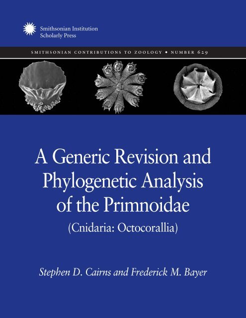
Cover images, left to right: Left side detail from stereo images <strong>of</strong> Figures 8l, 5b, <strong>and</strong> 16h.<br />
Published by Smithsonian Institution Scholarly Press<br />
P.O. Box 37012<br />
MRC 957<br />
Washington, D.C. 20013-7012<br />
www.scholarlypress.si.edu<br />
Library <strong>of</strong> Congress Cataloging-in-Publication Data<br />
Cairns, Stephen D. (Stephen Douglas), 1949–<br />
A generic revision <strong>and</strong> phylogenetic analysis <strong>of</strong> <strong>the</strong> <strong>Primnoidae</strong> (Cnidaria: Octocorallia) /<br />
Stephen D. Cairns <strong>and</strong> Frederick M. Bayer.<br />
p. cm. — (Smithsonian contributions to zoology ; no. 629)<br />
Includes bibliographical references <strong>and</strong> index.<br />
1. <strong>Primnoidae</strong>—Classifi cation. I. Bayer, Frederick M. II. Title.<br />
QL377.C6C336 2009<br />
593.6—dc22 2008043094<br />
∞ The paper used in this publication meets <strong>the</strong> minimum requirements <strong>of</strong> <strong>the</strong> American National<br />
St<strong>and</strong>ard for Permanence <strong>of</strong> Paper for Printed Library Materials Z39.48–1992.
Contents<br />
INTRODUCTION 1<br />
A Brief History <strong>of</strong> <strong>the</strong> <strong>Primnoidae</strong>, with Special Emphasis<br />
on <strong>the</strong> Higher Classifi cation 3<br />
Acknowledgments 5<br />
MATERIAL AND METHODS 5<br />
Material 5<br />
Descriptive Methodology 5<br />
<strong>Phylogenetic</strong> <strong>Analysis</strong> Methodology 5<br />
Remarks on <strong>the</strong> Choice <strong>of</strong> Out-Group <strong>and</strong> Evidence<br />
for Monophyly 5<br />
<strong>Analysis</strong> <strong>of</strong> Primnoid Genera <strong>and</strong> Characters Used 8<br />
RESULTS AND DISCUSSION OF CHARACTER EVOLUTION<br />
AND THE EVOLUTIONARY TREE 16<br />
Tree <strong>Analysis</strong> 16<br />
Character <strong>Analysis</strong> 17<br />
SYSTEMATIC ACCOUNTS 19<br />
Family <strong>Primnoidae</strong> Milne Edwards, 1857 19<br />
Artifi cial Indented Key to <strong>the</strong> Genera <strong>of</strong> <strong>Primnoidae</strong> 19<br />
1. Primnoeides Studer <strong>and</strong> Wright in Studer, 1887 23<br />
2. Ophidiogorgia Bayer, 1980 23<br />
3. Aglaoprimnoa Bayer, 1996 23<br />
4. Armadillogorgia Bayer, 1980 24<br />
5. Ainigmaptilon Dean, 1926 24<br />
6. Primnoella Gray, 1858 25<br />
7. Convexella Bayer, 1996 26<br />
8. Dicholaphis Kinoshita, 1907 26<br />
9. Callozostron Wright, 1885 32<br />
10. Arntzia López-González, Gili <strong>and</strong> Orejas, 2002 33<br />
11. Thouarella (Thouarella) Gray, 1870 33<br />
12. Thouarella (Euthouarella) Kükenthal, 1915 34
13. Thouarella (Diplocalyptra) Kinoshita, 1908 34<br />
14. Thouarella (Epithouarella) Kükenthal, 1915 35<br />
15. Metafannyella, new genus. 35<br />
16. Fannyella (Fannyella) Gray, 1872 35<br />
17. Fannyella (Scyphogorgia), new subgenus 36<br />
18. Fannyella (Cyathogorgia), new subgenus 36<br />
19. Onogorgia, new genus 37<br />
20. Pyrogorgia, new genus 37<br />
21. Amphilaphis Studer <strong>and</strong> Wright in Studer, 1887 38<br />
22. Mirostenella Bayer, 1988 38<br />
23. Acanthoprimnoa Cairns <strong>and</strong> Bayer, 2004 39<br />
24. Plumarella Gray, 1870 39<br />
25. Callogorgia Gray, 1858 40<br />
26. Fanellia Gray, 1870 40<br />
27. Paranarella Cairns, 2007 41<br />
28. Primnoa Lamouroux, 1812 41<br />
29. Australogorgia, new genus 42<br />
Australogorgia aldersladei, new species 42<br />
30. Narella Gray, 1870 43<br />
31. Arthrogorgia Kükenthal, 1908 43<br />
32. Paracalyptrophora Kinoshita, 1908 44<br />
33. Calyptrophora Gray, 1866 44<br />
34. Tokoprymno Bayer, 1996 45<br />
35. Parastenella Versluys, 1906 45<br />
36. C<strong>and</strong>idella Bayer, 1954 46<br />
37. Microprimnoa Bayer <strong>and</strong> Stefani, 1989 46<br />
38. Pterostenella Versluys, 1906 47<br />
39. Perissogorgia Bayer <strong>and</strong> Stefani, 1989 47<br />
40. Dasystenella Versluys, 1906 47<br />
41. Pseudoplumarella Kükenthal, 1915 48<br />
FIGURES 3–19 49<br />
REFERENCES 67<br />
INDEX 73
A <strong>Generic</strong> <strong>Revision</strong> <strong>and</strong> <strong>Phylogenetic</strong><br />
<strong>Analysis</strong> <strong>of</strong> <strong>the</strong> <strong>Primnoidae</strong><br />
(Cnidaria: Octocorallia)<br />
Stephen D. Cairns <strong>and</strong> † Frederick M. Bayer,<br />
Smithsonian Institution, Department <strong>of</strong> Invertebrate<br />
Zoology, P.O. Box 27012, MRC 163,<br />
Washington, D.C. 20013-7012, USA. Manuscript<br />
received 7 March 2008; accepted 25 June 2008.<br />
† Deceased 2 October 2007.<br />
INTRODUCTION<br />
Among <strong>the</strong> 44 families <strong>of</strong> Octocorallia (Williams <strong>and</strong> Cairns, 2005, updated),<br />
<strong>the</strong> <strong>Primnoidae</strong> ranks fourth in number <strong>of</strong> species (233/3200<br />
= 7.3%, see Tables 1, 4) <strong>and</strong> third in number <strong>of</strong> genera (36/340 =<br />
10.6%), resulting in an average <strong>of</strong> 6.5 species per genus. No confi<br />
rmed fossil species are known. Primnoids occur worldwide at depths <strong>of</strong> 8–<br />
5,850 m, although <strong>the</strong>y are most common at slope <strong>and</strong> upper abyssal depths<br />
(Table 1), <strong>the</strong> few shallow records being uncommon. The primnoids thus may<br />
be considered to be <strong>the</strong> quintessential deepwater octocoral family, only some<br />
pennatulaceans occurring in deeper water. It is tempting to speculate that <strong>the</strong><br />
modifi cation <strong>of</strong> <strong>the</strong>ir imbricate external calycular scales is <strong>the</strong> key adaptation<br />
for <strong>the</strong>ir success in deep water; however, two isidid subfamilies, many <strong>of</strong> which<br />
occur at shelf depths, also have very similar sclerite morphology.<br />
Aside from <strong>the</strong> innate taxonomic interest <strong>of</strong> <strong>the</strong> family <strong>and</strong> <strong>the</strong> beauty <strong>of</strong><br />
<strong>the</strong>ir calycular architecture, primnoids serve important ecological <strong>and</strong> geophysical<br />
roles. Because <strong>of</strong> <strong>the</strong>ir large size (e.g., Primnoa occurs up to 2 m in height<br />
<strong>and</strong> 7 m wide) <strong>and</strong> local abundance (sometimes occurring in large monospecifi<br />
c fi elds), some primnoids, such as Primnoa, Narella, <strong>and</strong> Callogorgia, form<br />
habitat for fi sh (Etnoyer <strong>and</strong> Morgan, 2005; Stone, 2006; Etnoyer <strong>and</strong> Warrick,<br />
2007) <strong>and</strong> o<strong>the</strong>r invertebrates (Krieger <strong>and</strong> Wing, 2002; Buhl-Mortensen<br />
<strong>and</strong> Mortensen, 2004, 2005), especially on seamounts (Cairns <strong>and</strong> Baco, 2007;<br />
Rogers et al., 2007; Cairns <strong>and</strong> Bayer, 2007 [2008]), <strong>and</strong> on deepwater coral<br />
banks (Cairns <strong>and</strong> Bayer, 2005). Also, because <strong>of</strong> <strong>the</strong>ir solid, layered axis <strong>and</strong><br />
purported long life span (Andrews et al., 2002; Risk et al., 2002), various isotopic<br />
analyses <strong>of</strong> <strong>the</strong>ir axes can be used to determine paleotemperatures (Heikoop<br />
et al., 2002; Sinclair et al., 2005; Sherwood et al., 2005).<br />
The following abbreviations are used in <strong>the</strong> text.<br />
Museums<br />
AM Australian Museum, Sydney<br />
BM The Natural History Museum, London<br />
MNHNP Muséum National d’Histoire Naturelle, Paris<br />
NTM Nor<strong>the</strong>rn Territories Museum, Darwin
2 SMITHSONIAN CONTRIBUTIONS TO ZOOLOGY<br />
TABLE 1. Distributional characteristics <strong>of</strong> <strong>the</strong> primnoid genera <strong>and</strong> subgenera. The number <strong>of</strong> valid species plus subtaxa may include<br />
subspecies, varieties, <strong>and</strong>/or forms (see Table 4 for complete list).<br />
Number <strong>of</strong><br />
Genus Distribution Depth Range (m) species + subtaxa<br />
Primnoeides Sou<strong>the</strong>rn Indian Ocean 400–558 1<br />
Ophidiogorgia Antarctica 27–426 2<br />
Aglaoprimnoa Subantarctic South America 70–686 1<br />
Armadillogorgia South Georgia, Scotia Sea 659–1,886 1<br />
Ainigmaptilon Antarctica to South Georgia 75–550 5<br />
Primnoella Western Atlantic, Australia, New Zeal<strong>and</strong> 8–1,249 10<br />
Convexella Antarctic, Kermadec, North Atlantic 12–5,850 5<br />
Dicholaphis Off Japan 731 1<br />
Callozostron Antarctic, New Zeal<strong>and</strong> 1,354–3,876 4<br />
Arntzia<br />
Thouarella<br />
Antarctic 64–604 1<br />
T. (Thouarella) Subantarctic, western Atlantic, North Pacifi c 60–1,005 14 + 2<br />
T. (Euthouarella) Indo–western Pacifi c, North Atlantic 256–1,644 10 + 1<br />
T. (Diplocalyptra) Off Japan 146 2<br />
T. (Epithouarella) Antarctic, subantarctic 106–686 3<br />
Metafannyella<br />
Fannyella<br />
Antarctic 265–1,280 4<br />
F. (Fannyella) Antarctic 46–852 2<br />
F. (Scyphogorgia) Antarctic 100–550 1<br />
F. (Cyathogorgia) Antarctic 55–485 1<br />
Onogorgia Antarctic 22–433 1<br />
Pyrogorgia Tierra del Fuego 384–511 1<br />
Amphilaphis Subantarctic, Galapagos, Antarctic, Hawaii 55–3,182 6<br />
Mirostenella Subantarctic 201–1,647 2<br />
Acanthoprimnoa Caribbean, Japan 45–686 4 + 1<br />
Plumarella Western Pacifi c, Patagonia, northwest Atlantic 10–1,914 21 + 4<br />
Callogorgia Indo-Pacifi c, North Atlantic 37–2,472 25 + 3<br />
Fanellia Western, central, <strong>and</strong> nor<strong>the</strong>rn Pacifi c 92–1,028 8<br />
Paranarella Northwest Atlantic 3,855 1<br />
Primnoa North Atlantic, North Pacifi c, subantarctic 9–1,020 4 + 1<br />
Australogorgia Off Tasmania 987 1<br />
Narella Cosmopolitan 55–4,594 38<br />
Arthrogorgia North Pacifi c 163–1,127 4<br />
Paracalyptrophora Western <strong>and</strong> central Pacifi c, North Atlantic 150–1,480 6<br />
Calyptrophora Pacifi c, western Atlantic 229–3,107 14 + 2<br />
Tokoprymno Subantarctic 549 1<br />
Parastenella Cosmopolitan , except east Atlantic 567–3,470 6<br />
C<strong>and</strong>idella Atlantic, central Pacifi c 378–2,165 4<br />
Microprimnoa New Caledonia 415 1<br />
Pterostenella Indo–western Pacifi c 60–75 2<br />
Perissogorgia New Caledonia 55–750 7<br />
Dasystenella Subantarctic South Atlantic 300–5,087 1<br />
Pseudoplumarella Eastern Australia 55–115 5<br />
Totals 8–5,850 233 + 14
NMNH National Museum <strong>of</strong> Natural History,<br />
Smithsonian, Washington, D.C.<br />
USNM United States National Museum (now <strong>the</strong><br />
NMNH)<br />
ZMA Zöologisch Museum, Amsterdam<br />
ZMB Zoologisches Museum, Berlin<br />
O<strong>the</strong>r Terms<br />
CI Consistency index<br />
Coel. Coelenterata (term prefaces catalog numbers<br />
associated with ZMA)<br />
L:W Ratio <strong>of</strong> length to width <strong>of</strong> a sclerite<br />
PAUP* <strong>Phylogenetic</strong> <strong>Analysis</strong> Using Parsimony<br />
RI Rescaled consistency index<br />
SEM Scanning electron microscopy<br />
A BRIEF HISTORY OF THE PRIMNOIDAE, WITH SPECIAL<br />
EMPHASIS ON THE HIGHER CLASSIFICATION<br />
The oldest described primnoid species was Gorgonia<br />
resedaeformis Gunnerus, 1763 (=Primnoa resedaeformis),<br />
whereas <strong>the</strong> earliest described primnoid genus was Primnoa<br />
Lamouroux, 1816.<br />
Although many have cited Gray (1858) as <strong>the</strong> author<br />
<strong>of</strong> <strong>the</strong> family <strong>Primnoidae</strong>, some even citing this work as<br />
published in 1857, his paper was, in fact, published on<br />
23 February 1858. However, one year earlier, Milne Edwards<br />
(1857:138) introduced <strong>the</strong> term Primnoacées to<br />
include two genera (Primnoa <strong>and</strong> Muricea). Primnoacées<br />
was termed an agèle by Milne Edwards, a category between<br />
subfamily <strong>and</strong> genus, thus ei<strong>the</strong>r a tribe or supergenus.<br />
We interpret this taxonomic level to be a tribe <strong>and</strong><br />
thus among <strong>the</strong> family group names, making it <strong>the</strong> earliest<br />
available name for <strong>the</strong> family <strong>Primnoidae</strong>. Gray (1858) referred<br />
to <strong>the</strong> family as Primnoadae (spelling later corrected<br />
to <strong>Primnoidae</strong> by Verrill, 1868) <strong>and</strong> included three genera:<br />
<strong>the</strong> type genus Primnoa <strong>and</strong> Callogorgia <strong>and</strong> Primnoella.<br />
Later, Gray (1870) included 13 genera in <strong>the</strong> family, many<br />
<strong>of</strong> which are now considered to be in different families,<br />
<strong>and</strong> established <strong>the</strong> family “Calyptrophoradae” for one<br />
genus <strong>and</strong> “Calligorgiadae” for seven genera now assigned<br />
to <strong>the</strong> <strong>Primnoidae</strong> <strong>and</strong> Ellisellidae. Verrill (1883) correctly<br />
eliminated some <strong>of</strong> <strong>the</strong> nonprimnoid genera from <strong>the</strong> family.<br />
In his comprehensive classifi cation <strong>of</strong> <strong>the</strong> Alcyonaria,<br />
Studer (1887) placed 12 genera in <strong>the</strong> family, all <strong>of</strong> which<br />
are still considered primnoids, <strong>and</strong> divided <strong>the</strong> genera into<br />
four subfamilies (Callozostrinae, Calyptrophorinae, Primnoinae,<br />
<strong>and</strong> Primnoeidinae) but did not <strong>of</strong>fer justifi cation<br />
for <strong>the</strong> subfamilies. The classifi cation <strong>of</strong> <strong>the</strong> 14 primnoid<br />
number 629 3<br />
genera in Wright <strong>and</strong> Studer’s (1889) Challenger Expedition<br />
report followed that <strong>of</strong> Studer (1887) but also included<br />
diagnoses <strong>of</strong> <strong>the</strong> four subfamilies: Callozostrinae<br />
was characterized by having a fl exible axis; Calyptrophorinae<br />
had a rigid axis <strong>and</strong> a small, fi xed number <strong>of</strong> annular<br />
body wall scales; Primnoinae had a rigid axis <strong>and</strong> a larger,<br />
variable number <strong>of</strong> body wall scales; <strong>and</strong> Primnoeidinae<br />
had a rigid axis but lacked an operculum. Except for Versluys<br />
(1906) <strong>and</strong> Kinoshita (see below), this subfamilial<br />
arrangement has been followed until recent times.<br />
Versluys (1906), in his beautifully illustrated revision<br />
<strong>of</strong> <strong>the</strong> Siboga (1902) primnoids, provided <strong>the</strong> most comprehensive<br />
exploration <strong>of</strong> this family to date. He provided<br />
extensive discussion about most <strong>of</strong> <strong>the</strong> characters used to<br />
differentiate primnoid genera <strong>and</strong> even suggested a preliminary<br />
evolutionary tree <strong>of</strong> <strong>the</strong> genera, creating a hypo<strong>the</strong>tical<br />
ancestral form (“Stammform”), which he called<br />
Proprimnoa, <strong>and</strong> <strong>the</strong>n choosing Primnoeides as <strong>the</strong> closest<br />
genus to <strong>the</strong> ancestral form. The ancestral form, as well<br />
as Primnoeides, was characterized by having all sclerites,<br />
both coenenchymal <strong>and</strong> body wall, <strong>of</strong> <strong>the</strong> same shape; a<br />
very poorly developed operculum; body wall scales not arranged<br />
in rows (except in <strong>the</strong> juvenile form); uniplanar pinnate<br />
colonies; calyces arranged in whorls <strong>of</strong> two to four;<br />
<strong>and</strong> two layers <strong>of</strong> coenenchymal sclerites. He concluded by<br />
listing <strong>the</strong> 67 known species <strong>and</strong> 11 known genera in <strong>the</strong><br />
family. Although Versluys followed Studer’s subfamilial<br />
classifi cation for <strong>the</strong> descriptive part <strong>of</strong> his paper, saying<br />
that he did not want to cause confusion by deviating from<br />
previous classifi cations (Versluys, 1906:158), in <strong>the</strong> second<br />
part <strong>of</strong> his paper, titled “Comparative <strong>Phylogenetic</strong><br />
Section,” he none<strong>the</strong>less proposed a different organization<br />
<strong>of</strong> <strong>the</strong> genera into fi ve subfamilies, two <strong>of</strong> <strong>the</strong>m new (see<br />
below).<br />
Subfamily 1: Primnoeidinae (Primnoeides): operculum<br />
poorly developed, body wall scales not in<br />
well- arranged rows.<br />
Subfamily 2: Primnoellinae, new subfamily (Primnoella):<br />
operculum <strong>and</strong> circumoperculum developed,<br />
adaxial side <strong>of</strong> body wall exposed.<br />
Subfamily 3: Thouarellinae, new subfamily (Thouarella,<br />
Amphilaphis, Plumarella, Stenella): eight<br />
rows <strong>of</strong> body wall scales but <strong>of</strong>ten indistinct <strong>and</strong><br />
reduced in size near base, none<strong>the</strong>less, body wall<br />
completely covered, operculum well developed.<br />
Subfamily 4: Primnoinae (Primnoa, Callogorgia,<br />
Stachyodes, <strong>and</strong> Calyptrophora): operculum present<br />
but no circumoperculum, body wall scales<br />
greatly reduced in number.
4 SMITHSONIAN CONTRIBUTIONS TO ZOOLOGY<br />
Subfamily 5: Callozostroninae (Callozostron): axis<br />
fl exible, eight rows <strong>of</strong> body wall scales, operculum<br />
present.<br />
Kinoshita was also very active at this time, publishing<br />
seven papers (Kinoshita, 1907, 1908a, 1908b, 1908c,<br />
1908d, 1908e, 1909) on <strong>the</strong> primnoids <strong>of</strong> Japan, as well as<br />
discussing general characteristics <strong>of</strong> <strong>the</strong> family. He was <strong>the</strong><br />
only author to adopt Versluys’ subfamilial organization <strong>of</strong><br />
<strong>the</strong> genera, but most <strong>of</strong> his papers were in Japanese, which<br />
limited <strong>the</strong>ir use to many taxonomists. English translations<br />
are available at <strong>the</strong> Smithsonian. At <strong>the</strong> conclusion<br />
<strong>of</strong> this series <strong>of</strong> papers, Kinoshita (1909) tabulated 118<br />
species in 13 genera. Much later, Utinomi (1979) resumed<br />
<strong>the</strong> tradition <strong>of</strong> research on Japanese primnoids.<br />
Nutting was also active in working on <strong>the</strong> primnoids<br />
collected by <strong>the</strong> USFWS Albatross in Pacifi c waters, reporting<br />
new primnoid species from <strong>the</strong> Hawaiian Isl<strong>and</strong>s (Nutting,<br />
1908), <strong>of</strong>f California (Nutting ,1909), <strong>and</strong> <strong>of</strong>f Japan<br />
<strong>and</strong> <strong>the</strong> Aleutian Isl<strong>and</strong>s (Nutting, 1912). And Thomson<br />
<strong>and</strong> Mackinnon (1911) reported 11 primnoid species (7<br />
new) from <strong>of</strong>f New South Wales, Australia, collected by<br />
<strong>the</strong> Thetis.<br />
Willi Kükenthal, undoubtedly <strong>the</strong> most prolifi c octocoral<br />
researcher <strong>of</strong> all time, published four papers on <strong>the</strong><br />
primnoids <strong>of</strong> Japan (Kükenthal, 1907, 1908; Kükenthal<br />
<strong>and</strong> Gorzawsky, 1908a, 1908b); he (Kükenthal, 1912)<br />
also reported <strong>the</strong> primnoids <strong>of</strong> <strong>the</strong> German South Polar<br />
Expedition <strong>of</strong> 1901–1903, making <strong>the</strong> six-year period <strong>of</strong><br />
1907–1912 an incredibly active one for <strong>the</strong> study <strong>of</strong> this<br />
family! Kükenthal (1915) later revised <strong>the</strong> family, providing<br />
keys to <strong>the</strong> genera <strong>and</strong> all 126 known species at that<br />
time. Perhaps on <strong>the</strong> basis <strong>of</strong> this rash <strong>of</strong> papers, Kükenthal<br />
(1915:142) stated that <strong>Primnoidae</strong> may be <strong>the</strong> most<br />
thoroughly investigated family in <strong>the</strong> order Gorgonacea, a<br />
perception that holds true even today.<br />
In his monumental work on <strong>the</strong> gorgonians <strong>of</strong> <strong>the</strong><br />
German deep-sea expedition <strong>of</strong> <strong>the</strong> Valdivia (1898–1899),<br />
Kükenthal (1919) devoted 185 pages to a revision <strong>of</strong> <strong>the</strong><br />
family <strong>Primnoidae</strong>, including descriptions <strong>of</strong> all species <strong>and</strong><br />
dichotomous as well as indented keys to all genera <strong>and</strong> <strong>the</strong><br />
138 species. His treatment <strong>of</strong> <strong>the</strong> <strong>Primnoidae</strong> in his l<strong>and</strong>mark<br />
revision <strong>of</strong> Gorgonaria (Kükenthal, 1924) is largely<br />
derivative <strong>of</strong> this earlier 1919 work. In general, Kükenthal<br />
followed <strong>the</strong> same higher classifi cation <strong>of</strong> <strong>the</strong> primnoid<br />
genera <strong>and</strong> subfamilies as suggested by Studer (1887).<br />
Kükenthal (1919) also rigorously analyzed Versluys’ phylogenetic<br />
analysis <strong>of</strong> <strong>the</strong> genera, strongly disagreeing with<br />
<strong>the</strong> interpretation <strong>of</strong> many <strong>of</strong> <strong>the</strong> characters he used (see<br />
below). For instance, although Kükenthal agreed with Versluys<br />
that two layers <strong>of</strong> coenenchymal sclerites <strong>and</strong> similar<br />
types <strong>of</strong> sclerites in <strong>the</strong> coenenchyme <strong>and</strong> calyx were primitive<br />
states, contrary to Versluys, he thought that calyces<br />
arranged in whorls, body wall scales not in rows, a rudimentary<br />
operculum, <strong>and</strong> unbranched colonies were derived<br />
states ra<strong>the</strong>r than primitive. He implied that Versluys did<br />
not give adequate explanation for his choice <strong>of</strong> ancestral<br />
states <strong>and</strong> <strong>the</strong>n went on to didactically list what he thought<br />
were <strong>the</strong> primitive states. Ironically, after this ra<strong>the</strong>r harsh<br />
criticism, Kükenthal agreed with Versluys that Primnoeides<br />
was <strong>the</strong> most primitive <strong>of</strong> <strong>the</strong> primnoid genera.<br />
O<strong>the</strong>r expeditionary reports that included <strong>the</strong> description<br />
<strong>of</strong> deepwater primnoids include Thomson <strong>and</strong> Richie<br />
(1906), <strong>the</strong> Scottish National Antarctic Expedition <strong>of</strong><br />
1903; Mol<strong>and</strong>er (1929), <strong>the</strong> Swedish Antarctic Expedition<br />
<strong>of</strong> 1901–1903; Aurivillius (1931), <strong>the</strong> gorgonians <strong>of</strong> Sixten<br />
Bock’s expedition to <strong>the</strong> Bonin Isl<strong>and</strong>s in 1914; Thomson<br />
<strong>and</strong> Rennet (1931), <strong>the</strong> Australasian Antarctic Expedition<br />
<strong>of</strong> 1911–1914; Deichmann (1936), <strong>the</strong> Blake expeditions<br />
in <strong>the</strong> western Atlantic; <strong>and</strong> Madsen (1944), <strong>the</strong> Danish<br />
Ingolf Expedition in <strong>the</strong> boreal North Atlantic.<br />
Subsequent to 1950, <strong>the</strong> major contributions to primnoid<br />
taxonomy were made by F. M. Bayer <strong>and</strong> coauthors<br />
(1950–2004, see References), <strong>and</strong> <strong>the</strong>y tend to concentrate<br />
on taxa from <strong>the</strong> western Atlantic <strong>and</strong> Antarctic. Most notable<br />
was his revision <strong>of</strong> <strong>the</strong> octocoral genera in <strong>the</strong> Treatise<br />
on Invertebrate Paleontology (Bayer, 1956), in which<br />
all primnoid genera were diagnosed <strong>and</strong> placed in four<br />
subfamilies, Ainigmaptilon being placed in a family <strong>of</strong><br />
its own. The four subfamilies were distinguished on only<br />
three characters: Primnoeidinae (one genus) had a poorly<br />
developed operculum, body wall scales not arranged in<br />
rows, <strong>and</strong> polyps that inclined toward <strong>the</strong> branch axis;<br />
Primnoinae (six genera) had a well-developed operculum,<br />
eight rows <strong>of</strong> body wall scales, <strong>and</strong> inclined polyps; Calyptrophorinae<br />
(three genera) had a well-developed operculum,<br />
two rows <strong>of</strong> body wall scales that wrapped around<br />
<strong>the</strong> polyp, <strong>and</strong> inclined polyps; <strong>and</strong> Callozostrinae (three<br />
genera) had a well-developed operculum, fi ve to eight rows<br />
<strong>of</strong> body wall scales, <strong>and</strong> polyps that stood perpendicular to<br />
<strong>the</strong> branch. Bayer (1956) was <strong>the</strong> last published subfamilial<br />
listing <strong>of</strong> <strong>the</strong> primnoid genera <strong>and</strong> thus <strong>the</strong> model that<br />
will be compared in <strong>the</strong> phylogenetic analysis, although<br />
not much evolved since <strong>the</strong> original model proposed by<br />
Studer (1887). Bayer (1961) also published <strong>the</strong> fi rst <strong>of</strong> his<br />
keys to <strong>the</strong> primnoid genera, this one an illustrated dichotomous<br />
key. His second key to <strong>the</strong> primnoid genera (Bayer,<br />
1981b) was part <strong>of</strong> a larger key to all octocoral genera.<br />
His third key (Bayer <strong>and</strong> Stefani, 1989) was in French <strong>and</strong><br />
was published in <strong>the</strong> context <strong>of</strong> <strong>the</strong> description <strong>of</strong> new<br />
primnoid species <strong>and</strong> genera from New Caledonia. Sig-
nifi cant morphological <strong>and</strong> historical remarks on <strong>the</strong> family<br />
are also made in Bayer (1982, 1988). A series <strong>of</strong> seven<br />
papers were published by Cairns <strong>and</strong> Bayer (2002, 2003,<br />
2004b, 2004c, 2005) <strong>and</strong> Cairns (2006, 2007a) that revise<br />
<strong>the</strong> primnoid species from <strong>the</strong> western Atlantic.<br />
Finally, a new primnoid genus was described by<br />
López-González et al. (2002) from <strong>the</strong> Sou<strong>the</strong>rn Ocean.<br />
A total <strong>of</strong> 233 valid primnoid species are now known<br />
(Table 4).<br />
ACKNOWLEDGMENTS<br />
We thank <strong>the</strong> following people, who loaned us specimens<br />
used in this study: Susanne Horner (Nor<strong>the</strong>rn Territories<br />
Museum, Darwin), for <strong>the</strong> type <strong>of</strong> Australogorgia<br />
<strong>and</strong> comparative circinisidinine samples, <strong>and</strong> Stephen Kaeble<br />
(Australian Museum, Sydney), for <strong>the</strong> types <strong>of</strong> Pseudoplumarella<br />
<strong>the</strong>tis <strong>and</strong> Dicholaphis delicatula. We are also<br />
most grateful to Phil Alderslade (retired) for discussions<br />
about potential out-groups <strong>and</strong> primnoid classifi cation, as<br />
well as for loaning us additional specimens <strong>of</strong> Australogorgia.<br />
Allen Collins (NOAA) was invaluable in helping with<br />
<strong>the</strong> phylogenetic analyses. Molly Ryan (Smithsonian Institution)<br />
drafted <strong>the</strong> phylogenetic trees (Figures 1, 2), Tim<br />
C<strong>of</strong>fer (Smithsonian Institution) composed <strong>the</strong> Photoshop<br />
plates, <strong>and</strong> Rosemarie Baron-Szabo (freelance translator)<br />
helped with German translation. Most <strong>of</strong> <strong>the</strong> SEM images<br />
were taken in <strong>the</strong> SEM Laboratory <strong>of</strong> <strong>the</strong> NMNH by <strong>the</strong><br />
second author over a period <strong>of</strong> 25 years.<br />
MATERIAL AND METHODS<br />
MATERIAL<br />
This study was based primarily on <strong>the</strong> collections <strong>of</strong><br />
<strong>the</strong> NMNH, which currently must be considered <strong>the</strong> most<br />
comprehensive in <strong>the</strong> world for this family. Of <strong>the</strong> 41<br />
higher taxa included in <strong>the</strong> revision, <strong>the</strong> NMNH contains<br />
representatives <strong>of</strong> <strong>the</strong> type species <strong>of</strong> 39 <strong>of</strong> <strong>the</strong>m, <strong>and</strong> 15<br />
<strong>of</strong> those 39 are represented by <strong>the</strong> type specimens <strong>of</strong> <strong>the</strong><br />
type species. The type <strong>of</strong> Pseudoplumarella (P. <strong>the</strong>tis) was<br />
borrowed from <strong>the</strong> AM. No original material <strong>of</strong> Dicholaphis<br />
Kinoshita, 1907 could be obtained; thus, that taxon<br />
is described <strong>and</strong> fi gured from <strong>the</strong> literature. Although emphasis<br />
was given to <strong>the</strong> type species in defi ning <strong>the</strong> genera<br />
<strong>and</strong> subgenera, as many species as possible were examined<br />
within each taxon to fully describe its variation <strong>and</strong> to<br />
properly code for phylogenetic analysis. Representatives<br />
<strong>of</strong> <strong>the</strong> out-group were obtained from <strong>the</strong> NTM.<br />
number 629 5<br />
Eogorgia Hickson, 1938, described from <strong>the</strong> Eocene<br />
<strong>of</strong> Mississippi, was tentatively placed in <strong>the</strong> <strong>Primnoidae</strong><br />
by Bayer (1956) solely on <strong>the</strong> basis <strong>of</strong> its size. Hickson<br />
had considered it to be a scleraxonian. On <strong>the</strong> basis <strong>of</strong><br />
its spindle- <strong>and</strong> needle-shaped sclerites <strong>and</strong> general lack<br />
<strong>of</strong> any o<strong>the</strong>r information, it is not considered to be a<br />
primnoid in this revision. Two putative fossil species <strong>of</strong><br />
Primnoa are listed in Table 4 but are considered highly<br />
doubtful for <strong>the</strong> family.<br />
DESCRIPTIVE METHODOLOGY<br />
Genera are arranged in an order <strong>of</strong> roughly less to more<br />
derived as determined from <strong>the</strong> cladogram. The generic synonymies<br />
include <strong>the</strong> original descriptions as well as o<strong>the</strong>r<br />
references that include useful descriptive or illustrative information,<br />
with special mention <strong>of</strong> any keys to <strong>the</strong> generic<br />
or specifi c levels. <strong>Generic</strong> diagnoses are telegraphic in style<br />
<strong>and</strong> consistent in order <strong>of</strong> characters to facilitate comparison<br />
among genera. The deposition <strong>of</strong> <strong>the</strong> type specimens <strong>of</strong><br />
<strong>the</strong> type species are given when known. The terminology<br />
for <strong>the</strong> calyces <strong>and</strong> sclerites follows that developed by Versluys<br />
(1906) as modifi ed by Bayer et al. (1983).<br />
With primnoids, it is important to be able to distinguish<br />
<strong>and</strong> count <strong>the</strong> scales on various rows on a calyx,<br />
especially those on <strong>the</strong> adaxial side. This requires careful<br />
dissection <strong>of</strong> a calyx from <strong>the</strong> branch in order to see <strong>the</strong><br />
inner (=adaxial) face. Distinguishing scale rows is most<br />
easily accomplished by mounting entire calyces for SEM<br />
photography, stereo views being most helpful in viewing<br />
scales, especially those <strong>of</strong> <strong>the</strong> operculum <strong>and</strong> marginal<br />
scales, which are sometimes similar in shape <strong>and</strong> size.<br />
However, if an SEM view is not practical, one may allow<br />
<strong>the</strong> calyx to dry, mount it on a small piece <strong>of</strong> clay on a<br />
glass slide (for stability), <strong>and</strong> <strong>the</strong>n progressively touch <strong>the</strong><br />
calyx with a fi ne-tipped colored marker. The ink <strong>of</strong> <strong>the</strong><br />
marker will provide excellent contrast, allowing one to see<br />
<strong>the</strong> outlines <strong>and</strong> texture <strong>of</strong> <strong>the</strong> scales. One can even mark<br />
specifi c scales prior to dissolution by bleach <strong>and</strong> thus trace<br />
specifi c scales for later description.<br />
PHYLOGENETIC ANALYSIS METHODOLOGY<br />
Remarks on <strong>the</strong> Choice <strong>of</strong> Out-Group<br />
<strong>and</strong> Evidence for Monophyly<br />
The <strong>Primnoidae</strong> is one <strong>of</strong> fi ve gorgonacean families<br />
constituting <strong>the</strong> suborder Calcaxonia, a suborder characterized<br />
by having a solid (nonloculated) axis composed <strong>of</strong><br />
calcium carbonate that is embedded in a gorgonin matrix,
6 SMITHSONIAN CONTRIBUTIONS TO ZOOLOGY<br />
<strong>the</strong> latter giving <strong>the</strong> branch a variable degree <strong>of</strong> fl exibility.<br />
Two <strong>of</strong> <strong>the</strong> o<strong>the</strong>r four calcaxonian families, <strong>the</strong> Ellisellidae<br />
<strong>and</strong> Ifalukellidae, have very different sclerite compositions<br />
(i.e., spindles, dumbbells, <strong>and</strong> small discs), which easily<br />
distinguish <strong>the</strong>m from <strong>the</strong> <strong>Primnoidae</strong>. One <strong>of</strong> <strong>the</strong> o<strong>the</strong>r<br />
calcaxonian families, <strong>the</strong> Chrysogorgiidae, has many<br />
similarities with <strong>the</strong> primnoids, including <strong>the</strong> possession<br />
<strong>of</strong> scales in <strong>the</strong>ir sclerite complement; a fl exible, metalliccolored<br />
axis; <strong>and</strong> an axis composed <strong>of</strong> concentric layers.<br />
However, <strong>the</strong> chrysogorgiid sclerites do not form an external<br />
squamous protective armor as <strong>the</strong>y do in all primnoids,<br />
many chrysogorgiids have rod-shaped body wall<br />
sclerites (which do not occur in primnoids), <strong>and</strong> <strong>the</strong> crystal<br />
orientation in <strong>the</strong> chrysogorgiid scales is longitudinal,<br />
which produces a circular extinction pattern under polarized<br />
light (Figure 3c), whereas <strong>the</strong> crystal orientation <strong>of</strong> all<br />
primnoids is radial, which leads to a distinctive cruciform<br />
extinction pattern. The remaining calcaxonian family, <strong>the</strong><br />
Isididae, also has many similarities with <strong>the</strong> <strong>Primnoidae</strong>,<br />
particularly <strong>the</strong> subfamilies Mopseinae <strong>and</strong> Circinisidinae,<br />
both <strong>of</strong> which have external squamous protective scales<br />
very similar to that <strong>of</strong> <strong>the</strong> primnoids. Indeed, it would appear<br />
that <strong>the</strong> two families underwent separate but parallel<br />
adaptive radiations, solving all <strong>the</strong> problems <strong>of</strong> having external<br />
plate-like armor, but <strong>of</strong>ten in slightly different ways<br />
(Alderslade, 1998). Traditionally, <strong>the</strong> isidids were differentiated<br />
from <strong>the</strong> o<strong>the</strong>r calcaxonians by <strong>the</strong>ir jointed axis,<br />
<strong>the</strong>ir axes composed <strong>of</strong> rigid, solid calcareous nodes joined<br />
toge<strong>the</strong>r with slightly fl exible gorgonin internodes. However,<br />
Bayer (1988) described a jointed primnoid, Mirostenella,<br />
which seemed to bridge <strong>the</strong> gap between isidids<br />
<strong>and</strong> primnoids or at least to blur <strong>the</strong> distinction. Bayer<br />
(1988) did not make any phylogenetic speculations regarding<br />
this anomalous genus, but later, in an unpublished<br />
manuscript, he described a second species <strong>of</strong> Mirostenella<br />
that did not have a jointed axis, implying that having joints<br />
was not even <strong>of</strong> generic importance. Fur<strong>the</strong>rmore, Bayer<br />
(1988) pointed out that Kükenthal (1919) hypo<strong>the</strong>sized<br />
that <strong>the</strong> jointed isidid axis may have evolved four times,<br />
thus minimizing its potential for phylogenetic signal. Isidids<br />
also differ from primnoids in a fundamental way in<br />
<strong>the</strong> structure <strong>of</strong> <strong>the</strong>ir axis (Table 2, character 24), which<br />
is composed <strong>of</strong> radial crystallization, not longitudinal<br />
(Bayer, 1955, 1961). Lesser differences between <strong>the</strong> isidids<br />
<strong>and</strong> primnoids are that <strong>the</strong> isidids have large tentacular<br />
sclerites (Table 2, character 25), sometimes have opercular<br />
octants composed <strong>of</strong> multiple sclerites (Table 2, character<br />
26), <strong>and</strong> have rooted heads in <strong>the</strong> coenenchyme (Table 2,<br />
character 27). A fi nal major character that had been used<br />
to distinguish <strong>the</strong> <strong>Primnoidae</strong> from all o<strong>the</strong>r genera was<br />
<strong>the</strong> cruciform extinction pattern <strong>of</strong> its scales in polarized<br />
light, which results from radial crystal orientation. However,<br />
Alderslade (Nor<strong>the</strong>rn Territories Museum, Darwin,<br />
personal communication, 2007) affi rms that genera <strong>of</strong> <strong>the</strong><br />
isidid subfamily Circinisidinae also yield a cruciform pattern,<br />
which would make <strong>the</strong>m <strong>the</strong> most similar subfamily<br />
to <strong>the</strong> primnoids <strong>and</strong> thus a logical choice for an out-group.<br />
The Circinisidinae consists <strong>of</strong> seven genera, all restricted<br />
to sou<strong>the</strong>rn Australia <strong>and</strong> New Zeal<strong>and</strong> at depths <strong>of</strong> 12–<br />
146 m. Instead <strong>of</strong> choosing one genus as an out-group, as<br />
that genus may have evolved in various ways, <strong>the</strong> entire<br />
subfamily was used as <strong>the</strong> out-group. Characteristics <strong>of</strong><br />
all seven genera were coded for <strong>the</strong> phylogenetic analysis,<br />
even though that meant using multiple states for many <strong>of</strong><br />
<strong>the</strong> characters. None<strong>the</strong>less, it is interesting to note that<br />
<strong>the</strong> circinisidinine genus Plexipomisis is remarkably similar<br />
to <strong>the</strong> least derived primnoid genus, as determined by<br />
Versluys (1906), Kükenthal (1919), <strong>and</strong> our phylogenetic<br />
analysis. Also, Alderslade (pers. comm., 2007), in examining<br />
an unidentifi ed Primnoeides-like primnoid, stated that<br />
“if it had an isidid axis <strong>the</strong>n it would fi t perfectly in <strong>the</strong><br />
Circinisidinae.”<br />
It is reassuring to know that <strong>of</strong> <strong>the</strong> few molecular studies<br />
that have included primnoid genera (Berntson et al.,<br />
1999, 2001; McFadden et al., 2006), which were based<br />
on18S nuclear <strong>and</strong> msh1 <strong>and</strong> ND2 mt markers, <strong>the</strong> isidids<br />
(albeit <strong>of</strong> <strong>the</strong> subfamily Keratoisidinae) were always <strong>the</strong><br />
sister group <strong>of</strong> <strong>the</strong> primnoids. Fur<strong>the</strong>rmore, <strong>the</strong> McFadden<br />
et al. (2006) analysis showed <strong>the</strong> Chrysogorgiidae to be sister<br />
to <strong>the</strong> <strong>Primnoidae</strong> + Isididae, consistent with <strong>the</strong> morphological<br />
analysis presented above. The phylogenetic tree<br />
presented by McFadden et al. (2006) also supported <strong>the</strong><br />
monophyly <strong>of</strong> <strong>the</strong> <strong>Primnoidae</strong> (based on four genera) but<br />
showed <strong>the</strong> suborder Calcaxonia to be paraphyletic with<br />
<strong>the</strong> order Pennatulacea, implying that <strong>the</strong> calcaxonian family<br />
Ellisellidae was <strong>the</strong> sister group <strong>of</strong> <strong>the</strong> pennatulids.<br />
In summary, an assertion <strong>of</strong> <strong>the</strong> monophyly <strong>of</strong> <strong>the</strong><br />
<strong>Primnoidae</strong> is based on a combination <strong>of</strong> morphological<br />
characters, i.e., a solid axis having concentric layers<br />
<strong>of</strong> calcifi ed material embedded in gorgonin, calyces <strong>and</strong><br />
branches heavily armored with imbricating scales, a cruciform<br />
extinction pattern <strong>of</strong> <strong>the</strong> scales, longitudinal crystallization<br />
<strong>of</strong> <strong>the</strong> branch axes, lack <strong>of</strong> tentacular sclerites,<br />
polyps <strong>of</strong>ten arranged in whorls, <strong>and</strong> opercular scales<br />
<strong>of</strong>ten keeled, as well as molecular evidence based on two<br />
mitochondrial genes. Although few <strong>of</strong> <strong>the</strong> morphological<br />
characters are unique to <strong>the</strong> family, <strong>the</strong> combination <strong>of</strong><br />
characters defi nes <strong>the</strong> family. As more characters are compared<br />
between Isididae <strong>and</strong> <strong>Primnoidae</strong>, <strong>the</strong> gap between<br />
<strong>the</strong>se two families seems to decrease, but it would appear
number 629 7<br />
TABLE 2. Characters <strong>and</strong> character states used in <strong>the</strong> phylogenetic analysis <strong>of</strong> <strong>the</strong> primnoid genera; CI = consistency index for each<br />
character per <strong>the</strong> 50% majority rule tree.<br />
Character Character state<br />
Morphology <strong>of</strong> <strong>the</strong> Colony<br />
1. Colony shape 0, unbranched (fl agelliform) (Figure 3h); 1, dichotomous (planar); 2, dichotomous (lyriform); 3, dichotomous<br />
(bushy); 4, dichotomous (sparse); 5, sympodial; 6, pinnate (opposite); 7, pinnate (alternate); 8, bottlebrush<br />
(CI = 0.368)<br />
2. Shape <strong>of</strong> base 0, discoidal or dendritic (attached); 1, funnel shaped (free) (CI = 1)<br />
3. Branch nodes 0, present; 1, absent (CI = 0.5)<br />
Morphology <strong>of</strong> Calyces<br />
4. Basal fusion <strong>of</strong> calyces 0, not fused (Figure 17j); 1, fused (Figure 3i) (CI = 0.33)<br />
5. Coordination <strong>of</strong> polyps 0, isolated (irregular, on all sides but without order) (Figure 6g); 1, spirals; 2, biserial (alternate or opposite)<br />
(Figure 11k); 3, paired (Figure 17a); 4, in whorls (verticillate) (Figures 9j, 10a); 5, on leaves (Figure 4p)<br />
(CI = 0.545)<br />
6. Proximity <strong>of</strong> calyces to stem 0, inclined (Figure 5c) to perpendicular (Figure 17a); 1, appressed (Figure 10a, d); 2, adnate (Figure 4b)<br />
(CI = 0.222)<br />
7. Orientation <strong>of</strong> calyces 0, up (distad) (Figure 15i); 1, perpendicular (Figure 17a); 2, down (proximad) (Figure14g) (CI = 0.333)<br />
8. Operculum<br />
Morphology <strong>of</strong> Sclerites<br />
Operculars<br />
0, absent (or poorly developed) (Figure 3a); 1, present (Figure 16h) (CI = 1)<br />
9. Distal inner surface <strong>of</strong><br />
0, tuberculate (Figure 12g); 1, smooth (Figure 5f); 2, single medial keel (Figure 15g); 3, multiridged (Figure<br />
opercular scales<br />
12g); 4, spinose (Figure 11c) (CI = 0.4)<br />
10. Correspondence <strong>of</strong> opercular 0, correspond (Figures 6o, 7i); 1, no correspondence (e.g., may not be same number <strong>of</strong> marginals <strong>and</strong><br />
<strong>and</strong> marginal scales<br />
operculars) (Figures 16h, 17i); 2, regular <strong>of</strong>fset (Figure 16a) (CI = 0.67)<br />
11. Number <strong>of</strong> marginals<br />
Marginals (= Circumoperculars, Buccals)<br />
0, seven; 1, eight (Figure 6o); 2, more than eight (Figure 3s); 3, six (Figure 13s); 4, fi ve (Figure 18j); 5, four<br />
(Figure 16h); 6, two (Figure 14g) (CI = 0.714)<br />
12. Circumoperculum 0, present (Figure 7i); 1, absent (Figure 18j) (CI = 0.2)<br />
13. Distal margin <strong>of</strong> marginal 0, rounded (Figure 13m) or straight; 1, pectinate (ctenate) (Figure 11g–h); 2, serrate (Figure 11s); 3, pointed<br />
scales<br />
(Figure 5h–i); 4, spinose (Figures 5p, 8k) (CI = 0.2)<br />
Body Wall Sclerites<br />
14. Body wall sclerite imbrication 0, imbricate (usually thin) (Figures 3e, 4q); 1, mosaic (thick) (Figure 17a) (CI = 1)<br />
15. Body wall sclerite shape 0, elliptical (Figure 16e), oval (Figure 3e), or rectangular (Figure 18e); 1, triangular (Figure 4m–n); 2, polygonal<br />
(fi gure 17e–f); 3, sickle shaped (Figure 4e); 4, ascus shaped (Figure 8l) (CI = 0.8).<br />
16. Number <strong>of</strong> longitudinal rows <strong>of</strong> 0, not arranged in rows as adult; 1, 8 (Figure 3s); 2, 7; 3, 6 (Figure 8b); 4, 5 (Figure 18i); 5, 3; 6, 2 (fi gure 14g);<br />
body wall scales<br />
7, 1 (CI = 0.7)<br />
17. Coverage <strong>of</strong> adaxial body wall 0, naked (Figure 4r) or few vestigial sclerites (Figures. 8b, 13i); 1, narrow bare strip (Figure 6a); 2, completely<br />
covered (Figures 3a, 5a) (CI = 0.33)<br />
18. Number <strong>of</strong> abaxial rows <strong>of</strong> 0, two rows (Figure 4q); 1, one row (Figure 41a) (CI = 1)<br />
body wall scales<br />
19. Number <strong>of</strong> scales in each abaxial<br />
body wall row or<br />
on <strong>the</strong> abaxial face<br />
0, variable (usually >5) (Figure 12e); 1, fi xed (3 or 4) (Figure 14b); 2, fi xed (5) (Figure 13b); 3, fi xed (2)<br />
(Figure 15a) (CI = 1)<br />
continued
8 SMITHSONIAN CONTRIBUTIONS TO ZOOLOGY<br />
TABLE 2. (Continued)<br />
Character Character state<br />
20. Fusion <strong>of</strong> body wall sclerites 0, not fused (Figure 16i); 1, fused ab- <strong>and</strong> adaxially into continuous rings (Figure 15k–m) (CI = 1)<br />
21. External sculpture <strong>of</strong><br />
0, smooth (Figure 3f); 1, granular (Figure 18l); 2, longitudinal or radiating ridges (Figure 10b–c); 3, nodular<br />
body wall sclerites<br />
(Figure 11i); 4, spiny (Figure 14f); 5, two types (transverse ridge separating smooth distal portion from<br />
tuberculate proximal portion, <strong>the</strong> ascus scale) (Figure 8l); 6, tuberculate ridges (Figure 12j) (CI = 0.429).<br />
22. Infrabasals 0, none; 1, one pair (Figure 15i); 2, two or more pairs (Figure 14g) (CI = 1)<br />
23. Number <strong>of</strong> coenenchymal<br />
layers<br />
that <strong>the</strong> genera <strong>of</strong> <strong>the</strong> subfamily Circinisidinae are morphologically<br />
closest to <strong>the</strong> primnoids, <strong>and</strong> <strong>the</strong> isidid family<br />
to which it belongs is customarily considered to be <strong>the</strong><br />
sister group to <strong>the</strong> <strong>Primnoidae</strong>.<br />
<strong>Analysis</strong> <strong>of</strong> Primnoid Genera <strong>and</strong> <strong>the</strong> Characters Used<br />
<strong>Phylogenetic</strong> trees were generated using <strong>the</strong> principle<br />
<strong>of</strong> parsimony implemented in PAUP* (Beta version 4b10;<br />
Sw<strong>of</strong>ford, 1998). Characters were coded as binary variables<br />
or as multistate characters as discussed below (Table<br />
2). All multistate characters were treated as unordered.<br />
If a genus contained species with two states <strong>of</strong> a character,<br />
<strong>the</strong> state <strong>of</strong> <strong>the</strong> type species was used in <strong>the</strong> analysis,<br />
but ei<strong>the</strong>r <strong>the</strong> data matrix (Table 3) was footnoted to indicate<br />
<strong>the</strong> o<strong>the</strong>r state(s), or <strong>the</strong> o<strong>the</strong>r states are listed in<br />
<strong>the</strong> generic diagnoses. In <strong>the</strong> rare case (i.e., character 21<br />
<strong>of</strong> Ainigmaptilon <strong>and</strong> many characters <strong>of</strong> <strong>the</strong> out-group)<br />
when <strong>the</strong> type species was polymorphic for a character,<br />
both states were included in <strong>the</strong> data matrix. A question<br />
mark was used in only two instances (character 2 <strong>of</strong> Australogorgia<br />
<strong>and</strong> character 7 <strong>of</strong> Paranarella) to indicate<br />
that <strong>the</strong> character state was unknown; dashes were used<br />
to indicate that a character was inapplicable to that taxon.<br />
A heuristic search was carried out using 117 r<strong>and</strong>om addition<br />
sequences followed by branch swapping using <strong>the</strong><br />
tree bisection-reconnection algorithm. Characters were<br />
optimized using <strong>the</strong> Acctran algorithm. The 50% majority<br />
Coenenchymal Sclerites<br />
0, two layers (inner layer composed <strong>of</strong> small tuberculate spheroids that compose <strong>the</strong> walls <strong>of</strong> longitudinal stem<br />
canals) (Figure 4x–y); 1, one layer <strong>of</strong> scales (CI = 0.2)<br />
24. Axis calcifi cation pattern<br />
Characters Used to Distinguish <strong>the</strong> Out-group<br />
0, radial; 1, longitudinal (CI = 1)<br />
25. Large tentacular sclerites 0, present; 1, absent (CI = 1)<br />
26. Operculum or anthopoma 0, eight or more scales; 1, only eight scales (CI = 1).<br />
27. Coenenchymal sclerites 0, scales <strong>and</strong> rooted heads; 1, scales <strong>and</strong> tuberculate spheroids (CI = 1)<br />
rule tree is shown in Figures 1 <strong>and</strong> 2), <strong>the</strong> former showing<br />
<strong>the</strong> percentage <strong>of</strong> most parsimonious trees containing each<br />
node as well as bootstrap support. Bootstrap indices were<br />
calculated from 2000 modifi ed replicate searches. The<br />
phylogenetic <strong>and</strong> character analyses were based on <strong>the</strong><br />
50% majority rule tree (Figures 1, 2). For each bootstrap<br />
replicate, 10 heuristic searches were conducted, with a<br />
maximum <strong>of</strong> 1000 most parsimonious trees held for each.<br />
This approach served to accelerate <strong>the</strong> bootstrap analysis,<br />
which took nearly three days to complete. Twenty-seven<br />
characters, consisting <strong>of</strong> 94 character states, were employed<br />
in <strong>the</strong> analysis. The characters used in <strong>the</strong> analysis<br />
include all those that have traditionally been used to defi ne<br />
<strong>and</strong> distinguish genera (see above) as well as some characters<br />
not used before (e.g., characters 7, 13, 23), realizing<br />
that some <strong>of</strong> <strong>the</strong>se characters may occur in two states in<br />
a single genus. The following characters were used in <strong>the</strong><br />
analysis (Table 2), with specifi c character states noted in<br />
<strong>the</strong> discussion <strong>of</strong> each character.<br />
1. Colony Shape. Primnoids may occur<br />
in eight types <strong>of</strong> branching patterns, which lead to eight<br />
colony shapes (Table 2); a ninth pattern (sympodial, state<br />
5) was included to fully score <strong>the</strong> out-group. Kükenthal<br />
(1919:312–314) did not consider this character to be signifi<br />
cant at <strong>the</strong> generic level, <strong>and</strong> it is true that many genera<br />
have more than one colony shape (Table 3). However,<br />
colony shape is one <strong>of</strong> <strong>the</strong> most easily distinguished characters<br />
<strong>of</strong> a specimen <strong>and</strong> is thus <strong>of</strong>ten used in <strong>the</strong> key to
TABLE 3. Character matrix used in phylogenetic analysis, as defi ned by Table 2. A question mark indicates <strong>the</strong> state was unknown; a dash indicates <strong>the</strong> character was<br />
inapplicable. If more than one state is present in <strong>the</strong> genus, state <strong>of</strong> <strong>the</strong> type species is given, <strong>and</strong> that character is footnoted.<br />
Characters<br />
Taxon 1 2 3 4 5 6 7 8 9 10 11 12 13 14 15 16 17 18 19 20 21 22 23 24 25 26 27<br />
number 629 9<br />
Circinisidinae (outgroup) 0–5 0 0 0 0,2 0–2 0,1 0,1 0 0,1 0–2 0,1 0,2 0 0 0,2 0–2 0 0 0 0–2 0 0 0 0 0 0<br />
Primnoeides 6 0 1 0 4 0 0 0a 1 0 1 0 0 0 0 0b 2 – 0 0 0 0 0 1 1 1 1<br />
Ophidiogorgia 0 0 1 1 4 1 0 0 – – 2 1 0 0 0 0 1 – 0 0 1 0 0 1 1 1 1<br />
Aglaoprimnoa 4 0 1 0 4 1 0 0 2 0 2 0 0 0 0 0b 2 – 0 0 2 0 0 1 1 1 1<br />
Armadillogorgia 4 0 1 0 4 2 0 0 – – – – – 0 3 0c 2 0 0 0 0 0 0 1 1 1 1<br />
Ainigmaptilon 0 1 1 0 5 1 0 1 1 0 1 1 0 0 1 0d 2 – 0 0 0,4 0 1 1 1 1 1<br />
Primnoella 0e 0 1 0 4 1 0 1 1 0 1 0 0 0 0 1 0 0 0 0 0 0 0 1 1 1 1<br />
Convexella 0e 0 1 0 4 0 0 1 1 0 1 0 3 0 0 1 2 0 0 0 0 0 0 1 1 1 1<br />
Dicholaphis 1 0 1 0 0 0 0 1 1 0 1 1 3 0 0 1 2 0 0 0 0 0 0 1 1 1 1<br />
Callozostron 0e 0 1 1 4 0 1 1 1 0 1 1 4 0 0 1 2 0 0 0 0 0 1 1 1 1 1<br />
Arntzia 0 0 1 1 4 0 1 1 3 0 1 1 0 0 0 1 1 0 0 0 0 0 0 1 1 1 1<br />
Thouarella (Thouarella) 8e 0 1 0 0 0 0 1 3f 0 1g 0 4 0 0 3 2 0 0 0 2 0 0 1 1 1 1<br />
T. (Euthouarella) 7e 0 1 0 4 0 0 1 2 0 1g 0 4 0 0 3 2 0 0 0 2 0 0 1 1 1 1<br />
T. (Diplocalyptra) 1e 0 1 0 4 0 1 1 2 0 1g 0 4 0 0 3 2 0 0 0 0 0 0 1 1 1 1<br />
T. (Epithouarella) 8 0 1 0 0 0 0 1 2 0 1g 0 3 0 0 3 2 0 0 0 2 0 0 1 1 1 1<br />
Metafannyella 1 0 1 0 4 1 0 1 2 0 1 0 0 0 4 1 2 0 0 0 0 0 0 1 1 1 1<br />
Fannyella (Fannyella) 2 0 1 0 4 1 0 1 3 0 1 0 3 0 4 3 0 0 0 0 5 0 0 1 1 1 1<br />
F. (Scyphogorgia) 8 0 1 0 4 1 0 1 3 0 1 0 4 0 4 1 2 0 0 0 5 0 0 1 1 1 1<br />
F. (Cyathogorgia) 1e 0 1 0 4 1 0 1 3 0 1 0 4 0 4 1 2 0 0 0 5 0 0 1 1 1 1<br />
Onogorgia 0 0 1 1 4 1 0 1 3 0 1 0 3 0 4 1 2 0 0 0 5 0 0 1 1 1 1<br />
Pyrogorgia 2 0 1 0 4 1 0 1 3 0 1 0 0 0 0 1 2 0 0 0 2 0 0 1 1 1 1<br />
Amphilaphis 7 0 1 0 1 0 0 1 3 0 1g 0 3 0 0 1 2 0 0 0 2 0 0 1 1 1 1<br />
Mirostenella 1e 0 0 0 4 0 0 1 1 0 1 1 4 0 0 1 2 0 0 0 0 0 1 1 1 1 1<br />
Acanthoprimnoa 7e 0 1 0 2 0 0 1 4 0 1 1 4h 0 0 1 2 0 0 0 3 0 1 1 1 1 1<br />
Plumarella 7 0 1 0 2 1 0 1 1 0 1 1 2 0 0 1 2 0 0 0 1 0 1 1 1 1 1<br />
Callogorgia 7e 0 1 0 4 1 0 1 3 0 1 1 0 0 0 1 0 0 0 0 1 0 1 1 1 1 1<br />
Fanellia 1e 0 1 0 4 1 0 1 3 0 1 1 0 0 0 1 0 0 0 0 6 0 0 1 1 1 1<br />
Paranarella 1 0 1 0 4 1 ? 1 2 0 1 1 0 0 0 6i 0 0 2 0 1 0 1 1 1 1 1<br />
Primnoa 3 0 1 0 0 1 2 1 2 0 1 0 0 0 0 3 0 0 0 0 1 0 1 1 1 1 1<br />
Australogorgia 1 ? 1 0 6 1 2 1 2 1 3 1 0 0 0 6 0 0 1 0 1 0 1 1 1 1 1<br />
Narella 1e 0 1 0 4 1 2 1 2 1 5i 1 0 0 0 6i 0 0 1 0 4 0 1 1 1 1 1<br />
Arthrogorgia 7e 0 1 0 4 1 2 1 2 1 6 1 4 0 0 6 0 0 3 0 1 2 1 1 1 1 1<br />
Paracalyptrophora 1 0 1 0 4 1 2 1 2 1 6 1 4 0 0 6 0k 0 3 0 0 1 1 1 1 1 1<br />
Calyptrophora 1e 0 1 0 4 1 0l 1 2 1 6 1 4 0 0 6m 0 0m 3 1 0 1 1 1 1 1 1<br />
Tokoprymno 3 0 1 0 2 0 1 1 2 0 1 1 0 0 0 1 2 0 0 0 2 0 1 1 1 1 1<br />
Parastenella 1 0 1 0 4 0 1 1 2 2 1 1 4n 0 0 4 2 0 0 0 1 0 1 1 1 1 1<br />
continued
10 SMITHSONIAN CONTRIBUTIONS TO ZOOLOGY<br />
TABLE 3. (Continued)<br />
Characters<br />
Taxon 1 2 3 4 5 6 7 8 9 10 11 12 13 14 15 16 17 18 19 20 21 22 23 24 25 26 27<br />
C<strong>and</strong>idella 1e 0 1 0 4 0 1 1 2 1 5 1 0 0 0 0 2 – – 0 1 0 1 1 1 1 1<br />
Microprimnoa 4 0 1 0 3 0 1 1o 1 1 5 1 0 1 2 0 2 – – 0 2 0 1 1 1 1 1<br />
Pterostenella 7 0 1 0 4 0 0 1 2 1 4 1 3 0 0 2 2 0 0 0 1 0 1 1 1 1 1<br />
Perissogorgia 0e 0 1 0 4 1 0 1 2 1 4 1 2 0 0 7 0 1 0 0 4 0 1 1 1 1 1<br />
Dasystenella 8 0 1 0 4 0 0 1 2 1 4 1 4 0 0 4 2 1 0 0 1 0 1 1 1 1 1<br />
Pseudoplumarella 7 0 1 0 2 2 0 1 2p 1 4 1 0 0 0 5 2 1 0 0 0 0 0 1 1 1 1<br />
h A. pectinata has a pectinate tip (state 1).<br />
a Operculum present but rudimentary.<br />
i Vestigial adaxial “rows” not counted.<br />
b Early in ontogeny, body wall scales arranged in eight rows (state 1), but in adult <strong>the</strong> row<br />
j<br />
Adaxial buccals counted.<br />
k<br />
The type species has a pair <strong>of</strong> adaxial buccals <strong>and</strong> thus does not have a naked adaxial side.<br />
structure is lost (state 0).<br />
c Body wall scales in rows on abaxial side but not adaxial side <strong>of</strong> calyx.<br />
l The type species <strong>and</strong> most species have state 0 but two species have state 2.<br />
m Two body wall rows present but inseparably fused.<br />
n Distal tip both spinose <strong>and</strong> fl uted.<br />
o Only fi ve or six <strong>of</strong> <strong>the</strong> opercular scales visible when operculum closed.<br />
d<br />
Body wall scales not in rows on type species (state 0) but arranged in eight rows in o<strong>the</strong>r<br />
species (state 1).<br />
e<br />
Colony shape <strong>of</strong> types species indicated, but o<strong>the</strong>r shapes may be present in o<strong>the</strong>r nontype<br />
species (see text).<br />
f<br />
Opercular keel not present in T. diadema (state 1); o<strong>the</strong>rwise, having a foliate keel (state 2).<br />
p Opercular locking mechanism unique.<br />
g The eight marginals are arranged in two crowns.
78<br />
90<br />
Grade A<br />
95<br />
53<br />
84<br />
Clade B<br />
99<br />
100<br />
Clade C<br />
95<br />
71<br />
Clade E<br />
84<br />
Clade D<br />
100<br />
100<br />
Clade Cʹʹ<br />
100<br />
number 629 11<br />
Circinisidinae (OG)<br />
eAinigmaptilon<br />
aPseudoplumarella<br />
aDasystenella<br />
bPerissogorgia<br />
aPterostenella<br />
Microprimnoa<br />
dC<strong>and</strong>idella<br />
dParastenella<br />
dTokoprymno<br />
aPrimnoa<br />
bCalyptrophora<br />
bParacalyptrophora<br />
bArthrogorgia<br />
bNarella<br />
bAustralogorgia<br />
bParanarella<br />
aCallogorgia<br />
aFanellia<br />
aPlumarella<br />
aAcanthoprimnoa<br />
aMirostenella<br />
aOnogorgia<br />
aFannyella<br />
aPyrogorgia<br />
aAmphilaphis<br />
aF.(Scyphogorgia)<br />
aF.(Cyathogorgia)<br />
aMetafannyella<br />
aTh. (Diplocalyptra)<br />
aThouarella<br />
aTh. (Epithouarella)<br />
aTh. (Euthouarella)<br />
aArntzia<br />
dCallozostron<br />
aDicholaphis<br />
aConvexella<br />
aPrimnoella<br />
cArmadillogorgia<br />
cAglaoprimnoa<br />
cPrimnoeides<br />
cOphidiogorgia<br />
FIGURE 1. Fifty percent majority rule cladogram <strong>of</strong> <strong>the</strong> primnoid genera, showing clades discussed in text. Percentage <strong>of</strong> <strong>the</strong> most parsimonious<br />
trees that contain that node is listed above <strong>the</strong> line, <strong>and</strong> bootstrap values are below <strong>the</strong> line. Genera are prefaced with a letter referring to <strong>the</strong> traditional<br />
subfamilial groupings: a, Primnoinae; b, Calyptrophorinae; c, Primnoeidinae; d, Callozostrinae; <strong>and</strong> e, Ainigmaptilinae. Microprimnoa<br />
had never been assigned to a subfamily.<br />
99<br />
95<br />
100<br />
Clade Cʹ<br />
100<br />
100<br />
95<br />
100<br />
96<br />
52<br />
51<br />
99<br />
100<br />
100<br />
51<br />
100<br />
70<br />
99<br />
100<br />
100<br />
69
12 SMITHSONIAN CONTRIBUTIONS TO ZOOLOGY<br />
8<br />
1<br />
16 1<br />
1<br />
6<br />
3<br />
0<br />
2<br />
1<br />
5 9 24 25 26 27<br />
02 0 0 0 0 0<br />
5 12 15 23<br />
5 1 1 1<br />
6<br />
1<br />
1 1<br />
12 1<br />
13 4<br />
6 13<br />
0 3<br />
1<br />
4<br />
1 5 9 14 15 21<br />
4 3 1 1 2 2<br />
11 13 19 22<br />
6 4 3 1<br />
23<br />
1<br />
9<br />
2<br />
9<br />
2<br />
11<br />
2<br />
11 12<br />
2 1<br />
21<br />
1<br />
9 2<br />
13 0<br />
12<br />
0<br />
17<br />
1<br />
5<br />
0<br />
21<br />
1<br />
1 6 13 16 17 21<br />
0 1 2 7 0 4<br />
16 4<br />
10 1<br />
6 1<br />
17 0<br />
6 1<br />
15 4<br />
13<br />
3<br />
17<br />
0<br />
10<br />
1<br />
Circinisidinae (OG)<br />
eAinigmaptilon<br />
aPseudoplumarella<br />
aDasystenella<br />
bPerissogorgia<br />
aPterostenella<br />
Microprimnoa<br />
dC<strong>and</strong>idella<br />
dParastenella<br />
dTokoprymno<br />
aPrimnoa<br />
bCalyptrophora<br />
bParacalyptrophora<br />
bArthrogorgia<br />
bNarella<br />
bAustralogorgia<br />
bParanarella<br />
aCallogorgia<br />
aFanellia<br />
aPlumarella<br />
aAcanthoprimnoa<br />
aMirostenella<br />
aOnogorgia<br />
aFannyella<br />
aPyrogorgia<br />
aAmphilaphis<br />
aF.(Scyphogorgia)<br />
aF.(Cyathogorgia)<br />
aMetafannyella<br />
aTh. (Diplocalyptra)<br />
aThouarella<br />
aTh. (Epithouarella)<br />
aTh. (Euthouarella)<br />
aArntzia<br />
dCallozostron<br />
aDicholaphis<br />
aConvexella<br />
aPrimnoella<br />
cArmadillogorgia<br />
cAglaoprimnoa<br />
cPrimnoeides<br />
cOphidiogorgia<br />
FIGURE 2. Fifty percent majority rule cladogram showing all character state changes (see Tables 2 <strong>and</strong> 3). Character numbers are above (or to<br />
<strong>the</strong> left <strong>of</strong>) lines, <strong>and</strong> character state are below (or to <strong>the</strong> right <strong>of</strong>) lines.<br />
1<br />
3<br />
7 2<br />
16 6<br />
1<br />
7<br />
5<br />
2<br />
9 3<br />
21 5<br />
1<br />
0<br />
16<br />
4<br />
5<br />
2<br />
16<br />
3<br />
4 7<br />
1 1<br />
1 0<br />
13 3<br />
5 6 16 21 23<br />
2 2 5 0 0<br />
1 7<br />
11 4<br />
9<br />
3<br />
1<br />
7<br />
11 21<br />
5 4<br />
5<br />
6<br />
4<br />
1<br />
18 1 13<br />
1 8 4<br />
13<br />
3<br />
16<br />
3<br />
11 16<br />
5 0<br />
7<br />
10 13<br />
0 4<br />
7 21<br />
1 2<br />
1 5 12 16<br />
3 0 0 3<br />
7 20<br />
21<br />
0<br />
0 1<br />
1011 19<br />
1 3 1<br />
13<br />
0<br />
1<br />
1<br />
2<br />
1<br />
8<br />
1 21<br />
7 2<br />
22<br />
2<br />
1<br />
7<br />
21 23<br />
6 0<br />
6 13<br />
1 2<br />
16 17<br />
3 0<br />
13<br />
15 21 0<br />
0 2 6 1 5<br />
0 7 1<br />
1<br />
8<br />
7<br />
1<br />
5<br />
0<br />
9<br />
3<br />
13<br />
3<br />
9 13 17<br />
3 0 1<br />
23<br />
1<br />
6 15<br />
2 3<br />
21<br />
2
<strong>the</strong> genera, even though some genera must be keyed twice<br />
to accommodate multiple branching patterns, resulting in<br />
different colony shapes. Fur<strong>the</strong>rmore, branching pattern is<br />
<strong>of</strong>ten consistent at <strong>the</strong> generic level; thus, it was included<br />
if only to investigate <strong>the</strong> distribution <strong>of</strong> <strong>the</strong> states on <strong>the</strong><br />
phylogenetic tree. The out-group has genera with six <strong>of</strong> <strong>the</strong><br />
nine growth forms, <strong>the</strong> only forms not included being pinnate<br />
(states 6, 7) <strong>and</strong> bottlebrush (state 8). Versluys (1906)<br />
considered <strong>the</strong> alternate pinnate (state 7) colony to be <strong>the</strong><br />
primitive state, but Kükenthal (1919) doubted that supposition,<br />
noting that it was a very rare form among primnoids,<br />
but did not <strong>of</strong>fer an alternative ancestral form.<br />
2. Shape <strong>of</strong> Base. The base <strong>of</strong> all primnoid<br />
genera but Ainigmaptilon, as well as <strong>the</strong> base <strong>of</strong> <strong>the</strong><br />
out-group taxon, is fi rmly attached through a discoidal<br />
encrustation or dendritic rootlets (state 0), <strong>the</strong> base <strong>of</strong> Ainigmaptilon<br />
being funnel shaped (state 1), adapted to enclose<br />
s<strong>of</strong>t mud, like a pennatulacean. This autapomorphic<br />
character state (funnel shaped) was included only to indicate<br />
<strong>the</strong> derived position <strong>of</strong> Ainigmaptilon with respect to<br />
<strong>the</strong> out-group.<br />
3. Branch Nodes. The calcifi ed axis <strong>of</strong> all<br />
primnoid genera is continuous, except for <strong>the</strong> axes <strong>of</strong><br />
Mirostenella <strong>and</strong> all taxa within <strong>the</strong> out-group, in which<br />
<strong>the</strong> calcifi ed axis is interrupted by organic nodes at points<br />
<strong>of</strong> bifurcation. The presence <strong>of</strong> nodes (state 0) in Mirostenella<br />
was once considered as a possible transition<br />
between isidids <strong>and</strong> primnoids (Bayer, 1988), which is discussed<br />
above. This autapomorphic character was included<br />
in <strong>the</strong> analysis to allow for potential affi nity with <strong>the</strong> outgroup<br />
taxon.<br />
4. Basal Fusion <strong>of</strong> Calyces. In four primnoid<br />
genera <strong>the</strong> bases <strong>of</strong> <strong>the</strong> calyces within each whorl are<br />
intimately joined or fused toge<strong>the</strong>r (state 1), sometimes<br />
forming a common atrium for eggs. At fi rst this was scored<br />
as one <strong>of</strong> several character states <strong>of</strong> character 5 (coordination<br />
<strong>of</strong> polyps), as it could be envisioned as a transition<br />
from a whorled arrangement to <strong>the</strong> leaf arrangement <strong>of</strong><br />
character 5, but a more conservative scoring would separate<br />
this character. None <strong>of</strong> <strong>the</strong> out-group genera are arranged<br />
in whorls <strong>and</strong> thus none can be fused.<br />
5. Coordination <strong>of</strong> Polyps. The polyps/<br />
calyces <strong>of</strong> primnoids may be arranged in six different confi<br />
gurations (Table 2), ranging from irregular (state 0) to<br />
whorls (state 4) <strong>and</strong> even concentrated on specialized leaflike<br />
structures (state 5) as in <strong>the</strong> pennatulaceans. Although<br />
Kükenthal (1919) used this character for species-level discrimination<br />
<strong>and</strong> even “species group” discrimination, he<br />
did not believe that it was useful for higher-level classifi cation<br />
because <strong>of</strong> variation within genera. However, we have<br />
number 629 13<br />
found this character to be very useful in <strong>the</strong> key as well as<br />
phylogenetic analysis, perhaps because many <strong>of</strong> <strong>the</strong> genera<br />
that previously contained dual-character states have been<br />
split into smaller genera for which <strong>the</strong> states are uniform.<br />
The condition <strong>of</strong> <strong>the</strong> out-group is isolated (state 0) or biserial<br />
(state 2), implying that <strong>the</strong> more complex states <strong>of</strong><br />
paired (state 3), whorled (state 4), <strong>and</strong> in leaves (state 5)<br />
are derived (but see Results). Versluys (1906) considered<br />
<strong>the</strong> whorled state to be primitive, <strong>the</strong> whorls having only<br />
two to four calyces; however, Kükenthal (1919) strongly<br />
implied <strong>the</strong> presence <strong>of</strong> whorls was an advanced state associated<br />
with unbranched colonies.<br />
6. Proximity <strong>of</strong> Calyces to Stem. The<br />
calyces <strong>of</strong> some primnoid genera st<strong>and</strong> perpendicular to<br />
<strong>the</strong> branch or are only slightly inclined (state 0) distally or<br />
(more rarely) proximally. Such calyces are usually cylindrical<br />
<strong>and</strong> have a uniform number <strong>of</strong> body wall scale rows<br />
on all sides <strong>of</strong> <strong>the</strong> calyx. In o<strong>the</strong>r genera, <strong>the</strong> calyces bend<br />
close to <strong>the</strong> branch, such that <strong>the</strong>ir adaxial side is almost<br />
touching <strong>the</strong> branch coenenchyme; <strong>the</strong>se calyces are said<br />
to be appressed (state 1). In appressed calyces <strong>the</strong> adaxial<br />
body wall row <strong>of</strong> scales are sometimes reduced in size or<br />
not present at all, an adaptation to <strong>the</strong> smaller surface area<br />
presented by <strong>the</strong> bent calyx <strong>and</strong> <strong>the</strong> apparent lack <strong>of</strong> <strong>the</strong><br />
need <strong>of</strong> protective scales for a body region that is appressed<br />
to <strong>the</strong> branch. In <strong>the</strong> third possibility (adnate, state 2), <strong>the</strong><br />
calyces are so strongly wedded to <strong>the</strong> branch coenenchyme<br />
that essentially, <strong>the</strong>re is no adaxial side to <strong>the</strong> calyx <strong>and</strong><br />
thus no adaxial scales ei<strong>the</strong>r. Kükenthal (1919) considered<br />
this character to have great classifi catory signifi cance <strong>and</strong><br />
used it as a basis for subfamilial classifi cation. Genera in<br />
<strong>the</strong> out-group have all three variations.<br />
7. Orientation <strong>of</strong> Calyces. As mentioned<br />
for character 6, if <strong>the</strong> calyces are inclined, appressed, or adnate,<br />
<strong>the</strong>re is <strong>the</strong> possibility that <strong>the</strong> calyces are directed up<br />
(state 0; distad) or down (state 2; proximad). Perpendicular<br />
(state 1) orientation is also a possibility. The calyces<br />
<strong>of</strong> <strong>the</strong> out-group genera are oriented up or perpendicular,<br />
never downward.<br />
8. Operculum. In most primnoid genera each<br />
<strong>of</strong> <strong>the</strong> distalmost eight scales <strong>of</strong> every calyx is highly modifi<br />
ed into an isosceles triangle shape, which, when <strong>the</strong> calyx<br />
is closed, forms a tight-fi tting, impervious, conical operculum<br />
that covers <strong>the</strong> aboral tentacular region <strong>of</strong> <strong>the</strong> polyp<br />
(state 1). The opercular scales are usually larger than <strong>the</strong><br />
adjacent marginals <strong>and</strong> differently shaped, easily distinguished<br />
from all o<strong>the</strong>r sclerites <strong>of</strong> <strong>the</strong> calyx. The opercular<br />
scales usually decrease in size from <strong>the</strong> ab- to <strong>the</strong> adaxial<br />
position. However, in some genera <strong>the</strong> distalmost sclerites<br />
are similar to <strong>the</strong> marginals in shape <strong>and</strong> size, <strong>and</strong>
14 SMITHSONIAN CONTRIBUTIONS TO ZOOLOGY<br />
although <strong>the</strong>y may fold over <strong>the</strong> polyp, <strong>the</strong>y are not differentiated<br />
as distinctive opercular scales, nor do <strong>the</strong>y form<br />
a distinctive operculum (state 0). The presence or absence<br />
<strong>of</strong> an operculum is one <strong>of</strong> <strong>the</strong> characters that most have<br />
used to help distinguish subfamilies. The out-group may<br />
have both states. Versluys (1906) considered <strong>the</strong> lack <strong>of</strong><br />
an operculum in Primnoeides to be primitive, whereas<br />
Kükenthal (1919) disagreed, considering that genus to<br />
simply have a rudimentary operculum.<br />
9. Distal Inner Surface <strong>of</strong> Opercular<br />
Scales. The inner basal region <strong>of</strong> all opercular<br />
scales bears numerous small complex tubercles, as is <strong>the</strong><br />
case <strong>of</strong> <strong>the</strong> inner side <strong>of</strong> most primnoid scales. The tubercles<br />
probably function as anchors, securing <strong>the</strong> sclerite in<br />
<strong>the</strong> underlying mesogloea. However, <strong>the</strong> inner distal half<br />
<strong>of</strong> opercular scales is <strong>of</strong>ten longitudinally convex as well<br />
as bearing a prominent keel (state 2) or ridge along that<br />
convexity. The musculature required for closing <strong>the</strong> opercular<br />
apparatus attaches to this keel or ridge, <strong>the</strong> extent<br />
<strong>and</strong> height <strong>of</strong> <strong>the</strong> keel presumably refl ecting <strong>the</strong> strength<br />
<strong>of</strong> <strong>the</strong> attached musculature. When <strong>the</strong> operculum closes<br />
over <strong>the</strong> polyp, <strong>the</strong>se keels also tend to come toge<strong>the</strong>r,<br />
guiding <strong>the</strong> operculars into place. O<strong>the</strong>r forms <strong>of</strong> internal<br />
sculpture include multiple ridges (state 3), a uniformly spinose<br />
(state 4) surface, or a smooth (state 1) surface. The<br />
out-group is unique in having tubercles (state 0) on both<br />
proximal <strong>and</strong> distal ends.<br />
10. Correspondence <strong>of</strong> Opercular <strong>and</strong><br />
Marginal Scales. In all primnoid <strong>and</strong> isidid<br />
genera that have opercula, <strong>the</strong> operculum consists <strong>of</strong> eight<br />
scales or, in <strong>the</strong> case <strong>of</strong> some isidids, seven or eight anthopoma<br />
octant regions (Alderslade, 1998). In most cases<br />
<strong>the</strong>se eight scales are directly aligned (correspond, state 0)<br />
with eight subadjacent marginal scales; however, in those<br />
genera with fewer than eight marginal scales <strong>the</strong>re cannot<br />
be a 1:1 correspondence (state 1). Fur<strong>the</strong>rmore, in one<br />
genus (Parastenella), <strong>the</strong> marginal scales are <strong>of</strong>fset (state<br />
2) from <strong>the</strong> operculars, each overlapping half <strong>of</strong> two adjacent<br />
opercular scales. The out-group has two <strong>of</strong> <strong>the</strong>se<br />
states, lacking <strong>the</strong> <strong>of</strong>fset state.<br />
11. Number <strong>of</strong> Marginals. As mentioned<br />
for character 10, many primnoid genera have eight<br />
marginal scales (state 1), which is probably <strong>the</strong> ancestral<br />
state <strong>of</strong> this character. However, some genera have more<br />
or less than eight marginals, <strong>and</strong> a group <strong>of</strong> genera (Figure<br />
1: clade D) is characterized by having only two marginal<br />
scales (state 6), which, in this case, are termed <strong>the</strong><br />
buccal scales. Marginal scales are also sometimes called<br />
circumopercular scales (see character 12). The out-group<br />
genera may have more than eight (state 2), eight (state 1),<br />
or seven (state 0) marginals, <strong>the</strong> latter state being unique<br />
to <strong>the</strong> out-group.<br />
12. Circumoperculum. In some primnoid<br />
genera <strong>the</strong> marginal scales are capable <strong>of</strong> folding over <strong>the</strong><br />
opercular scales, or at least over <strong>the</strong> bases <strong>of</strong> <strong>the</strong> opercular<br />
scales, providing additional protection for <strong>the</strong> vulnerable<br />
distal end <strong>of</strong> <strong>the</strong> polyp. This infolding, <strong>of</strong> usually eight<br />
marginals, was termed <strong>the</strong> circumoperculum (state 0) by<br />
Kükenthal (1919:329). This character is used as a higherlevel<br />
discriminating character in <strong>the</strong> key to <strong>the</strong> genera in<br />
this report. In o<strong>the</strong>r genera, <strong>the</strong> marginal scales do not fold<br />
over <strong>the</strong> operculum (state 1). The out-group taxa have<br />
both states. Versluys (1906) considered <strong>the</strong> circumoperculum<br />
to be primitive, whereas Kükenthal (1919) considered<br />
it to be advanced.<br />
13. Distal Margin <strong>of</strong> Marginal Scales.<br />
The distal margin <strong>of</strong> <strong>the</strong> marginal scales has a variety <strong>of</strong><br />
shapes (Table 2), ranging from straight (state 0) to prominently<br />
spinose (state 4); <strong>the</strong>se shapes are usually consistent<br />
at <strong>the</strong> genus level. The architecture <strong>of</strong> this region probably<br />
has some relation to <strong>the</strong> amount <strong>of</strong> protection afforded to<br />
<strong>the</strong> polyp within <strong>the</strong> calyx, as does <strong>the</strong> circumoperculum.<br />
The genera in <strong>the</strong> out-group taxa have only two <strong>of</strong> <strong>the</strong>se<br />
states: straight (state 0) <strong>and</strong> serrate (state 2).<br />
14. Body Wall Sclerite Imbrication. In<br />
<strong>the</strong> out-group <strong>and</strong> all primnoid genera but Microprimnoa<br />
<strong>the</strong> body wall scales are relatively thin <strong>and</strong> arranged in an<br />
imbricate (state 0) fashion, <strong>the</strong> free end being distal. However,<br />
in Microprimnoa <strong>the</strong> body wall scales are extremely<br />
thick <strong>and</strong> placed in a mosaic (state 1) arrangement (not<br />
imbricate). This autapomorphic character state (mosaic)<br />
was included only to indicate <strong>the</strong> derived position <strong>of</strong> Microprimnoa<br />
with respect to <strong>the</strong> out-group.<br />
15. Body Wall Sclerite Shape. Most<br />
primnoid genera <strong>and</strong> <strong>the</strong> out-group have body wall scales<br />
that are elliptical, oval, or somewhat rectangular in shape,<br />
which are all considered to be <strong>the</strong> same state, called elliptical<br />
(state 0); however, several genera have uniquely shaped<br />
body wall scales (Table 2). Except for <strong>the</strong> ascus-shaped<br />
scales (state 4) <strong>of</strong> Fannyella <strong>and</strong> related genera (Figure 1:<br />
clade C″), <strong>the</strong> shape <strong>of</strong> <strong>the</strong> body wall scales is autapomorphic<br />
<strong>and</strong> does not contribute to phylogenetic structure. In<br />
general, Kükenthal (1919) suggested that body wall sclerite<br />
shape, number, <strong>and</strong> arrangement are important at <strong>the</strong><br />
species <strong>and</strong> generic levels; <strong>the</strong>se attributes have been divided<br />
into characters 15–19 in our analysis.<br />
16. Number <strong>of</strong> Longitudinal Rows <strong>of</strong><br />
Body Wall Scales. Many primnoid genera have<br />
eight longitudinal rows <strong>of</strong> body wall scales (state 1), but<br />
in genera in which <strong>the</strong> calyces become appressed or ad-
nate to <strong>the</strong> branches, <strong>the</strong>re is a tendency to have fewer<br />
<strong>and</strong>/or smaller scales on <strong>the</strong> adaxial side <strong>of</strong> <strong>the</strong> polyp (e.g.,<br />
Thouarella) or to completely eliminate <strong>the</strong> adaxial rows<br />
(e.g., Narella), resulting in six rows (state 3) or less. In<br />
<strong>the</strong> latter case, <strong>the</strong>re results a partially unprotected adaxial<br />
region (see character 17). Some genera (e.g., Figure 1,<br />
clade D) have an even far<strong>the</strong>r reduction to a small, fi xed<br />
number (two to fi ve) <strong>of</strong> scales in only two rows (state 6),<br />
<strong>and</strong> <strong>the</strong> calyces <strong>of</strong> one genus (Perissogorgia) are reduced<br />
to only one row <strong>of</strong> scales (state 7). On <strong>the</strong> o<strong>the</strong>r h<strong>and</strong>, in<br />
some genera <strong>the</strong> scales are not arranged in rows but are<br />
small <strong>and</strong> appear to occur r<strong>and</strong>omly (state 0). In <strong>the</strong> case<br />
<strong>of</strong> Aglaoprimnoa <strong>and</strong> Primnoeides, however, this r<strong>and</strong>om<br />
stage ontogenetically proceeds through a stage having<br />
eight longitudinal rows, which is probably <strong>the</strong> ancestral<br />
state for <strong>the</strong> primnoids. A similar reduction in number <strong>of</strong><br />
longitudinal rows in response to polyp bending is documented<br />
for <strong>the</strong> out-group isidids (Alderslade, 1998). The<br />
number <strong>of</strong> longitudinal rows <strong>of</strong> scales was considered to<br />
be <strong>of</strong> great classifi catory value by Kükenthal (1919) <strong>and</strong><br />
has been used by most authors to help distinguish subfamilies<br />
<strong>and</strong> defi ne <strong>the</strong> genera. Unfortunately, it is one <strong>of</strong><br />
<strong>the</strong> characters that is hardest to observe, in that a calyx<br />
must be carefully dissected from <strong>the</strong> branch in order to<br />
examine <strong>the</strong> adaxial side <strong>and</strong> <strong>the</strong>n <strong>the</strong> longitudinal rows<br />
must be carefully analyzed. Stereo SEM views are <strong>of</strong> great<br />
help. Versluys (1906) considers <strong>the</strong> lack <strong>of</strong> row structure<br />
(state 0) to be primitive, whereas Kükenthal (1919) considered<br />
eight body wall rows (state 1) to be <strong>the</strong> primitive<br />
state, <strong>the</strong> unordered state proceeding from <strong>the</strong> octameral<br />
one, <strong>and</strong> he viewed <strong>the</strong> fewer number <strong>of</strong> rows as a reduction<br />
from eight.<br />
17. Coverage <strong>of</strong> Adaxial Body Wall.<br />
As mentioned above (characters 6 <strong>and</strong> 16), when <strong>the</strong> calyx<br />
is appressed or adnate to <strong>the</strong> branch, <strong>the</strong> adaxial body<br />
wall is reduced in surface area <strong>and</strong> may not need a covering<br />
<strong>of</strong> sclerites because <strong>of</strong> its proximity to <strong>the</strong> branch,<br />
which would also allow greater fl exibility for bending. If<br />
<strong>the</strong> calyx is not completely covered (state 2) with scales, it<br />
may be completely bare (state 0) or be covered with a r<strong>and</strong>om<br />
covering <strong>of</strong> very small scales or, in two cases, reveal<br />
a narrow bare strip (state 1). All <strong>of</strong> <strong>the</strong>se adaptations are<br />
present in <strong>the</strong> out-group. Kükenthal (1919) considered <strong>the</strong><br />
modifi cation <strong>of</strong> <strong>the</strong> adaxial face by lacking scales or having<br />
modifi ed scales to be a derived state.<br />
18. Number <strong>of</strong> Abaxial Rows <strong>of</strong> Body<br />
Wall Scales. In most primnoid genera <strong>the</strong>re are<br />
two (state 0) abaxial rows <strong>of</strong> body wall scales, which are<br />
relatively easily visible in abaxial view <strong>of</strong> a calyx in situ. In<br />
several genera, <strong>the</strong> abaxial rows are reduced to one (state<br />
number 629 15<br />
1), <strong>and</strong> in those genera without row structure, this state is<br />
entered with a dash. Most genera in <strong>the</strong> out-group do not<br />
have scales arranged in rows, but when <strong>the</strong>y are, <strong>the</strong>re are<br />
two abaxial rows.<br />
19. Number <strong>of</strong> Scales in Each Abaxial<br />
Body Wall Row or on <strong>the</strong> Abaxial<br />
Face. Most primnoid genera, as well as <strong>the</strong> outgroup<br />
genera, have a variable (state 0) number <strong>of</strong> scales in<br />
each abaxial body wall row: usually over 5 <strong>and</strong> up to 70.<br />
Included in this tabulation are genera that may not have<br />
<strong>the</strong>ir scales ordered in rows but none<strong>the</strong>less have over fi ve<br />
tiers <strong>of</strong> scales on <strong>the</strong> abaxial face <strong>of</strong> <strong>the</strong> calyx. In three genera<br />
(Table 3) <strong>the</strong>re is a fi xed number <strong>of</strong> two (state 3) scales<br />
in each abaxial row, in one genus (Narella) <strong>the</strong>re are three<br />
or four scales per row (usually 3, state 1), <strong>and</strong> in one genus<br />
(Paranarella) <strong>the</strong>re are fi ve scales per row (state 2).<br />
20. Fusion <strong>of</strong> Body Wall Sclerites. In<br />
<strong>the</strong> out-group <strong>and</strong> all primnoid genera except for Calyptrophora,<br />
all body wall scales are independent <strong>of</strong> one<br />
ano<strong>the</strong>r (state 0), but in Calyptrophora both pairs <strong>of</strong><br />
body wall scales (buccal <strong>and</strong> basal) are inseparably fused<br />
on both <strong>the</strong> ab- <strong>and</strong> adaxial sides <strong>of</strong> <strong>the</strong> calyx, forming<br />
two ring-shaped structures that do not disarticulate after<br />
bleaching. The sclerites <strong>of</strong> <strong>the</strong> genus have been prosaically<br />
described by Deichmann (1936:172) as “joined angularly<br />
to each o<strong>the</strong>r like <strong>the</strong> elbows <strong>of</strong> a stove pipe downward<br />
bent.” This autapomorphic character state (fused, state 1)<br />
was included only to indicate <strong>the</strong> derived position <strong>of</strong> Calyptrophora<br />
with respect to <strong>the</strong> out-group.<br />
21. External Sculpture <strong>of</strong> Body Wall<br />
Sclerites. The outer surface <strong>of</strong> <strong>the</strong> body wall<br />
scales are ei<strong>the</strong>r smooth (state 0) or sculptured in various<br />
ways (Table 2), this character usually being consistent<br />
within a genus. If granular (state 1) or ridged (state 2),<br />
<strong>the</strong> granules <strong>and</strong> ridges appear to radiate from a central<br />
nucleus located near <strong>the</strong> center <strong>of</strong> <strong>the</strong> scale. The out-group<br />
taxa have three <strong>of</strong> <strong>the</strong> seven known ornamentations.<br />
22. Infrabasals. Some primnoid genera have<br />
a pair (state 1) <strong>of</strong> distinctive crescent-shaped sclerites at<br />
<strong>the</strong> base <strong>of</strong> <strong>the</strong> calyx that appear to help anchor <strong>the</strong> calyx<br />
to <strong>the</strong> coenenchymal sclerites. These sclerites are called infrabasals.<br />
In one genus (Arthrogorgia) <strong>the</strong>re are multiple<br />
pairs (state 2) <strong>of</strong> <strong>the</strong>se scales, but in most genera as well as<br />
<strong>the</strong> out-group, <strong>the</strong>y do not occur at all (state 0).<br />
23. Number <strong>of</strong> Coenenchymal Layers.<br />
Many primnoid genera have only one layer <strong>of</strong> coenenchymal<br />
sclerites (state 1), <strong>the</strong> combined layer <strong>of</strong> sclerites <strong>and</strong><br />
underlying tissue being quite thin. In o<strong>the</strong>r genera <strong>the</strong>re is<br />
a second underlying component <strong>of</strong> sclerites (state 0) consisting<br />
<strong>of</strong> longitudinal tracks <strong>of</strong> small irregularly shaped
16 SMITHSONIAN CONTRIBUTIONS TO ZOOLOGY<br />
tuberculate spheroids, which separate <strong>the</strong> stem canals that<br />
run along <strong>the</strong> branch axis (Bayer, 1980). The out-group<br />
also appears to have this secondary layer. The shape <strong>and</strong><br />
size <strong>of</strong> <strong>the</strong> outer coenenchymal scales, while useful at species<br />
differentiation, were found to be too variable to be<br />
used as a genus-level character; however, a different type<br />
is found in <strong>the</strong> out-group (see character 29). Both Versluys<br />
(1906) <strong>and</strong> Kükenthal (1919) considered <strong>the</strong> two-layer<br />
coenenchyme to be ancestral.<br />
24. Axis Calcification Pattern. As<br />
Bayer (1955) described <strong>and</strong> illustrated, a cross section <strong>of</strong><br />
<strong>the</strong> axis <strong>of</strong> a primnoid reveals a concentric pattern <strong>of</strong> calcifi<br />
cation resulting from longitudinal crystallization (state<br />
1), whereas an axial cross section <strong>of</strong> an isidid reveals a<br />
radial pattern, with wedge-shaped sectors <strong>of</strong> crystals radiating<br />
outward from <strong>the</strong> center <strong>of</strong> <strong>the</strong> branch (state 0).<br />
25. Large Tentacular Sclerites. The<br />
genera <strong>of</strong> <strong>the</strong> out-group all have large crescent-shaped tentacular<br />
sclerites (state 0) that give rigidity <strong>and</strong> protection<br />
to <strong>the</strong> tentacles (Alderslade, 1998), whereas <strong>the</strong>re are no<br />
large sclerites in <strong>the</strong> tentacles <strong>of</strong> primnoids (state 1).<br />
26. Operculum or Anthopoma. When<br />
primnoid genera have an operculum, it is composed <strong>of</strong><br />
eight discrete opercular scales (state 1). However, in <strong>the</strong><br />
out-group, each homologous region corresponding to an<br />
opercular scale <strong>of</strong> <strong>the</strong> operculum, called an octant <strong>of</strong> <strong>the</strong><br />
anthopoma (Alderslade, 1998), may be composed <strong>of</strong> one<br />
or a series <strong>of</strong> smaller crescent- to club-shaped sclerites<br />
(state 0).<br />
27. Coenenchymal Sclerites. The coenenchymal<br />
sclerites <strong>of</strong> <strong>the</strong> primnoids consist <strong>of</strong> thin scales<br />
or thick plates (Microprimnoa) or, sometimes, an inner<br />
layer <strong>of</strong> tuberculate spheroids (state 1). It is common in<br />
<strong>the</strong> out-group to also have rooted heads as a component<br />
(state 0).<br />
Few phylogenetic analyses <strong>of</strong> octocorallian taxa have<br />
been performed on <strong>the</strong> basis <strong>of</strong> morphological characters.<br />
Those that we know <strong>of</strong> include Williams (1993, 1995,<br />
1997) on various pennatulacean genera <strong>and</strong> Sánchez<br />
(2005) on <strong>the</strong> species <strong>of</strong> <strong>the</strong> Pargorgiidae.<br />
RESULTS AND DISCUSSION OF CHARACTER<br />
EVOLUTION AND THE EVOLUTIONARY TREE<br />
TREE ANALYSIS<br />
The unweighted analysis <strong>of</strong> <strong>the</strong> data matrix (Table 3)<br />
yielded 137,408 equally most parsimonious trees, <strong>and</strong>,<br />
not unexpectedly, <strong>the</strong> strict consensus tree <strong>of</strong> those trees<br />
shows little resolution. Thus, for <strong>the</strong> purposes <strong>of</strong> establishing<br />
working relationships among taxa <strong>and</strong> subsequent<br />
hypo<strong>the</strong>ses <strong>of</strong> character evolution, a 50% majority rule<br />
consensus tree was used (Figures 1, 2). This tree has a<br />
length <strong>of</strong> 144 steps, CI = 0.472, <strong>and</strong> RI = 0.642. Figure 1<br />
also shows bootstrap support for several branches, based<br />
on 2000 replicates, <strong>and</strong> indicates some <strong>of</strong> <strong>the</strong> clades referred<br />
to in <strong>the</strong> text. Figure 2 indicates all <strong>of</strong> <strong>the</strong> inferred<br />
character state changes. Both Figures 1 <strong>and</strong> 2 are used as<br />
<strong>the</strong> basis for <strong>the</strong> following discussion <strong>of</strong> phylogeny <strong>and</strong><br />
character analysis with <strong>the</strong> caveat that <strong>the</strong> character state<br />
transformations are not necessarily optimal since <strong>the</strong> analysis<br />
is based on only a subset <strong>of</strong> <strong>the</strong> data, i.e., <strong>the</strong> 50%<br />
majority rule tree.<br />
The 50% majority rule consensus tree does not lend<br />
strong support to <strong>the</strong> fi ve traditionally established subfamilies,<br />
ei<strong>the</strong>r <strong>the</strong> classifi cation <strong>of</strong> Wright <strong>and</strong> Studer (1889)<br />
as amended by Kükenthal (1919) <strong>and</strong> Bayer (1956) or<br />
<strong>the</strong> alternate classifi cation <strong>of</strong> Versluys (1906), but it does<br />
show deep structure <strong>and</strong> groups some genera in clades that<br />
are close to <strong>the</strong> composition <strong>of</strong> some <strong>of</strong> <strong>the</strong> subfamilies as<br />
well as showing evidence <strong>of</strong> o<strong>the</strong>r potential classifi catory<br />
groupings. For instance, <strong>the</strong> four genera <strong>of</strong> <strong>the</strong> subfamily<br />
Primnoeidinae, including that genus Versluys (1906) <strong>and</strong><br />
Kükenthal (1919) thought to be <strong>the</strong> most plesiomorphic,<br />
Primnoeides, group toge<strong>the</strong>r (Figure 1: grade A, four genera<br />
prefaced with <strong>the</strong> letter c) in a basal polychotomy with <strong>the</strong><br />
out-group. They share <strong>the</strong> characteristics <strong>of</strong> having a poor<br />
or absent operculum <strong>and</strong> body wall scales not arranged<br />
in rows. Primnoeides shows <strong>the</strong> shortest patristic distance<br />
from <strong>the</strong> out-group (nine steps), justifying Versluys’ use <strong>of</strong><br />
that genus as <strong>the</strong> most primitive primnoid. It is interesting<br />
to note that <strong>the</strong> genus having <strong>the</strong> second shortest patristic<br />
distance to <strong>the</strong> out-group (10 steps), Primnoella was <strong>the</strong><br />
second genus derived by Versluys (1906:155), although<br />
<strong>the</strong>re is little resemblance <strong>of</strong> our tree to <strong>the</strong> rest <strong>of</strong> his evolutionary<br />
hypo<strong>the</strong>sis. This clade <strong>and</strong> <strong>the</strong> earliest diverging<br />
genus among <strong>the</strong> rest <strong>of</strong> <strong>the</strong> primnoids, Ainigmaptilon, are<br />
all restricted to <strong>the</strong> Antarctic, suggesting that <strong>the</strong> origin <strong>of</strong><br />
<strong>the</strong> <strong>Primnoidae</strong> may have been from that region.<br />
Ainigmaptilon, constituting <strong>the</strong> subfamily Ainigmaptilinae,<br />
is indicated to be derived just after <strong>the</strong> basal polychotomy<br />
but is also in a very isolated position, probably<br />
justifying a separate subfamilial placement. Unique features<br />
<strong>of</strong> that family are its discoidal base <strong>and</strong> placement <strong>of</strong><br />
calyces on polyp leaves.<br />
The Callozostrinae (Figure 1: four genera prefaced<br />
with <strong>the</strong> letter d), which had been grouped on <strong>the</strong> basis<br />
that its calyces are oriented perpendicular to <strong>the</strong> branch<br />
axis, does not form a natural grouping; thus, contrary to
Kükenthal’s (1919) suggestion that it be used as <strong>the</strong> basis<br />
for subfamilial differentiation, this character appears to<br />
have little taxonomic weight <strong>and</strong> no classifi cation value.<br />
The Calyptrophorinae (Figure 1: clade D, six genera<br />
prefaced with <strong>the</strong> letter b <strong>and</strong> one prefaced with <strong>the</strong><br />
letter a) form a cohesive unit defi ned by having calyces<br />
that point downward <strong>and</strong> having only two rows <strong>of</strong> body<br />
wall scales. Primnoa is indicated to be a part <strong>of</strong> this clade,<br />
which is consistent with its close morphological affi nity,<br />
some species <strong>of</strong> Primnoa having a “Narella”-type polyp<br />
(Cairns <strong>and</strong> Bayer, 2005). The placement <strong>of</strong> Perissogorgia<br />
in <strong>the</strong> adjacent clade E may well be correct.<br />
The remaining subfamily, Primnoinae (24 genera<br />
prefaced with letter <strong>the</strong> letter a), is a highly polyphyletic<br />
<strong>and</strong> unnatural grouping, but none<strong>the</strong>less, several clades<br />
appear to have cohesion among <strong>the</strong>se genera. These genera<br />
are roughly divided in half depending on whe<strong>the</strong>r <strong>the</strong><br />
coenenchyme occurs in two layers (clade C) or in one layer<br />
(clade B). Within clade C, clade C′ contains Thouarella<br />
<strong>and</strong> its subgenera, characterized by having only six rows<br />
<strong>of</strong> body wall scales, <strong>and</strong> clade C″ contains Fannyella <strong>and</strong><br />
its subgenera <strong>and</strong> associated genera, characterized by having<br />
<strong>the</strong> ascus-type body wall scale. Most <strong>of</strong> <strong>the</strong> genera in<br />
clade C occur in <strong>the</strong> Antarctic, especially those in clade<br />
C″, whereas some genera in clade C′ also occur in <strong>the</strong> Atlantic<br />
<strong>and</strong> Pacifi c. Clade B, on <strong>the</strong> o<strong>the</strong>r h<strong>and</strong>, contains<br />
few genera that occur in <strong>the</strong> Antarctic, most being found<br />
in <strong>the</strong> Atlantic <strong>and</strong> Indo-Pacifi c, two genera (Parastenella<br />
<strong>and</strong> Narella) being cosmopolitan. Clade B contains clade<br />
D (see above) <strong>and</strong> also clade E, consisting <strong>of</strong> seven genera<br />
characterized by <strong>the</strong> lack <strong>of</strong> a correspondence between<br />
marginal <strong>and</strong> opercular scales. Within this group is Microprimnoa,<br />
<strong>the</strong> only genus that previously could not be<br />
placed in a subfamily.<br />
Although some subfamilies <strong>and</strong> grouping are defi ned<br />
by <strong>the</strong> consensus tree, we do not recommend basing a<br />
classifi cation on it, as <strong>the</strong>re is still much work needed on<br />
character analysis <strong>and</strong> interpretation, not to mention corroboration<br />
by molecular methods. For now, we recommend<br />
not using subfamilial divisions since <strong>the</strong> traditional<br />
groupings are not supported by our phylogenetic hypo<strong>the</strong>sis<br />
<strong>and</strong> our hypo<strong>the</strong>sis does not produce compelling evidence<br />
for homogeneously monophyletic clades.<br />
CHARACTER ANALYSIS<br />
Character 1 (Colony Shape). As Kükenthal<br />
(1919) suggested, colony shape <strong>and</strong> branching pattern<br />
are highly variable, sometimes even within a genus (Table<br />
3), <strong>and</strong> may not be <strong>of</strong> much use for determining phylog-<br />
number 629 17<br />
eny, its CI being only 0.368. The tree shows that <strong>the</strong> ancestral<br />
state (unbranched, state 0) may evolve into four o<strong>the</strong>r<br />
states including various dichotomous states (states 1, 2,<br />
<strong>and</strong> 4) <strong>and</strong> opposite pinnate (state 6), whereas <strong>the</strong> planar<br />
dichotomous state (state 1) may evolve into an equal number<br />
<strong>of</strong> variations, including alternate pinnate (state 7) <strong>and</strong><br />
o<strong>the</strong>r dichotomous forms (states 3 <strong>and</strong> 4). The most common<br />
transition in this character is planar dichotomous to<br />
alternate pinnate, which occurs fi ve times on <strong>the</strong> tree. The<br />
bottlebrush state (8) is derived three times: twice from alternate<br />
pinnate <strong>and</strong> once from planar dichotomous. Transitions<br />
in colony shape occur most commonly on stems<br />
leading to terminal taxa.<br />
Character 3 (Branch Nodes). The tree<br />
implies that, notwithst<strong>and</strong>ing <strong>the</strong> out-group having nodes,<br />
<strong>the</strong> ancestral state for <strong>the</strong> primnoids was lacking nodes<br />
(state 1), <strong>the</strong> single primnoid genus having nodes (state 0)<br />
being deeply buried in <strong>the</strong> tree without close affi nity to <strong>the</strong><br />
out-group.<br />
Character 4 (Basal Fusion <strong>of</strong> Calyces).<br />
Basal fusion gives little structure to <strong>the</strong> tree except to help<br />
unite Arntzia <strong>and</strong> Callozostron.<br />
Character 5 (Coordination <strong>of</strong> Polyps).<br />
The tree implies that, notwithst<strong>and</strong>ing <strong>the</strong> out-group having<br />
isolated <strong>and</strong> biserially arranged polyps, <strong>the</strong> ancestral<br />
state for <strong>the</strong> primnoids was in whorls (state 4), as Versluys<br />
(1906) suggested. The most parsimonious interpretation<br />
<strong>of</strong> <strong>the</strong> majority rule tree is that <strong>the</strong> whorled state gave rise<br />
to every o<strong>the</strong>r state, <strong>the</strong> biserial <strong>and</strong> isolated states, three<br />
times each. For <strong>the</strong> most part, this character does not give<br />
much structure to <strong>the</strong> tree, occurring mainly on stems<br />
leading to terminal taxa.<br />
Character 6 (Proximity <strong>of</strong> Calyces to<br />
Stem). The tree implies that state 1 (appressed) is<br />
ancestral in <strong>the</strong> primnoids. Although this character has a<br />
very low CI (0.222), showing many transitions between<br />
states 0 (inclined) <strong>and</strong> state 1, some <strong>of</strong> <strong>the</strong> changes help to<br />
support cladistic structure within clade B.<br />
Character 7 (Orientation <strong>of</strong> Calyces).<br />
This character is labile on <strong>the</strong> tree, with <strong>the</strong> upward directed<br />
calyces (state 0, ancestral) changing to perpendicular<br />
(state 1) four times, but <strong>the</strong> transition from upward<br />
(state 0) to downward (state 2) occurs only once <strong>and</strong> helps<br />
to defi ne clade D.<br />
Character 8 (Operculum). The presence<br />
<strong>of</strong> an operculum (state 1) serves as <strong>the</strong> unique synapomorphy<br />
that separates most <strong>of</strong> <strong>the</strong> primnoid genera from <strong>the</strong><br />
four least derived genera (clade A), all <strong>of</strong> which branch in<br />
a basal polytomy along with <strong>the</strong> out-group. The cladogram<br />
thus suggests that <strong>the</strong> operculum is a character that
18 SMITHSONIAN CONTRIBUTIONS TO ZOOLOGY<br />
was derived early in primnoid evolution <strong>and</strong> was never<br />
lost once evolved, having a robust CI <strong>of</strong> 1. Thus, Versluys<br />
(1906) was likely correct to have suggested that <strong>the</strong> lack <strong>of</strong><br />
an operculum was a primitive state.<br />
Character 9 (Distal Inner Surface <strong>of</strong><br />
Opercular Scales). The tree implies that, notwithst<strong>and</strong>ing<br />
<strong>the</strong> out-group having a tuberculate inner<br />
surface (state 0), <strong>the</strong> ancestral state for <strong>the</strong> primnoids was<br />
smooth (state 1); however, fairly early in <strong>the</strong> diversifi cation<br />
<strong>of</strong> <strong>the</strong> family, <strong>the</strong> smooth state changed to keeled<br />
(state 2), <strong>and</strong> <strong>the</strong> keels <strong>the</strong>n transformed to multiple ridges<br />
(state 3), both <strong>of</strong> <strong>the</strong>se transitions happening three convergent<br />
times. The keeled <strong>and</strong> spinose nature <strong>of</strong> <strong>the</strong> opercular<br />
scales helps to defi ne clade C.<br />
Character 10 (Correspondence <strong>of</strong><br />
Opercular <strong>and</strong> Marginal Scales). The<br />
cladistic hypo<strong>the</strong>sis suggests that correspondence <strong>of</strong> operculars<br />
<strong>and</strong> marginals (state 0) was <strong>the</strong> ancestral state, a<br />
state that twice lost this correspondence (state 1), <strong>and</strong> one<br />
taxon (Parastenella) evolved an <strong>of</strong>fset arrangement (state<br />
2) from <strong>the</strong> r<strong>and</strong>om state (state 1). The character has a<br />
fairly high CI <strong>of</strong> 0.667 <strong>and</strong> helps to defi ne clade E.<br />
Character 11 (Number <strong>of</strong> Marginals).<br />
The tree suggests that <strong>the</strong> ancestral number <strong>of</strong> primnoid<br />
marginals was eight (state 1), from which independent increases<br />
(state 2, occurring in <strong>the</strong> least derived genera) <strong>and</strong><br />
decreases <strong>of</strong> scales (states 3, 4, <strong>and</strong> 5) occurred. Fur<strong>the</strong>rmore,<br />
it suggests that <strong>the</strong> reduction to four (state 5) <strong>and</strong><br />
two (state 6) marginal scales proceeded through a stage<br />
having six marginals (state 3). The CI for this character is<br />
fairly high (0.714) <strong>and</strong> can be used to help justify groupings<br />
within clades E <strong>and</strong> D.<br />
Character 12 (Circumoperculum).<br />
The ancestral state is implied to be <strong>the</strong> presence <strong>of</strong> a circumoperculum<br />
(state 0), but this state is independently<br />
lost (state 1) three times <strong>and</strong> purportedly regained twice,<br />
contributing to a very low CI (0.200). None<strong>the</strong>less, <strong>the</strong><br />
presence <strong>of</strong> a circumoperculum helps to justify clade C,<br />
<strong>and</strong> its absence helps to differentiate Ainigmaptilon.<br />
Character 13 (Distal Margin <strong>of</strong> Marginal<br />
Scales). Although <strong>the</strong> ancestral state is<br />
considered to be rounded (state 0), <strong>the</strong> distribution <strong>of</strong> <strong>the</strong><br />
o<strong>the</strong>r states are quite labile/homoplastic, resulting in a low<br />
CI (0.2). In general, this character was <strong>of</strong> limited value in<br />
<strong>the</strong> analysis.<br />
Character 15 (Body Wall Sclerite<br />
Shape). Although <strong>the</strong> CI is high (0.8), most <strong>of</strong> <strong>the</strong><br />
character states (states 1, 2, <strong>and</strong> 3) are autapomorphic,<br />
so that <strong>the</strong> only real structure provided by this character<br />
is uniting <strong>the</strong> genera that had been split (clade C″) from<br />
Fannyella, i.e., those having an ascus-type body wall scale<br />
(state 4). Kükenthal’s (1919) suggestion that number <strong>of</strong><br />
body wall scale rows <strong>and</strong> scale shape <strong>and</strong> arrangement<br />
are important at <strong>the</strong> genus level is thus only partly accurate,<br />
with characters 15 <strong>and</strong> 17 being poorly supported<br />
but characters 16, 18, <strong>and</strong> 19 being useful in providing<br />
structure.<br />
Character 16 (Number <strong>of</strong> Longitudinal<br />
Rows <strong>of</strong> Body Wall Scales). The ancestral<br />
state is inferred to be that <strong>the</strong> body wall scales are not arranged<br />
in longitudinal rows (state 0), a condition shared<br />
with <strong>the</strong> primitive genera that stem from <strong>the</strong> basal polytomy,<br />
as Versluys (1906) suggested. Early in evolution,<br />
eight rows (state 1) becomes <strong>the</strong> st<strong>and</strong>ard, which <strong>the</strong>n goes<br />
through a series <strong>of</strong> reductions from eight to fi ve (state 4) or<br />
two (state 6) <strong>and</strong> <strong>the</strong>n from fi ve to one (state 7), two (state<br />
6), three (state 5), or six (state 3) <strong>and</strong> from two (state 6) to<br />
six (state 3). The CI is relatively high (0.7), showing just<br />
one reversal <strong>and</strong> one convergence. This character is useful<br />
in distinguishing several clades, such as clades C′, D, <strong>and</strong><br />
E, as well as Ainigmaptilon.<br />
Character 17 (Coverage <strong>of</strong> Adaxial<br />
Body Wall). The ancestral primnoid state is inferred<br />
to be completely covered (state 2), this state changing<br />
to naked (state 0) four times <strong>and</strong> to a bare strip (state<br />
1) twice. The only topological structure provided for <strong>the</strong><br />
tree is to reinforce monophyletic support for an unnamed<br />
clade consisting <strong>of</strong> clade D (= Calyptrophorinae) <strong>and</strong> three<br />
o<strong>the</strong>r taxa. O<strong>the</strong>rwise, <strong>the</strong> character state changes lead to<br />
terminal taxa <strong>and</strong> are spread throughout <strong>the</strong> tree.<br />
Character 18 (Number <strong>of</strong> Abaxial Rows<br />
<strong>of</strong> Body Wall Scales). The presumed ancestral<br />
state is to have two abaxial body wall rows <strong>of</strong> sclerites<br />
(state 0), this state changing only once to code for <strong>the</strong><br />
three genera having only one abaxial row (state 1); thus, it<br />
helps to distinguish genera in clade E. Its CI is 1.<br />
Character 19 (Number <strong>of</strong> Body Wall<br />
Scales on Abaxial Face). Most primnoid genera<br />
have a variable number <strong>of</strong> scales (usually over fi ve) in<br />
<strong>the</strong>ir abaxial rows, which is inferred from <strong>the</strong> cladogram<br />
to be <strong>the</strong> ancestral state (state 0). Within clade D (Calyptrophorinae)<br />
<strong>the</strong>re is a gradual reduction from a high variable<br />
number to a low fi xed number <strong>of</strong> three or four (state<br />
1) <strong>and</strong> <strong>the</strong>n to only two (state 3), <strong>and</strong> <strong>the</strong>re is an independent<br />
reduction for Parastenella to a fi xed number <strong>of</strong> fi ve<br />
(state 2). The CI for this character is 1.<br />
Character 21 (External Sculpture <strong>of</strong><br />
Body Wall Sclerites). The ancestral state<br />
is presumed to be smooth (state 0), a state shared with<br />
<strong>the</strong> out-group <strong>and</strong> many <strong>of</strong> <strong>the</strong> lesser derived genera. The
smooth state transforms to granular (state 1) from which<br />
<strong>the</strong> spiny (state 4), nodular (state 3), ridged (state 2), <strong>and</strong><br />
tuberculate ridge (state 6) states evolved; however, <strong>the</strong>re is<br />
rampant homoplasy among <strong>the</strong>se character states, resulting<br />
in a low CI (0.429). None<strong>the</strong>less, at least one <strong>of</strong> <strong>the</strong><br />
transitions to <strong>the</strong> ascus scale morphology helps to defi ne<br />
clade C″, <strong>the</strong> Fannyella-type genera.<br />
Character 22 (Infrabasals). The state<br />
<strong>of</strong> <strong>the</strong> out-group <strong>and</strong> most primnoid taxa, including <strong>the</strong><br />
ancestral state, is to lack infrabasal scales (state 0). But<br />
within clade D (Calyptrophorinae) <strong>the</strong>re is a progression<br />
from none (state 0) to one pair (state 1) to more than one<br />
pair (state 2), which gives some resolution within this<br />
clade. The CI is 1 for this character.<br />
Character 23 (Number <strong>of</strong> Coenenchymal<br />
Layers). The ancestral state as well as that<br />
<strong>of</strong> <strong>the</strong> out-group is to have two layers <strong>of</strong> sclerites (state<br />
0), as suggested by Versluys (1906). Although <strong>the</strong> CI for<br />
this character is low, it does serve to distinguish <strong>the</strong> two<br />
largest clades within <strong>the</strong> family (clades B <strong>and</strong> C) <strong>and</strong> is still<br />
considered to be signifi cant.<br />
Characters 2, 14, <strong>and</strong> 20 are autapomorphic <strong>and</strong> thus<br />
do not contribute to tree topology. Likewise, characters<br />
24–27 were included to show how <strong>the</strong> <strong>Primnoidae</strong> differ<br />
from <strong>the</strong> out-group <strong>and</strong> thus show evidence <strong>of</strong> <strong>the</strong> monophyly<br />
<strong>of</strong> <strong>the</strong> <strong>Primnoidae</strong>. They do not contribute to tree<br />
structure.<br />
In summary, <strong>the</strong> characters that show high CI <strong>and</strong>/or<br />
signifi cant support for clades are 8 (presence or absence<br />
<strong>of</strong> an operculum), 10 (correspondence <strong>of</strong> marginal <strong>and</strong><br />
opercular scales), 16 (<strong>the</strong> number <strong>of</strong> longitudinal body<br />
wall scales), 18 (<strong>the</strong> number <strong>of</strong> adaxial rows <strong>of</strong> body wall<br />
scales), <strong>and</strong> 19 (<strong>the</strong> number <strong>of</strong> scales in each body wall<br />
row). Characters <strong>of</strong> little value in illustrating generic affi<br />
nities include 1 (colony shape), 4 (basal fusion <strong>of</strong> calyces),<br />
6 (proximity <strong>of</strong> calyces to stem), 13 (distal margin <strong>of</strong><br />
marginal scales), <strong>and</strong> 21 (external sculpture <strong>of</strong> body wall<br />
scales).<br />
SYSTEMATIC ACCOUNT<br />
SUBCLASS OCTOCORALLIA<br />
ORDER GORGONACEA<br />
number 629 19<br />
SUBORDER CALCAXONIA GRASSHOFF, 1999<br />
FAMILY PRIMNOIDAE MILNE EDWARDS, 1857<br />
Diagnosis. Cross section <strong>of</strong> axis reveals undulating<br />
concentric layers <strong>of</strong> calcifi ed material embedded in<br />
gorgonin, resulting from a longitudinal (not radial) pattern<br />
<strong>of</strong> calcifi cation (Bayer, 1955:pls. 4e–f). This arrangement<br />
usually results in a longitudinally striate (or grooved)<br />
outer axial surface; axis solid <strong>and</strong> continuous (with only<br />
one exception, Mirostenella, which has a jointed axis),<br />
without a chambered central core. Colonies branched in a<br />
variety <strong>of</strong> manners or unbranched, usually fi rmly attached<br />
to substrate by a calcareous discoidal holdfast. Polyps<br />
nonretractile, <strong>the</strong> calyces occurring in a variety <strong>of</strong> arrangements<br />
<strong>and</strong> orientations, in one case (Ainigmaptilon) arranged<br />
in clusters on leaf-like structures. Polyps heavily<br />
armored with calcareous scales that are usually arranged<br />
in an imbricate manner: each calyx has eight triangular<br />
opercular scales forming a closeable operculum, a variable<br />
number <strong>of</strong> marginal (also called buccal or circumopercular)<br />
scales (usually eight), <strong>and</strong> a variable number <strong>of</strong> rows<br />
<strong>of</strong> longitudinal body wall scales (<strong>of</strong>ten eight). Scales distinguished<br />
by <strong>the</strong> cruciform extinction pattern (Figure 3c),<br />
revealed in polarized light (dark fi eld or crossed Nicol fi lter),<br />
that results from a radial crystal orientation (Bayer,<br />
1956:F221, fi g. 158-5). Coenenchymal sclerites also predominantly<br />
imbricate scales, but some genera also have<br />
an inner layer <strong>of</strong> tuberculate spheroids that compose <strong>the</strong><br />
walls <strong>of</strong> <strong>the</strong> longitudinal canals.<br />
Type genus. Primnoa Lamouroux, 1812.<br />
Distribution. Worldwide (especially common<br />
in Antarctic region), occurring predominantly at<br />
slope depths, but ranging from 8 to 5,850 m.<br />
ARTIFICIAL INDENTED KEY TO THE GENERA OF PRIMNOIDAE<br />
I. Polyps united in groups forming polyp leaves [Figure 4p] placed along axis as in some pennatulaceans: “Subfamily<br />
Ainigmaptilinae”: Ainigmaptilon<br />
II. Polyps individually distinct or united basally [Figure 3i], but not united in groups forming polyp leaves: “Subfamily<br />
Primnoinae”<br />
A. Polyps adnate [Figure 4b] to coenenchyme except for oral region.<br />
1. Colonies dichotomous, large, <strong>and</strong> robust, terminal branches long <strong>and</strong> fl exible; polyps large, arranged in closeset<br />
whorls, abaxial side covered by two rows [Figure 4a] <strong>of</strong> narrow, sickle-blade-shaped sclerites [Figure 4e];<br />
distalmost polyp scales not differentiated as operculum [Figure 4b]: Armadillogorgia
20 SMITHSONIAN CONTRIBUTIONS TO ZOOLOGY<br />
2. Colonies closely pinnate, slender <strong>and</strong> plumose, side branches short <strong>and</strong> stiff; polyps small, not in whorls, biserial,<br />
or in close spirals [Figure 19a], directed obliquely upward, abaxial side with only one longitudinal row<br />
<strong>of</strong> scales, adaxial side extremely short <strong>and</strong> adnate to coenenchyme, lacking scales below marginals; operculum<br />
well developed, tall, conical [Figure 19b], <strong>the</strong> triangular opercular scales fi tting closely toge<strong>the</strong>r [Figure 19c–e]:<br />
Pseudoplumarella<br />
B. Polyps not adnate to coenenchyme (e.g., appressed [Figure 10a], inclined [Figure 5c], or perpendicular [Figure 17a]).<br />
1. Polyps having sclerites in <strong>the</strong> form <strong>of</strong> thick plates [Figure 17e–g], not imbricating but closely fi tted as in mosaic<br />
[Figure 17a], not aligned in regular rows: Microprimnoa<br />
2. Polyps having sclerites in <strong>the</strong> form <strong>of</strong> scales [Figure 3f], thin or thick, clearly imbricating <strong>and</strong> aligned in regular<br />
rows [Figure 4q], at least on immature polyps; adaxial body wall rows may be missing [Figure 4r] or vestigial<br />
[Figure 8b].<br />
a. Polyps having sclerites aligned in fi ve to eight or more complete, well-developed rows on all sides <strong>of</strong> polyp<br />
or r<strong>and</strong>omly, at least on immature polyps, resulting in adaxial side <strong>of</strong> polyp being completely covered with<br />
scales [Figure 3a].<br />
1′. Sclerites <strong>of</strong> mature polyps in multiple (more than eight) rows [Figure 3s], <strong>the</strong> longitudinal alignment <strong>of</strong><br />
eight regular rows present only in immature polyps; distalmost scales not differentiated as well-organized<br />
operculum [Figure 3a].<br />
a′. Colonies robust, dichotomous; polyps large, curved inward toward axis; polyps arranged in whorls;<br />
numerous distal body scales with strong apical keel [Figure 3p]: Aglaoprimnoa<br />
b′. Colonies small, delicate, pinnate; polyps small, not curved inward; polyps arranged biserially (not in<br />
whorls) [Figure 3b]; distal body scales lack keel [Figure 3f] but close over retracted tentacles <strong>and</strong> mouth<br />
[Figure 3a]: Primnoeides<br />
2′. Sclerites <strong>of</strong> polyps in fi ve to eight longitudinal rows; distalmost scales differentiated as operculum [Figure 5p].<br />
a′. Marginal scales <strong>of</strong> polyps form a circumoperculum [Figure 7i] that folds over bases <strong>of</strong> opercular<br />
scales.<br />
1″. Outer surface <strong>of</strong> abaxial <strong>and</strong> lateral body scales, including marginals <strong>and</strong> submarginals, with a<br />
well-defi ned, transverse [Figure 9c], serrate [Figure 8l], spinose, or granular [Figure 7o] ridge extending<br />
across <strong>the</strong> greatest width <strong>of</strong> <strong>the</strong> sclerite, separating <strong>the</strong> exposed distal part from <strong>the</strong> proximal<br />
part covered by <strong>the</strong> distal margin <strong>of</strong> <strong>the</strong> next lower scale. The transverse ridge is continuous<br />
with lateral <strong>and</strong> distal margins <strong>and</strong> forms a shallow concavity (<strong>the</strong> ascus scale) on upper surface <strong>of</strong><br />
sclerite.<br />
a″. Exposed outer surface <strong>of</strong> body wall scales sculptured with a serrate or spinose transverse ridge<br />
[Figure 8l]; inner surface <strong>of</strong> opercular scales ridged [Figure 9m].<br />
1′′′. Colonies fl agelliform; marginal scales without apical spine: Onogorgia<br />
2′′′. Colonies branched; marginal <strong>and</strong> sometimes submarginal scales with a strong, smooth<br />
apical spine.<br />
a′′′. Colonies bottle-brush shaped, with numerous simple twigs arising from all sides <strong>of</strong><br />
main stems: Fannyella (Scyphogorgia), new subgenus<br />
b′′′. Colonies dichotomously to quasi-pinnately branched: Fannyella (Cyathogorgia), new<br />
subgenus<br />
b″. Exposed outer surface <strong>of</strong> body scales sculptured with low, smooth projections <strong>and</strong> distinguished<br />
from <strong>the</strong> covered portion by a transverse row <strong>of</strong> granules or tubercles along a more or<br />
less thickened boundary between exposed <strong>and</strong> concealed part <strong>of</strong> scale [Figure 7o]; inner surface<br />
<strong>of</strong> opercular scales with a strong apical keel most prominent on abaxial [Figure 7u] <strong>and</strong> outer<br />
laterals: Metafannyella, new genus<br />
2″. No distinct boundary separating exposed distal part <strong>of</strong> body scales from proximal part covered by<br />
scale below [Figure 6i].<br />
a″. Colonies fl agelliform (unbranched), sometimes unattached: Convexella<br />
b″. Colonies abundantly branched.<br />
1′′′. Calyces with eight rows <strong>of</strong> equal-sized body wall scales [Figure10d].
number 629 21<br />
a′′′. Branching dichotomous: Pyrogorgia, new genus<br />
b′′′. Branching pinnate: Amphilaphis<br />
2′′′. Calyces with eight rows <strong>of</strong> body wall scales, <strong>the</strong> two adaxial rows having smaller scales<br />
especially near calyx base, <strong>the</strong>se rows being covered by broadened adjacent inner laterals:<br />
Thouarella sensu lato<br />
a′′′. Marginal scales lacking or without short distal spine [Figure 7k]: Thourella (Epithouarella)<br />
b′′′. Marginals with well-developed distal spine [Figure 6p].<br />
1′′′′. Polyps arranged in pairs or whorls [Figure 7b].<br />
a′′′′. Colonies pinnate or bottlebrush: Thouarella (Euthouarella)<br />
b′′′′. Colonies dichotomous branched: Thouarella (Diplocalyptra)<br />
2′′′′. Polyps isolated [Figure 6g]: Thourella (Thouarella)<br />
b′. Marginal scales <strong>of</strong> polyps do not fold over bases <strong>of</strong> opercular scales [Figure 10s].<br />
1″. Colonies unbranched or very sparsely branched.<br />
a″. Polyps with eight marginal scales [Figure 5p]; polyps fused basally [Figure 5o]: Callozostron<br />
b″. Polyps with four marginal scales [Figure 16h]; polyp bases not fused [Figure 16i]: C<strong>and</strong>idella<br />
(in part)<br />
2″. Colonies abundantly branched.<br />
a″. Branching closely pinnate.<br />
1′′′. Polyps biserial [Figure 11k] or isolated, directed strongly upward; body scales in eight<br />
longitudinal rows; eight marginal scales [Figure 10s].<br />
a′′′. Distal edges <strong>of</strong> body wall scales spinose [Figure 10x–y] or serrate [Figure 11q–r]; inner<br />
face <strong>of</strong> sclerites tuberculate [Figure 10w, y]: Plumarella (in part)<br />
b′′′. Distal edges <strong>of</strong> body wall scales pectinate [Figure 11e–f]; inner faces <strong>of</strong> sclerites smooth<br />
[Figure 11j] (not tuberculate): Acanthoprimnoa (in part)<br />
2′′′. Polyps in whorls, directed weakly upward [Figure 17j].<br />
a′′′. Body scales in fi ve longitudinal rows [Figure 17h]; fi ve marginal scales [Figure 17i]:<br />
Pterostenella<br />
b′′′. Body scales in eight longitudinal rows; eight marginal scales [Figure 10s]: Mirostenella<br />
(in part)<br />
b″. Branching dichotomous or bottlebrush.<br />
1′′′. Eight marginal scales [Figure 10s].<br />
a′′′. Polyps arranged biserially [Figure 11k].<br />
1′′′′. Inner surface <strong>of</strong> operculars keeled [Figure 15u]; brood polyps common [Figure<br />
15q]: Tokoprymno<br />
2′′′′. Inner surface <strong>of</strong> operculars not keeled [Figure 11c]; brood polyps rare.<br />
a′′′′. Distal edges <strong>of</strong> body wall scales spinose or serrate [Figure 11q–r]; inner face<br />
<strong>of</strong> sclerites tuberculate [Figure 11r]: Plumarella (in part)<br />
b′′′′. Distal edges <strong>of</strong> body wall scales pectinate [Figure 11e–f]; inner faces <strong>of</strong> sclerites<br />
smooth [Figure 11c, j] (not tuberculate): Acanthoprimnoa (in part)<br />
b′′′. Polyps arranged in whorls [Figure 10s], pairs [Figure 16c], or isolated [Figure 5j].<br />
1′′′′. Polyps isolated [Figure 5j]: Dicholaphis<br />
2′′′′. Polyps arranged in pairs [Figure 16c] <strong>and</strong> whorls [Figure 10s].<br />
a′′′′. Marginal scales <strong>of</strong>fset from opercular scales [Figure 16a]; inner face <strong>of</strong> operculars<br />
keeled [Figure 16f]: Parastenella<br />
b′′′′. Marginal scales align with opercular scales [Figure 10s]; inner face <strong>of</strong> operculars<br />
smooth [Figure 10v]: Mirostenella (in part)<br />
2′′′. Four [Figure 16h] or fi ve [Figure 18j] marginal scales.<br />
a′′′. Four marginal scales [Figure 16h]; colonies uniplanar dichotomous: C<strong>and</strong>idella (in part)<br />
b′′′. Five marginal scales [Figure 18j]; colonies bottlebrush in shape: Dasystenella
22 SMITHSONIAN CONTRIBUTIONS TO ZOOLOGY<br />
b. Polyps having sclerites aligned in complete, well-developed rows only on abaxial <strong>and</strong> sometimes outer-lateral<br />
sides [Figure 4p], <strong>the</strong> adaxial side having a few [Figure 8b], small (vestigial) [Figures 13i, 14a] or no (naked)<br />
[Figures 4r, 15b] sclerites below <strong>the</strong> adaxial marginal scales.<br />
1′. Abaxial side <strong>of</strong> polyps protected by large scales [Figure 15i]; distalmost eight scales form a well- differentiated<br />
operculum [Figure 13s].<br />
a′. Polyps crowded irregularly around stems, not in regular whorls: Primnoa<br />
b′. Polyps arranged in pairs or whorls [Figure 14b] around stems.<br />
1″. Abaxial body wall scales arranged in a single longitudinal row <strong>of</strong> large scales [Figure 18b]:<br />
Perissogorgia<br />
2″. Abaxial body wall scales arranged in two or more longitudinal rows [Figure 15a].<br />
a″. Body wall scales in two rows [Figure 15a, i] (no adaxial or outer- <strong>and</strong> inner-lateral rows <strong>of</strong><br />
scales, although one pair <strong>of</strong> outer- <strong>and</strong> inner-lateral marginal scales may be present [Figure<br />
13a]): “Subfamily Calyptrophorinae”<br />
1′′′. Two pairs <strong>of</strong> large abaxial body wall scales [Figure 15a].<br />
a′′′. Both pairs <strong>of</strong> abaxial plates extend around body as a solid ring, <strong>the</strong> members inseparably<br />
fused along abaxial <strong>and</strong> adaxial symphysis [Figure 15k–m]: Calyptrophora<br />
b′′′. Abaxial plates extend around body but are not solidly fused along abaxial <strong>and</strong> adaxial<br />
symphysis [Figure 15b].<br />
1′′′′. One pair <strong>of</strong> infrabasal scales [Figure 15h]: Paracalyptrophora<br />
2′′′′. Two or more pairs <strong>of</strong> infrabasal scales [Figure 14h]: Arthrogorgia<br />
2′′′. Three [Figure 14c] to fi ve [Figure 13b] pairs <strong>of</strong> abaxial body wall scales.<br />
a′′′. Three [Figure 14c] or four pairs <strong>of</strong> abaxial scales enclose body; no inner- or outerlateral<br />
scales [Figure 14a].<br />
1′′′′. Six marginal scales [Figure 13s]; calyces arranged unilinearly: Australogorgia<br />
2′′′′. Four [Figure 14a] marginal scales (including two small adaxial scales); calyces<br />
arranged in downward pointing whorls [Figure 14b]: Narella<br />
b′′′. Five pairs <strong>of</strong> abaxial body wall scales [Figure 13b]; one pair <strong>of</strong> both inner- <strong>and</strong> outerlaterals<br />
present [Figure 13a]: Paranarella<br />
b″. Body scales in four, six [Figure 4r], or eight longitudinal rows.<br />
1′′′. Circumopercular scales (marginals) fold over bases <strong>of</strong> opercular scales [Figure 4r].<br />
a′′′. Colonies unbranched (fl agelliform) or weakly branched with long whip-like branches;<br />
body sclerites are thin, smooth [Figure 4t, u], imbricating scales: Primnoella<br />
b′′′. Colonies dichotomously branched (lyriform), end branches long; body sclerites are<br />
thick ascus scales [Figure 8c–d]: Fannyella (Fannyella)<br />
2′′′. Circumopercular scales (marginals) do not fold over bases <strong>of</strong> opercular scales [Figure<br />
12c, e].<br />
a′′′. Colonies unbranched; polyps st<strong>and</strong> perpendicular to branch: Arntzia<br />
b′′′. Colonies branched (pinnate, dichotomous); polyps appressed to branch [Figure 12a,<br />
e], inclined upward.<br />
1′′′′. Body scales sculptured with ridges, crests, or smooth granules [Figure 12c]; coenenchymal<br />
scales nearly smooth or sculptured by radial or anastomosing ridges;<br />
colonies mostly pinnate, plumose, rarely dichotomous: Callogorgia<br />
2′′′′. Body scales sculptured with fi nely serrate or tuberculate crests closely radiating<br />
from depositional center [Figure 12h, j–k]; coenenchymal scales sculptured with<br />
crowded tubercles; colonies mostly dichotomous, sometimes openly pinnate or<br />
quasi-dichotomous: Fanellia<br />
2′. Abaxial side <strong>of</strong> polyps covered by numerous small warty plates [Figure 3j–l] not aligned in regular longitudinal<br />
rows except in small, immature individuals; distalmost sclerites not differentiated as opercular scales<br />
[Figure 3g]: Ophidiogorgia
1. Primnoeides Studer <strong>and</strong> Wright<br />
in Studer, 1887<br />
FIGURE 3A–F<br />
Primnoeides Studer <strong>and</strong> Wright in Studer, 1887:52.—Bayer, 1956:F220;<br />
1961:292 [illustrated key to genus]; 1981b:934 [key to genus].—Bayer <strong>and</strong><br />
Stefani, 1989:455 [key to genus].—Williams, 1992:276.<br />
Primnoides Wright <strong>and</strong> Studer, 1889:90 [incorrect subsequent spelling].—<br />
Versluys, 1906:9.—Kükenthal, 1915:142, 144 [key to genus]; 1919:339;<br />
1924:253.<br />
Diagnosis. Colonies uniplanar, branching in<br />
an opposite, pinnate manner. Calyces arranged in pairs<br />
(Figure 3b) or whorls <strong>of</strong> three, <strong>the</strong> calyces inclined upward;<br />
brood chambers in base <strong>of</strong> calyx common. Rudimentary<br />
operculum present, composed <strong>of</strong> small rounded scales<br />
not keeled on inner surface, in <strong>the</strong> closed position usually<br />
completed overshadowed by adjacent marginals (Figure<br />
3a); operculars usually do not entirely cover <strong>the</strong> distal<br />
polyp. Body wall, marginal, <strong>and</strong> opercular scales similar in<br />
shape, becoming progressively smaller distally. Small calyces<br />
completely covered with eight longitudinal rows <strong>of</strong><br />
body wall scales, but larger calyces add additional scales<br />
in an irregular manner, resulting in nonlinear arrangement<br />
<strong>of</strong> body wall scales. Outer surface <strong>of</strong> scales smooth,<br />
inner surface bearing only sparse tubercles. Coenenchymal<br />
scales in two layers: outer layer consists <strong>of</strong> smooth, fl at,<br />
circular to elliptical scales; inner layer consists <strong>of</strong> small<br />
tuberculate spheroids (see Versluys, 1906:fi g. 4).<br />
Discussion. Primnoeides bears resemblance<br />
to Aglaoprimnoa but differs in branching pattern, having<br />
a tendency to form only pairs (not whorls) <strong>of</strong> calyces, <strong>and</strong><br />
in lacking keels on <strong>the</strong> opercular scales.<br />
Williams (1992) implied that a second, undescribed<br />
species in <strong>the</strong> genus exits from <strong>of</strong>f southwestern South Africa<br />
(400–450 m), but it has not yet been described.<br />
Distribution. Prince Edward Isl<strong>and</strong>, <strong>of</strong>f South<br />
Africa (Williams, 1992, <strong>and</strong> herein), Indian Ocean, 400–<br />
558 m. This infrequently collected genus is known only<br />
from <strong>the</strong> type locality <strong>and</strong> generalized localities from <strong>of</strong>f<br />
southwestern South Africa (Williams, 1992). Ano<strong>the</strong>r record<br />
<strong>of</strong> P. sertularoides is reported herein: Gilchrist station<br />
70: 33°16′13″S, 17°28′10″E (<strong>of</strong>f Cape Town, South Africa),<br />
413 m, 7 May 1920, USNM 58601 (ex BM 1933.12.1.9).<br />
Type Species. P. sertularoides Wright <strong>and</strong><br />
Studer, 1889, by subsequent monotypy. The holotype<br />
(in alcohol) is deposited at <strong>the</strong> BM (1889.05.27.62 <strong>and</strong><br />
number 629 23<br />
1922.1.21.1), <strong>and</strong> a fragment <strong>of</strong> <strong>the</strong> type is deposited at<br />
<strong>the</strong> ZMA (Coel. 3085) (van Soest, 1979).<br />
2. Ophidiogorgia Bayer, 1980<br />
FIGURE 3G–L<br />
Ophidiogorgia Bayer, 1980:223–225; 1981b:934 [key to genus].—Bayer<br />
<strong>and</strong> Stefani, 1989:455.—Bayer, 1996b:179–180.<br />
Diagnosis. Colonies unbranched (fl agelliform).<br />
Calyces arranged in whorls <strong>of</strong> up to 21, <strong>the</strong> polyp bases<br />
within a whorl <strong>of</strong>ten fused (Figure 3i); <strong>the</strong> strongly appressed<br />
calyces face upward. Operculum <strong>and</strong> distinctive marginals<br />
absent, all body wall scales being <strong>the</strong> same size <strong>and</strong> shape:<br />
small discoidal scales (Figure 3j–l). Body wall scales numerous<br />
but not arranged in longitudinal rows, except on young<br />
calyces, occurring as two imbricate layers on abaxial <strong>and</strong><br />
lateral body walls, leaving a narrow strip on <strong>the</strong> adaxial face<br />
bare. Coenenchymal scales in two layers, including an inner<br />
layer <strong>of</strong> tuberculate spheroids that separate <strong>the</strong> stem canals.<br />
All sclerites have a coarsely granular outer surface.<br />
Discussion. Ophidiogorgia is distinctive among<br />
<strong>the</strong> primnoids in having several characters found in few,<br />
if any o<strong>the</strong>r, primnoid genera (Table 3): i.e., multilayered<br />
body wall sclerites, no operculum, <strong>and</strong> nonaligned body<br />
wall scales (except in juvenile calyces). Bayer (1996b) suggested<br />
that Ophidiogorgia may have evolved from Primnoella<br />
since <strong>the</strong>y are morphologically similar: Primnoella has<br />
very reduced opercular scales, both genera have reduced adaxial<br />
sclerite coverage, <strong>and</strong> ontogenetically, Ophidiogorgia<br />
does have longitudinal rows <strong>of</strong> body wall scales in its early<br />
stages (Bayer, 1996b). None<strong>the</strong>less, Ophidiogorgia differs in<br />
having fused basal calyces, more than eight marginal scales,<br />
granular sculpture on its body wall scales, <strong>and</strong> multiple layers<br />
<strong>of</strong> sclerites on <strong>the</strong> body wall <strong>and</strong> coenenchyme.<br />
Distribution. Antarctic waters: Bouvet Isl<strong>and</strong>,<br />
South Orkney, continental Antarctica, 27–426 m.<br />
Type Species. O. paradoxa Bayer, 1980, by<br />
original designation. The holotype is deposited at <strong>the</strong><br />
USNM (58165).<br />
3. Aglaoprimnoa Bayer, 1996<br />
FIGURE 3M–S<br />
?Amphilaphis.—Stibane, 1987:25, pl. 1, fi g. 2.<br />
Aglaoprimnoa Bayer, 1996a:516.
24 SMITHSONIAN CONTRIBUTIONS TO ZOOLOGY<br />
Diagnosis. Colonies uniplanar, sparsely dichotomously<br />
branched, with long terminal branches. Calyces arranged<br />
in closely placed whorls <strong>of</strong> up to 10, <strong>the</strong> appressed<br />
calyces directed upward; calyces cylindrical to cone shaped;<br />
brood chambers common. Poorly developed, almost fl at<br />
operculum present; inner face <strong>of</strong> opercular scales longitudinally<br />
keeled. Marginal scales not differentiated from o<strong>the</strong>r<br />
body wall scales, folding over bases <strong>of</strong> opercular scales<br />
(Figure 3r). Small calyces covered with eight longitudinal<br />
rows <strong>of</strong> scales, <strong>the</strong> adaxial rows somewhat shorter; however,<br />
larger calyces intercalate additional body wall scales,<br />
making <strong>the</strong> longitudinal row structure indistinct <strong>and</strong> even<br />
appear to spiral (Figure 3s). Outer face <strong>of</strong> body wall scales<br />
smooth but with radiating ridges near distal edge, which<br />
interlock with similarly sized ridges on inner distal face <strong>of</strong><br />
<strong>the</strong> more proximal scale, preventing lateral displacement<br />
<strong>of</strong> scales as calyx opens <strong>and</strong> closes (Bayer, 1996a). Coen -<br />
enchymal scales in two layers: outermost layer consists <strong>of</strong><br />
highly concave, saucer-like plates (Figure 3q), <strong>the</strong> inner<br />
layer composed <strong>of</strong> small tuberculate spheroids (Figure 3m).<br />
Discussion. Bayer (1996a) stated that this<br />
genus does not have an operculum; however, calyces from<br />
<strong>the</strong> type series <strong>and</strong> even one fi gured by him (Bayer, 1996a:<br />
fi g. 10, bottom) show a distinct, if somewhat ill defi ned,<br />
operculum composed <strong>of</strong> eight triangular, keeled, opercular<br />
scales.<br />
Distribution. South Georgia <strong>and</strong> Burdwood<br />
Bank, 70–686 m.<br />
Type Species. A. stefanii Bayer, 1996a, by<br />
original designation. The holotype is deposited at <strong>the</strong><br />
USNM (81289).<br />
4. Armadillogorgia Bayer, 1980<br />
FIGURE 4A–G<br />
Armadillogorgia Bayer, 1980:217; 1981b:934 [key to genus].—Bayer <strong>and</strong><br />
Stefani, 1989:455 [key to genus].—Bayer, 1996a:527.<br />
Diagnosis. Colonies sparsely dichotomously<br />
branched. Calyces arranged in whorls <strong>of</strong> up to 20 calyces,<br />
facing upward in <strong>the</strong> holotype but downward in<br />
o<strong>the</strong>r specimens; adaxial side <strong>of</strong> calyces adnate to branch.<br />
Operculum not present nor are distinctive marginal scales;<br />
calycular aperture (mouth) a simple, unadorned pore (Figure<br />
4b). Polyp completely covered by sclerites, <strong>the</strong> abaxial<br />
body wall covered by two rows <strong>of</strong> up to 70 broad, smooth,<br />
sickle-shaped sclerites that meet in an alternating (zigzag)<br />
pattern on calyx midline (Figure 4a, e). Sclerites on adaxial<br />
<strong>and</strong> circumoral region scale-like but not arranged in rows<br />
or in any order. Coenenchyme composed <strong>of</strong> two layers <strong>of</strong><br />
sclerites, an outer layer <strong>of</strong> goblet-shaped sclerites (Figure<br />
4f–g) <strong>and</strong> an inner layer <strong>of</strong> tuberculate spheroids.<br />
Discussion. Although Bayer (1980) compared<br />
Armadillogorgia to certain species <strong>of</strong> Primnoella, it<br />
is diffi cult to even consider its placement within <strong>the</strong> family<br />
<strong>Primnoidae</strong>. It lacks opercular <strong>and</strong> marginal scales, its<br />
body wall sclerites are not scales but a uniquely shaped<br />
sickle-like sclerite, its lateral <strong>and</strong> adaxial sclerites are not<br />
arranged in rows whereas <strong>the</strong> abaxial rows have up to 70<br />
sclerites, <strong>and</strong> its sclerites do not demonstrate <strong>the</strong> characteristic<br />
cruciform extinction pattern under crossed Nicols.<br />
If this is a primnoid, it is a very highly derived genus.<br />
Distribution. South Georgia, Scotia Sea,<br />
659–1,886 m.<br />
Type Species. A. cya<strong>the</strong>lla Bayer, 1980, by<br />
original designation. The holotype is deposited at <strong>the</strong><br />
USNM (58166).<br />
5. Ainigmaptilon Dean, 1926<br />
FIGURE 4H–P<br />
Ainigmaptilon Dean, 1926:337.—Carlgren, 1943:1–7.—Bayer, 1950:295–<br />
296 [key to species]; 1956:F222; 1981b:936 [key to genus].—Bayer <strong>and</strong> Stefani,<br />
1989:455 [key to genus].—Bayer, 1996b:151.<br />
Lycurus Mol<strong>and</strong>er, 1929:66–70.<br />
Diagnosis. Colonies unbranched (fl agelliform),<br />
<strong>the</strong> stem terminating proximally in a funnel-shaped<br />
base, which, when fi lled with mud, functions as an anchor<br />
in s<strong>of</strong>t sediment. Calyces arranged in terminal clusters <strong>of</strong><br />
up to 30 on unsupported, simple, or branched fl eshy lobes,<br />
called polyp leaves (Figure 4o, p) by previous authors.<br />
Polyp leaves arranged in an alternate biserially manner on<br />
<strong>the</strong> branch. Well-developed operculum present, each opercular<br />
having an extremely long, smooth apical spine (Figure<br />
4h–j), so long that <strong>the</strong>y cannot close over <strong>the</strong> tentacles<br />
<strong>of</strong> <strong>the</strong> polyp; inner face <strong>of</strong> operculars not keeled. Eight<br />
marginal scales present but not differentiated from o<strong>the</strong>r<br />
body wall scales, <strong>the</strong> latter arranged r<strong>and</strong>omly (type species)<br />
or in eight longitudinal rows (e.g., A. edisto). Body<br />
wall scales irregular in shape or triangular shaped, with a<br />
broad, notched distal end (arrowhead shaped; Figure 4m–<br />
n), having a spiny or smooth outer surface. Coenenchymal<br />
plates on branch surface as well as leaf surface fusiform in<br />
shape, having an L:W <strong>of</strong> up to 10; outer surface smooth,<br />
inner surface tuberculate; no inner layer <strong>of</strong> specialized coenenchymal<br />
sclerites.
Discussion. Ainigmaptilon has such an unusual<br />
mixture <strong>of</strong> characters that Dean (1926:337) remarked<br />
that it “differs so extraordinarily from any o<strong>the</strong>r<br />
Alcyonarian so far described that one fi nds it diffi cult<br />
to assign it to even any <strong>of</strong> <strong>the</strong> fi ve large orders.” She<br />
was referring not only to <strong>the</strong> leaf-like arrangement <strong>of</strong> <strong>the</strong><br />
polyps <strong>and</strong> axial canal structure, which allied it to <strong>the</strong><br />
order Pennatulacea, but also to <strong>the</strong> scale-like sclerites<br />
<strong>and</strong> lack <strong>of</strong> siphonozooids, which allied it more to <strong>the</strong><br />
family <strong>Primnoidae</strong> <strong>of</strong> <strong>the</strong> order Gorgonacea. She fi nally<br />
provisionally placed <strong>the</strong> new genus in <strong>the</strong> pennatulacean,<br />
with affi nities to <strong>the</strong> Virgulariidae. Perhaps unaware <strong>of</strong><br />
Dean’s work, Mol<strong>and</strong>er (1929) described two new species<br />
with similar morphology, but after going through<br />
much <strong>the</strong> same soul-searching as Dean, he concluded<br />
that his two species belonged in a new primnoid genus<br />
(Lycurus) <strong>and</strong> new subfamily (Lycurinae), having affi nities<br />
to <strong>the</strong> primnoid Callozostron. Hickson (1930) also<br />
favored a placement <strong>of</strong> Ainigmaptilon as a primnoid, but<br />
Thomson <strong>and</strong> Rennet (1931) reverted to Dean’s logic<br />
in placing it as a pennatulacean genus “Incertae sedis.”<br />
Carlgren (1943) described yet a fourth species, synonymized<br />
Ainigmaptilon <strong>and</strong> Lycurus, <strong>and</strong> raised Mol<strong>and</strong>er’s<br />
subfamily to family rank: Ainigmaptilonidae, with<br />
suggested affi nities to <strong>the</strong> <strong>Primnoidae</strong>. Bayer (1950) described<br />
<strong>the</strong> fi fth species (Table 4) <strong>and</strong> maintained <strong>the</strong><br />
taxon as a separate family (but corrected <strong>the</strong> spelling <strong>of</strong><br />
<strong>the</strong> family name to Ainigmaptilidae), as he (Bayer, 1956)<br />
did in <strong>the</strong> Treatise revision; however, in Bayer (1981b)<br />
<strong>and</strong> subsequent papers (see synonymy above), without<br />
explanation he considered Ainigmaptilon as a genus in<br />
<strong>the</strong> <strong>Primnoidae</strong>. Bayer (1996b) compared Ainigmaptilon<br />
to Callozostron, summarized in <strong>the</strong> discussion <strong>of</strong> <strong>the</strong> latter<br />
genus herein.<br />
The crystal orientation <strong>of</strong> <strong>the</strong> scales <strong>of</strong> Ainigmaptilon<br />
edisto is radial, producing <strong>the</strong> characteristic primnoid<br />
cruciform extinction pattern under crossed Nicols. The<br />
position <strong>of</strong> polyps on a leaf-like structure <strong>and</strong> <strong>the</strong> funnelshaped<br />
base are unique characters for this genus.<br />
Distribution. Antarctica to South Georgia,<br />
75–550 m.<br />
Type Species. Ainigmaptilon: A. haswelli<br />
Dean, 1926, by monotypy. Type reputed to be deposited<br />
at <strong>the</strong> University Museum, Sydney. The type species is best<br />
described by Dean (1926); Thomson <strong>and</strong> Rennet (1931)<br />
inexplicably repeated her description with virtually no<br />
changes, using Dean’s fi gures as well.<br />
Lycurus: L. virgularoides Mol<strong>and</strong>er, 1929, here designated.<br />
The type is reputedly deposited at <strong>the</strong> Stockholm<br />
Museum <strong>of</strong> Natural History.<br />
6. Primnoella Gray, 1858<br />
FIGURE 4Q–Z<br />
number 629 25<br />
Primnoella Gray, 1858:286.—Studer, 1887:51 [in part].—Wright <strong>and</strong> Studer,<br />
1889:80–83 [in part: group Convexae in part <strong>and</strong> group Carinatae].—Versluys,<br />
1906:48–51 [in part: group Carinatae].—Kükenthal, 1908:12–13 [in<br />
part: Compressae]; 1912:311–320 [in part]; 1915:143, 148–149 [in part:<br />
Compressae, key to genus <strong>and</strong> species]; 1919:384–387 [in part: key to species];<br />
1924:279–281 [in part: key to genus <strong>and</strong> species].—Bayer, 1956:F220<br />
[in part]; 1961 [illustrated key to genus]; 1981b:not 936 [= Convexella],<br />
938 [key to genus].—Bayer <strong>and</strong> Stefani, 1989:455 [key to genus].—Bayer,<br />
1996b:165–167 [not Caligorgia gracilis Thomson <strong>and</strong> Mackinnon, ra<strong>the</strong>r<br />
Caligorgia laevis; not P. gracilis Mol<strong>and</strong>er, 1929].—Cairns, 2006:162–163<br />
[key to west Atlantic species].<br />
Callirrhabdos Philippi, 1894:211–213.<br />
Diagnosis. Colonies usually unbranched (fl agelliform),<br />
although two species display sparse dichotomous<br />
branching. Calyces arranged in whorls <strong>of</strong> up to 15,<br />
each calyx strongly fl attened in cross section <strong>and</strong> strongly<br />
appressed to branch, facing upward. An inconspicuous<br />
operculum is present, <strong>of</strong>ten hidden from view by infolded<br />
marginal scales (Figure 4r); operculars smooth on inner<br />
surface (not keeled or ridged). Polyps protected by two abaxial<br />
rows <strong>of</strong> large body wall scales (Figure 4q), which are<br />
only scales visible in abaxial view; up to 32 scales occur<br />
per abaxial row, which alternate in position with <strong>the</strong>ir adjacent<br />
scales. Shorter, less conspicuous inner- <strong>and</strong> outerlateral<br />
longitudinal scale rows usually present, as well as<br />
some pairs <strong>of</strong> vestigial adaxial scales, resulting in six or<br />
eight longitudinal body wall scale rows. Lateral <strong>and</strong> adaxial<br />
scales much smaller than abaxials, resulting in a naked<br />
adaxial body wall (Figure 4r). Coenenchymal scales in two<br />
layers, including an inner layer <strong>of</strong> tuberculate spheroids<br />
(Figure 4x–y) that compose <strong>the</strong> walls <strong>of</strong> <strong>the</strong> longitudinal<br />
stem canals.<br />
Discussion. Species from at least three genera<br />
were at one time grouped in Primnoella, <strong>the</strong>se subgroups<br />
recognized as early as 1889 by Wright <strong>and</strong> Studer,<br />
but not formally <strong>and</strong> nomenclaturally recognized until<br />
1996 (Convexella <strong>of</strong> Bayer, 1996b) <strong>and</strong> 2002 (Arntzia<br />
<strong>of</strong> López-González et al., 2002). As explained more fully<br />
by Bayer (1996b) <strong>and</strong> Cairns (2006), Primnoella differs<br />
from Convexella in having calyces fl attened in cross section,<br />
body wall scales <strong>of</strong> different sizes (<strong>the</strong> abaxial scales<br />
being by far <strong>the</strong> largest <strong>and</strong> only ones visible in abaxial<br />
view), a naked adaxial body wall region, <strong>and</strong> appressed<br />
calyces (not inclined). Primnoella differs from Arntzia in
26 SMITHSONIAN CONTRIBUTIONS TO ZOOLOGY<br />
having compressed polyps, nonfused basally fused polyps,<br />
a smooth inner opercular surface, <strong>and</strong> a circumoperculum<br />
that folds over <strong>the</strong> operculars (Table 3). The 10 species<br />
currently attributed to Primnoella are listed in Table 4.<br />
Characters used to distinguish species include number <strong>of</strong><br />
polyps per whorl, number <strong>of</strong> abaxial body wall scales,<br />
number <strong>of</strong> adaxial body wall scales, size <strong>and</strong> shape <strong>of</strong><br />
marginal scales, <strong>and</strong> size <strong>of</strong> polyps (Cairns, 2006).<br />
The generic placement <strong>of</strong> P. antarctica Kükenthal, 1907<br />
is problematic. Kükenthal (1919) placed it in <strong>the</strong> Convexae<br />
section <strong>of</strong> <strong>the</strong> genus Primnoella (later called Convexella by<br />
Bayer, 1996b) because <strong>of</strong> its round-in-cross-section calyces,<br />
but Bayer removed it from that genus in 1996 because<br />
it had a bare adaxial body wall. He suggested an affi nity<br />
with Ophidiogorgia but did not elaborate. Because it appears<br />
to have a distinct, if small, operculum, <strong>and</strong> its body<br />
scales are arranged in longitudinal rows, it does not seem<br />
to belong in Ophidiogorgia. Short <strong>of</strong> describing a separate<br />
genus for this species, we suggest a tentative placement in<br />
Primnoella, but differing from that genus in having a calyx<br />
round in cross section <strong>and</strong> thus having four body wall scale<br />
rows visible in abaxial view.<br />
Distribution. Lesser Antilles, entire eastern<br />
<strong>and</strong> southwestern coasts <strong>of</strong> South America, sou<strong>the</strong>astern<br />
Australia, New Zeal<strong>and</strong>, Tasmania, 8–1,249 m (Cairns,<br />
2006).<br />
Type Species. Primnoella: Primnoa australasiae<br />
Gray, 1850, by monotypy. Dry syntypes deposited at<br />
<strong>the</strong> BM (1850.1.21.1, 6, <strong>and</strong> 10; 1983.3.2.13–14) (Bayer,<br />
1996b).<br />
Callirrhabdos: C. chilensis Philippi, 1894, by monotypy.<br />
Type not located.<br />
7. Convexella Bayer, 1996<br />
FIGURE 5A–I<br />
Primnoella.—Studer, 1887:51 [in part].—Wright <strong>and</strong> Studer, 1889:83–84<br />
[in part: Convexae].—Versluys, 1906:51 [in part: Convexae].—Kükenthal,<br />
1908:12–13 [in part: Convexae]; 1915:148 [in part: Convexae, in part, key<br />
to genus <strong>and</strong> species]; 1919:384–386 [in part: Convexae, in part, key to species];<br />
1924:279–280 [in part].—Madsen, 1944:39.—Bayer, 1956:F220 [in<br />
part]; 1981b:936, not 938 [= Primnoella] [key to genus].—Bayer <strong>and</strong> Stefani,<br />
1989:455 [in part: key to genus].<br />
Convexella Bayer, 1996b:171–180.<br />
Diagnosis. Colonies usually unbranched (fl agelliform)<br />
or sparsely dichotomously branched. Calyces<br />
arranged in whorls <strong>of</strong> up to 15 (Figure 5b), each calyx<br />
round in cross section <strong>and</strong> inclined upward or perpendicular<br />
to axis. An inconspicuous operculum consisting<br />
<strong>of</strong> 6–10 opercular scales is present (Figure 5a, f–g), <strong>of</strong>ten<br />
hidden from view by <strong>the</strong> eight infolded marginal (= circumopercular)<br />
scales; operculars smooth on inner surface.<br />
Marginals arranged in two circles <strong>of</strong> four that alternate<br />
with one ano<strong>the</strong>r, <strong>the</strong> circumference <strong>of</strong> <strong>the</strong> distal polyp<br />
not being large enough to accommodate eight marginal<br />
scales. Polyps protected by eight complete longitudinal<br />
rows <strong>of</strong> body wall scales, four <strong>of</strong> which can be seen in abaxial<br />
view. Body wall scales not paired but alternate with<br />
those <strong>of</strong> adjacent rows, appearing to spiral around calyx.<br />
Coenenchymal scales in two layers: outer layer composed<br />
<strong>of</strong> smooth, rounded scales; inner layer composed <strong>of</strong> small,<br />
irregularly shaped tuberculate rods.<br />
Discussion. As mentioned in <strong>the</strong> discussion<br />
<strong>of</strong> <strong>the</strong> genus Primnoella as well as in <strong>the</strong> publications <strong>of</strong><br />
Bayer (1996b) <strong>and</strong> Cairns (2006), species now placed<br />
in <strong>the</strong> genus Convexella had been placed in <strong>the</strong> species<br />
group “Convexae” <strong>of</strong> Primnoella. Bayer (1996b) fi nally<br />
separated fi ve species (Table 4) from Primnoella to form<br />
<strong>the</strong> basis <strong>of</strong> Convexella, <strong>the</strong> latter differing in having calyces<br />
round in cross section, eight rows <strong>of</strong> equal-sized body<br />
wall scales <strong>and</strong> thus no naked adaxial region, four rows<br />
<strong>of</strong> body wall visible in abaxial view, <strong>and</strong> calyces inclined<br />
(not appressed) upward. The species are briefl y reviewed<br />
by Bayer (1996b).<br />
Distribution. Antarctic, subantarctic South<br />
America, Kermadec Trench, North Atlantic, 12–5,850 m,<br />
C. krampi being <strong>the</strong> deepest known primnoid.<br />
Type Species. Primnoella magelhaenica<br />
Studer, 1879, by original designation. Type assumed to be<br />
deposited at <strong>the</strong> ZMB. The type species is well described<br />
<strong>and</strong> copiously illustrated by Bayer (1996b).<br />
8. Dicholaphis Kinoshita, 1907<br />
FIGURE 5J–N<br />
Dicholaphis Kinoshita, 1907:230–231; 1908a [27 Jul]:24–27 [best description<br />
<strong>of</strong> genus <strong>and</strong> species]; 1908c [15 Nov]:57 [key to genus, in Japanese];<br />
1908d [15 Dec]:524 [in Japanese]; 1908e:50 [key to genus].<br />
Primnoella.—Kükenthal, 1919:404 [in part: P. delicata]; 1924:279, 287.<br />
Diagnosis. Colonies branch in a regular, planar,<br />
dichotomous manner. Calyces occur without order<br />
on branches (not in pairs, whorls, or rows) <strong>and</strong> are inclined<br />
upward. A tall, well-developed operculum present,<br />
<strong>the</strong> operculars smooth on <strong>the</strong>ir distal inner face (not
number 629 27<br />
TABLE 4. List <strong>of</strong> <strong>the</strong> 36 genera, 7 subgenera, 233 species, 4 subspecies, <strong>and</strong> 10 forms or varieties <strong>of</strong> <strong>the</strong> <strong>Primnoidae</strong> Milne Edwards,<br />
1857, including purported junior synonyms <strong>and</strong> junior homonyms. Type species are indicated by an asterisk <strong>and</strong> listed fi rst, o<strong>the</strong>r species<br />
are listed chronologically; a plus sign (+) indicates a fossil species; a dash precedes a form, variety, or subspecies. Order <strong>of</strong> genera<br />
refl ects cladogram (Figures 1, 2).<br />
Genera <strong>and</strong> subgenera Species, subspecies, <strong>and</strong> forms<br />
Primnoeides Studer <strong>and</strong> Wright in Studer, 1887 *P. sertularoides Wright <strong>and</strong> Studer, 1889<br />
Ophidiogorgia Bayer, 1980 *O. paradoxa Bayer, 1980<br />
O. kuekenthali (Gravier, 1913)<br />
Aglaoprimnoa Bayer, 1996a *A. stefanii Bayer, 1996a<br />
Armadillogorgia Bayer, 1980 *A. cya<strong>the</strong>lla Bayer, 1980<br />
Ainigmaptilon Dean, 1926 (= Lycurus Mol<strong>and</strong>er, 1929) *A. haswelli Dean, 1926<br />
A. antarcticum (Mol<strong>and</strong>er, 1929)<br />
A. virgularoides (Mol<strong>and</strong>er, 1929)<br />
A. wallini Carlgren, 1943<br />
A. edisto Bayer, 1950<br />
Primnoella Gray, 1858 (= Callirrhabdos Philippi, 1894) *P. australasiae (Gray, 1850)<br />
P. divaricata (Studer, 1879)<br />
P. distans Studer, 1879<br />
P. gr<strong>and</strong>isquamis Wright <strong>and</strong> Studer, 1889<br />
P. chilensis (Philippi, 1894)<br />
= P. philippii Aurivillius, 1931<br />
= P. biserialis Wright <strong>and</strong> Studer, 1889<br />
P. scotiae Thomson <strong>and</strong> Richie, 1906<br />
= P. compressa Kükenthal, 1908<br />
?P. antarctica Kükenthal, 1907<br />
P. delicatissima Kükenthal, 1909<br />
P. laevis (Thomson <strong>and</strong> Mackinnon, 1911)<br />
P. polita Deichmann, 1936<br />
Convexella Bayer, 1996b *C. magelhaenica (Studer, 1879)<br />
= Primnoella fl agellum Studer, 1878<br />
=? P. vanhoeffeni Kükenthal, 1909<br />
C. murrayi (Wright <strong>and</strong> Studer, 1889)<br />
C. divergens (Hickson, 1907)<br />
C. jungerseni (Madsen, 1944)<br />
C. krampi (Madsen, 1956)<br />
Dicholaphis Kinoshita, 1907 *D. delicata Kinoshita, 1907<br />
Callozostron Wright, 1885 *C. mirabile Wright, 1885<br />
= C. horridum Kükenthal, 1909<br />
C. carlottae Kükenthal, 1909<br />
C. acanthodes Bayer, 1996b<br />
C. diplodiadema Bayer, 1996b<br />
Arntzia López-González, Gili <strong>and</strong> Orejas, 2002 *A. gracilis (Mol<strong>and</strong>er, 1929)<br />
Thouarella (Thouarella) Gray, 1870 (= Rhopalonella Roule, 1908; *T. antarctica (Valenciennes, 1846)<br />
= Primnodendron Nutting, 1912; = Parathouarella Kükenthal, 1915) T. variabilis typica Wright <strong>and</strong> Studer, 1889<br />
– var. brevispinosa Wright <strong>and</strong> Studer, 1889<br />
– var. gracilis Wright <strong>and</strong> Studer, 1889<br />
T. koellikeri Wright <strong>and</strong> Studer, 1889<br />
T. brucei (Thomson <strong>and</strong> Richie, 1906)<br />
T. versluysi Kükenthal, 1907<br />
T. striata Kükenthal, 1907<br />
T. pendulina (Roule, 1908)<br />
T. clavata Kükenthal, 1908<br />
T. hicksoni Thomson, J.S., 1911<br />
T. alternata Nutting, 1912<br />
= P. attenuate Kükenthal, 1924:301 (misspelling <strong>of</strong> alternata)<br />
continued
28 SMITHSONIAN CONTRIBUTIONS TO ZOOLOGY<br />
TABLE 4. (Continued)<br />
Genera <strong>and</strong> subgenera Species, subspecies, <strong>and</strong> forms<br />
T. recta Nutting, 1912<br />
T. superba (Nutting, 1912)<br />
T. bipinnata Cairns, 2006<br />
T. diadema Cairns, 2006<br />
Thouarella (Euthouarella) Kükenthal, 1915 *T. hilgendorfi (Studer, 1878)<br />
– T. h. forma plumatilis Aurivillius, 1931<br />
T. moseleyi Wright <strong>and</strong> Studer, 1889<br />
T. laxa Versluys, 1906<br />
T. tydemani Versluys, 1906<br />
= ?Hookerella pulchella Gray, 1870<br />
T. typica Kinoshita, 1907<br />
T. fl abellata Kükenthal, 1907<br />
T. carinata Kükenthal, 1908<br />
T. tenuisquamis Kükenthal, 1908<br />
= T. regularis Kükenthal, 1907 (junior homonym)<br />
?T. longispinosa Kükenthal,1912<br />
T. grassh<strong>of</strong>fi Cairns, 2006<br />
Thouarella (Diplocalyptra) Kinoshita, 1908e *D. coronata Kinoshita, 1908e<br />
D. parva Kinoshita, 1908e<br />
Thouarella (Epithouarella) Kükenthal, 1915 *T. crenelata Kükenthal, 1907<br />
T. affi nis Wright <strong>and</strong> Studer, 1889<br />
T. chilensis Kükenthal, 1908<br />
Metafannyella, new genus *F. lepidota (Bayer, 1998), new combination<br />
F. eos (Bayer, 1998), new combination<br />
F. aurora (Bayer, 1998), new combination<br />
F. mawsoni (Bayer, 1998), new combination<br />
Fannyella (F.) ss. Gray, 1872 (= Ascolepis Thomson <strong>and</strong> Rennet, 1931) *F. rossii Gray, 1872<br />
=?Caligorgia antarctica Kükenthal, 1909<br />
= Ascolepis splendens Thomson <strong>and</strong> Rennet, 1931<br />
F. kuekenthali (Mol<strong>and</strong>er, 1929)<br />
Fannyella (Scyphogorgia), new subgenus *F. liouvillei (Gravier, 1913)<br />
= Thouarella abies Broch, 1965<br />
Fannyella (Cyathogorgia), new subgenus *F. spinosa (Thomson <strong>and</strong> Rennet, 1931)<br />
– F. spinosa “intermediate form” Bayer, 1998<br />
Onogorgia, new genus *O. nodosa (Mol<strong>and</strong>er, 1929), new combination<br />
= Primnoella vanhoefeni sensu Kükenthal, 1912<br />
Pyrogorgia, new genus *P. lemnos (Bayer, 1998), new combination<br />
Amphilaphis Studer <strong>and</strong> Wright in Studer, 1887 *A. regularis Wright <strong>and</strong> Studer, 1889<br />
A. abietina Studer, 1894<br />
A. biserialis (Nutting, 1908)<br />
A. plumacea Thomson <strong>and</strong> Mackinnon, 1911<br />
A. dispersa (Kükenthal, 1912)<br />
A. gr<strong>and</strong>ifl ora (Kükenthal, 1912)<br />
Mirostenella Bayer, 1988 *M. articulata Bayer, 1988<br />
?M. delicatula (Thomson <strong>and</strong> Rennet, 1931), new combination<br />
Acanthoprimnoa Cairns <strong>and</strong> Bayer, 2004c *A. goesi (Aurivillius, 1931)<br />
A. cristata (Kükenthal <strong>and</strong> Gorzawsky, 1908a)<br />
= P. carinata Kinoshita, 1908a<br />
A. serta typica (Kükenthal <strong>and</strong> Gorzawsky, 1908a)<br />
– A. s. squamosa (Kükenthal <strong>and</strong> Gorzawsky, 1908a)<br />
A. pectinata Cairns <strong>and</strong> Bayer, 2004c<br />
continued
TABLE 4. (Continued)<br />
Genera <strong>and</strong> subgenera Species, subspecies, <strong>and</strong> forms<br />
Plumarella Gray, 1870 *P. penna (Lamarck, 1815)<br />
P. pourtalesii (Verrill, 1883)<br />
– P. p. forma robusta Deichmann, 1936<br />
– P. p. forma obtusa Cairns <strong>and</strong> Bayer, 2004c<br />
P. delicatissima Wright <strong>and</strong> Studer, 1889<br />
P. fl abellata Versluys, 1906<br />
P. spinosa typica Kinoshita, 1907<br />
– P. s. brevispina Kükenthal, 1919<br />
P. acuminata Kinoshita, 1908a<br />
P. alba Kinoshita, 1908a<br />
P. d<strong>of</strong>l eini Kükenthal <strong>and</strong> Gorzawsky, 1908a<br />
– P. d. var. boninensis Aurivillius, 1931<br />
P. gracilis Kinoshita, 1908a<br />
P. lata Kükenthal <strong>and</strong> Gorzawsky, 1908a<br />
P. longispina Kinoshita, 1908a<br />
P. rigida Kükenthal <strong>and</strong> Gorzawsky, 1908a<br />
P. dentata Thomson <strong>and</strong> Russell, 1910<br />
P. laevis Thomson <strong>and</strong> Mackinnon, 1911<br />
P. adhaerans Nutting, 1912<br />
P. spicata Nutting, 1912<br />
P. aurea (Deichmann, 1936)<br />
P. pellucida Cairns <strong>and</strong> Bayer, 2004c<br />
P. laxiramosa Cairns <strong>and</strong> Bayer, 2004c<br />
P. dichotoma Cairns <strong>and</strong> Bayer, 2004c<br />
P. aculeata Cairns <strong>and</strong> Bayer, 2004c<br />
number 629 29<br />
Callogorgia Gray, 1858 (= Xiphocella Gray, 1870; *C. verticillata (Pallas, 1766)<br />
= Callicella Gray, 1870; = Caligorgia sensu – C. v. var. grimaldii (Studer, 1890)<br />
Wright <strong>and</strong> Studer, 1889: spelling variation) C. fl abellum typica (Ehrenberg, 1834)<br />
= Primnoa fl abellum Ehrenberg, 1834<br />
– C. f. gr<strong>and</strong>is Kükenthal <strong>and</strong> Gorzawsky, 1908a<br />
C. elegans (Gray, 1870)<br />
C. ventilabrum (Studer, 1878)<br />
C. modesta (Studer, 1879)<br />
C. sertosa (Wright <strong>and</strong> Studer, 1889)<br />
C. gracilis (Milne Edwards <strong>and</strong> Haime, 1857)<br />
C. versluysi (Thomson, 1905)<br />
C. dubia (Thomson <strong>and</strong> Henderson, 1906)<br />
C. indica Versluys, 1906<br />
C. robusta (Versluys, 1906)<br />
C. joubini (Versluys, 1906)<br />
C. pennacea (Versluys, 1906)<br />
C. minuta (Versluys, 1906)<br />
C. affi nis (Versluys, 1906)<br />
C. similis (Versluys, 1906)<br />
C. formosa Kükenthal, 1907<br />
= Primnoella indica Kükenthal, 1907 (junior secondary homonym:<br />
P. indica Verrill, 1906)<br />
C. gilberti (Nutting, 1908)<br />
C. ramosa (Kükenthal <strong>and</strong> Gorzawsky, 1908a)<br />
C. laevis (Thomson <strong>and</strong> Mackinnon, 1911)<br />
C. kinoshitae Kükenthal, 1913<br />
C. cristata Aurivillius, 1931<br />
= C. weltneri (Versluys, 1906)<br />
C. chariessa Bayer, 1982<br />
C. linguimaris Cairns <strong>and</strong> Bayer, 2003<br />
C. americana americana Cairns <strong>and</strong> Bayer, 2003<br />
– C. a. delta Cairns <strong>and</strong> Bayer, 2003<br />
continued
30 SMITHSONIAN CONTRIBUTIONS TO ZOOLOGY<br />
TABLE 4. (Continued)<br />
Genera <strong>and</strong> subgenera Species, subspecies, <strong>and</strong> forms<br />
Fanellia Gray, 1870 *F. compressa (Verrill, 1865)<br />
F. tuberculata (Versluys, 1906)<br />
= Caligorgia aspera Kinoshita, 1908a<br />
= C. pseud<strong>of</strong>l abellum Bayer, 1949<br />
F. granulosa (Kinoshita, 1907)<br />
F. fraseri (Hickson, 1915)<br />
F. corymbosa Bayer, 1982<br />
F. medialis Bayer <strong>and</strong> Stefani, 1989<br />
F. euthyeia Bayer <strong>and</strong> Stefani, 1989<br />
F. korema Bayer <strong>and</strong> Stefani, 1989<br />
Paranarella Cairns, 2007a *P. watlingi Cairns, 2007a<br />
Primnoa Lamouroux, 1812 P. resedaeformis typica (Gunnerus, 1763)<br />
= Gorgonia reseda Pallas, 1766<br />
= *Gorgonia lepadifera Linnaeus, 1767<br />
P. pacifi ca Kinoshita, 1907<br />
– P. pacifi ca var. willeyi (Hickson, 1915)<br />
Not +P. costata Nielsen, 1913 (Early Paleocene, Denmark)<br />
Not +P. gracilis Nielsen, 1925 (= Epiphaxum auloporoides (Lonsdale,<br />
1850) Early Paleocene, Denmark)<br />
P. notialis Cairns <strong>and</strong> Bayer, 2005<br />
P. wingi Cairns <strong>and</strong> Bayer, 2005<br />
Australogorgia, new genus *A. aldersladei, new species<br />
Narella Gray, 1870 (= Stachyodes Wright <strong>and</strong> Studer in Studer, 1887; *N. regularis (Duchassaing <strong>and</strong> Michelotti, 1860)<br />
not Stachyodes Bargatzky, 1881 (a stromatoporoid); = Calypterinus N. allmani (Wright <strong>and</strong> Studer, 1889)<br />
Wright <strong>and</strong> Studer in Studer, 1887) N. ambigua (Studer, 1894)<br />
N. clavata (Versluys, 1906)<br />
N. horrida (Versluys, 1906)<br />
N. obscura (Versluys, 1906)<br />
N. orientalis (Versluys, 1906)<br />
N. parva (Versluys, 1906)<br />
N. dichotoma (Versluys, 1906)<br />
= N. nuttingi Bayer, 1997<br />
N. studeri (Versluys, 1906:94) (nomen novum for S. regularis Wright <strong>and</strong><br />
Studer, 1889)<br />
N. gr<strong>and</strong>ifl ora (Kükenthal, 1907)<br />
N. biannulata (Kinoshita, 1907)<br />
N. irregularis (Kinoshita, 1907)<br />
N. megalepis (Kinoshita, 1908a)<br />
N. compressa (Kinoshita, 1908a)<br />
N. bowersi (Nutting, 1908)<br />
N. versluysi (Hickson, 1909)<br />
= ?N. elegans Tixier-Durivault <strong>and</strong> Lafargue, 1968<br />
N. gilchristi (Thomson, 1911)<br />
= S. capensis Thomson, 1917<br />
= Stachyodes “gilberti” Kükenthal, 1919 (misspelling <strong>of</strong> gilchristi)<br />
N. gaussi (Kükenthal, 1912)<br />
N. bellissima (Kükenthal, 1915)<br />
N. japonensis (Aurivillius, 1931)<br />
N. laxa Deichmann, 1936<br />
N. paucifl ora Deichmann, 1936<br />
N. leilae Bayer, 1951<br />
N. ornata Bayer, 1995<br />
N. spectabilis Cairns <strong>and</strong> Bayer, 2003<br />
N. alvinae Cairns <strong>and</strong> Bayer, 2003<br />
N. bayeri Cairns <strong>and</strong> Baco, 2007<br />
continued
TABLE 4. (Continued)<br />
Genera <strong>and</strong> subgenera Species, subspecies, <strong>and</strong> forms<br />
N. arbuscula Cairns <strong>and</strong> Baco, 2007<br />
N. cristata Cairns <strong>and</strong> Baco, 2007<br />
N. abyssalis Cairns <strong>and</strong> Baco, 2007<br />
N. alaskensis Cairns <strong>and</strong> Baco, 2007<br />
N. hawaiinensis Cairns <strong>and</strong> Bayer, 2007 [2008]<br />
N. gigas Cairns <strong>and</strong> Bayer, 2007 [2008]<br />
N. muzikae Cairns <strong>and</strong> Bayer, 2007 [2008]<br />
N. vermifera Cairns <strong>and</strong> Bayer, 2007 [2008]<br />
N. macrocalyx Cairns <strong>and</strong> Bayer, 2007 [2008]<br />
N. alata Cairns <strong>and</strong> Bayer, 2007 [2008]<br />
Arthrogorgia Kükenthal in Kükenthal <strong>and</strong> Gorzawsky, 1908a *A. ijimai (Kinoshita, 1907)<br />
= A. membranacea Kükenthal <strong>and</strong> Gorzawsky, 1908a<br />
A. kinoshitai Bayer, 1952<br />
A. otsukai Bayer, 1952<br />
A. utinomii Bayer, 1996c<br />
Paracalyptrophora Kinoshita, 1908a *P. kerberti (Versluys, 1906)<br />
P. josephinae (Lindström, 1877)<br />
P. mariae (Versluys, 1906)<br />
P. duplex Cairns <strong>and</strong> Bayer, 2004b<br />
P. simplex Cairns <strong>and</strong> Bayer, 2004b<br />
P. carinata Cairns <strong>and</strong> Bayer, 2004b<br />
Calyptrophora Gray, 1866a japonica complex<br />
“Species group 1” (sensu Bayer, 2001)<br />
*C. japonica Gray, 1866<br />
– form A Versluys, 1906<br />
– form C Versluys, 1906<br />
C. clarki Bayer, 1951<br />
= C. japonica form B Versluys, 1906<br />
= C. japonica <strong>of</strong> Nutting, 1908<br />
C. juliae Bayer, 1952<br />
C. spinosa Pasternak, 1984<br />
C. microdentata Pasternak, 1985<br />
= C. pillsburyae Bayer, 2001<br />
C. gerdae Bayer, 2001<br />
C. antilla Bayer, 2001<br />
C. clinata Cairns, 2007a<br />
C. bayeri Cairns, 2007b<br />
C. laevispinosa Cairns, 2007b<br />
“Species group 2” (sensu Bayer, 2001)<br />
C. trilepis (Pourtal s, 1868)<br />
C. angularis (Nutting, 1908)<br />
wyvillei complex<br />
C. wyvillei Wright, 1885<br />
C. agassizii Studer, 1894<br />
= C. versluysi Nutting, 1908<br />
Tokoprymno Bayer, 1996a *T. maia Bayer, 1996a<br />
Parastenella Versluys, 1906 *P. doederleini (Wright <strong>and</strong> Studer, 1889)<br />
P. spinosa (Wright <strong>and</strong> Studer, 1889)<br />
P. ramosa (Studer, 1894)<br />
P. atlantica Cairns, 2007a<br />
P. gymnogaster Cairns, 2007b<br />
P. pacifi ca Cairns, 2007b<br />
C<strong>and</strong>idella Bayer, 1954 (nomen novum for Stenella Gray, 1870, *C. imbricata (Johnson, 1862)<br />
not Gray, 1866b) C. johnsoni (Wright <strong>and</strong> Studer, 1889)<br />
number 629 31<br />
continued
32 SMITHSONIAN CONTRIBUTIONS TO ZOOLOGY<br />
TABLE 4. (Continued)<br />
Genera <strong>and</strong> subgenera Species, subspecies, <strong>and</strong> forms<br />
keeled). Eight marginals present, <strong>the</strong> abaxial <strong>and</strong> lateral<br />
marginals pointed, <strong>the</strong> adaxial marginals smaller <strong>and</strong> not<br />
pointed. Polyps completely covered with eight longitudinal<br />
rows <strong>of</strong> body wall scales, <strong>the</strong> rows becoming disorganized<br />
near base <strong>of</strong> each calyx. Outer surface <strong>of</strong> most<br />
sclerites smooth. Coenenchymal scales in two layers:<br />
outer layer composed <strong>of</strong> smooth scales (Figure 5m–n);<br />
inner layer composed <strong>of</strong> small irregularly shaped tuberculate<br />
spheroids.<br />
Discussion. Kükenthal (1924) inadvertently<br />
synonymized this monotypic genus with both Primnoella<br />
<strong>and</strong> Thouarella, nei<strong>the</strong>r <strong>of</strong> which it appears to be (see<br />
Key). Except for <strong>the</strong> description <strong>of</strong> ano<strong>the</strong>r species <strong>of</strong> Dicholaphis<br />
from Macquarie Isl<strong>and</strong> (Thomson <strong>and</strong> Rennet,<br />
1931), which is closer to Mirostenella than Dicholaphis,<br />
<strong>the</strong> genus has not been discussed subsequent to 1924. It<br />
is treated herein as a separate genus for <strong>the</strong> purpose <strong>of</strong><br />
this revision <strong>and</strong> <strong>the</strong> phylogenetic analysis. No specimen<br />
<strong>of</strong> this genus was available for study; thus, this description<br />
is from <strong>the</strong> literature.<br />
Distribution. Sagami Bay, Japan, 731 m.<br />
Type Species. D. delicata Kinoshita, 1907, by<br />
monotypy. The type is deposited at <strong>the</strong> University Museum<br />
<strong>of</strong> <strong>the</strong> University <strong>of</strong> Tokyo (G-226).<br />
C. gigantea (Wright <strong>and</strong> Studer, 1889)<br />
C. helminthophora (Nutting, 1908)<br />
Microprimnoa Bayer <strong>and</strong> Stefani, 1989 *M. diabathra Bayer <strong>and</strong> Stefani, 1989<br />
Pterostenella Versluys, 1906 *P. plumatilis (Milne Edwards, 1857)<br />
P. anatole Bayer <strong>and</strong> Stefani, 1989<br />
Perissogorgia Bayer <strong>and</strong> Stefani, 1989 *P. viridis Bayer <strong>and</strong> Stefani, 1989<br />
P. petasus Bayer <strong>and</strong> Stefani, 1989<br />
P. colossus Bayer <strong>and</strong> Stefani, 1989<br />
P. bythia Bayer <strong>and</strong> Stefani, 1989<br />
P. vitrea Bayer <strong>and</strong> Stefani, 1989<br />
P. monile Bayer <strong>and</strong> Stefani, 1989<br />
P. penna Bayer <strong>and</strong> Stefani, 1989<br />
Dasystenella Versluys, 1906 *D. acanthina (Wright <strong>and</strong> Studer, 1889)<br />
Pseudoplumarella Kükenthal, 1915 *P. <strong>the</strong>tis (Thomson <strong>and</strong> MacKinnon, 1911)<br />
P. corruscans (Thomson <strong>and</strong> Mackinnon, 1911)<br />
P. fi licoides (Thomson <strong>and</strong> Mackinnon, 1911)<br />
P. versluysi (Thomson <strong>and</strong> Mackinnon, 1911)<br />
P. echidna Bayer, 1981a<br />
9. Callozostron Wright, 1885<br />
FIGURE 5O–X<br />
Callozostron Wright, 1885:690–691.—Studer, 1887:48.—Wright <strong>and</strong><br />
Studer, 1889:48.—Versluys, 1906:124.—Kükenthal, 1912:328–339;<br />
1915:152 [key to genus <strong>and</strong> species]; 1919:449–450 [key to genus <strong>and</strong> species];<br />
1924:306 [key to genus <strong>and</strong> species].—Bayer, 1961:295 [illustrated<br />
key to genus]; 1981b:936 [key to genus].—Bayer <strong>and</strong> Stefani, 1989:455<br />
[key to genus].—Bayer, 1996b:151–152 [revision <strong>of</strong> genus, key to species].<br />
Diagnosis. Colonies unbranched (type species)<br />
or sparsely dichotomously branched. Calyces arranged<br />
in whorls <strong>of</strong> up to 14, <strong>the</strong> bases <strong>of</strong> adjacent calyces<br />
within a whorl <strong>of</strong>ten fused, but not as leaves; calyces st<strong>and</strong><br />
perpendicular to branch or slightly inclined upward. Welldeveloped<br />
operculum present, <strong>the</strong> operculars having a<br />
smooth inner face. Eight marginal scales present, at least<br />
four <strong>of</strong> which have extremely long, smooth apical spines<br />
that do not fold over <strong>the</strong> operculum (Figure 5p, s–t); a<br />
second ring <strong>of</strong> spined submarginals may also be present.<br />
Polyp covered with eight longitudinal rows <strong>of</strong> body wall<br />
scales; outer surface <strong>of</strong> scales smooth. Coenenchymal<br />
scales arranged in one layer.
Discussion. Four species are known in this<br />
genus (Table 4), all <strong>of</strong> which are restricted to <strong>the</strong> Antarctic<br />
<strong>and</strong> subantarctic regions. Bayer (1996b) noted <strong>the</strong> resemblance<br />
<strong>of</strong> Callozostron to Ainigmaptilon, particularly<br />
with regard to <strong>the</strong> basal fusion <strong>of</strong> <strong>the</strong>ir calyces; however,<br />
whereas some polyps <strong>of</strong> Callozostron are basally united,<br />
as is <strong>the</strong> case <strong>of</strong> a few o<strong>the</strong>r primnoid genera (e.g., Arntzia,<br />
Onogorgia, Ophidiogorgia), <strong>the</strong> calyces <strong>of</strong> Ainigmaptilon<br />
are supported on distinct, fl eshy, leaf-like structures. Fur<strong>the</strong>rmore,<br />
<strong>the</strong> marginal scales <strong>of</strong> Callozostron are much<br />
larger than those in Ainigmaptilon <strong>and</strong> have prominent<br />
apical spines. Finally, Callozostron has a typical infolding<br />
operculum, whereas <strong>the</strong> opercular scales <strong>of</strong> Ainigmaptilon<br />
bear such long apical spines that <strong>the</strong>y cannot fold over<br />
<strong>the</strong> tentacles as most opercula do. Bayer (1996b:151–152,<br />
200) also compared Callozostron to Convexella (as Primnoella<br />
magelhaenica), Callozostron differing from that<br />
genus in having fused basal calyces <strong>and</strong> spinose marginal<br />
scales that do not fold over <strong>the</strong> operculum.<br />
Distribution. Antarctic, South Georgia,<br />
New Zeal<strong>and</strong>, 1,354–3,876 m.<br />
Type Species. C. mirabile Wright <strong>and</strong> Studer,<br />
1889, by monotypy. The type is deposited at <strong>the</strong> BM<br />
(1889.5.27.33). The type species was recently redescribed<br />
<strong>and</strong> illustrated by Bayer (1996b).<br />
10. Arntzia López-González,<br />
Gili <strong>and</strong> Orejas, 2002<br />
FIGURE 6A–F<br />
Primnoella.—Mol<strong>and</strong>er, 1929:63 [in part].—Bayer, 1966:179 [in part:<br />
discussion].<br />
Arntzia López-González, Gili <strong>and</strong> Orejas, 2002:385.<br />
Diagnosis. Colonies unbranched (fl agelliform).<br />
Calyces arranged in whorls <strong>of</strong> up to 22, <strong>the</strong> calyces<br />
inclined upward or oriented perpendicular to axis <strong>and</strong>,<br />
within each whorl, united at <strong>the</strong>ir bases. Brood chamber<br />
associated with <strong>the</strong> fused basal ring in each whorl. Polyps<br />
fl eshy, a thin layer <strong>of</strong> tissue covering <strong>the</strong> calyces <strong>and</strong><br />
branch, partially obscuring <strong>the</strong> underlying sclerites; calyces<br />
slightly fl attened, as in Primnoella. Well-developed operculum<br />
present, each opercular scale having one or more ridges<br />
on its inner surface. Eight marginal scales do not overlap<br />
<strong>the</strong> operculars, <strong>the</strong> adaxial marginals smaller than <strong>the</strong> rest.<br />
Polyps protected by eight rows <strong>of</strong> 8–11 thin, smooth, rectangular<br />
scales, <strong>the</strong> adaxial scales smaller <strong>and</strong> in shorter<br />
rows (Figure 6a), resulting in a bare strip on lower adaxial<br />
number 629 33<br />
polyp wall. Coenenchymal scales in two layers: outer layer<br />
composed <strong>of</strong> smooth scales; inner layer composed <strong>of</strong> small<br />
irregularly shaped tuberculate spheroids.<br />
Discussion. Although originally placed in<br />
Primnoella, Bayer (1996b) excluded P. gracilis from that<br />
genus but did not suggest an alternative. López-González<br />
et al. (2002) eventually established a new genus for it. Although<br />
<strong>the</strong> calyces are slightly fl attened, Arntzia differs<br />
from Primnoella primarily in having marginal scales that<br />
do not overlap or override <strong>the</strong> bases <strong>of</strong> <strong>the</strong> operculars. It<br />
also differs in having fused calycular bases, nonappressed<br />
calyces, <strong>and</strong> internally ridged opercular scales. Fur<strong>the</strong>rmore,<br />
it seems to be unique among <strong>the</strong> primnoids in having<br />
a thin layer <strong>of</strong> tissue covering its scales.<br />
Distribution. Weddell Sea, Scotia Arc, <strong>and</strong><br />
Ross Sea, 64–604 m.<br />
Type Species. Primnoella gracilis Mol<strong>and</strong>er,<br />
1929, by original designation. The holotype is deposited<br />
at <strong>the</strong> Stockholm Museum <strong>of</strong> Natural History (1140).<br />
11. Thouarella (Thouarella) Gray, 1870<br />
FIGURE 6G–L<br />
Primnoa.—Valenciennes, 1846 [in part].<br />
Thouarella Gray, 1870:45.—Studer, 1887:50.—Wright <strong>and</strong> Studer, 1889:<br />
xlix, 59–61 [in part].—Versluys, 1906:22–24 [in part: “Isolirte <strong>and</strong> Antarctic<br />
Gruppe”].—Kükenthal, 1908:10–11 [in part]; 1912:292–311 [in part: “antarctica<br />
<strong>and</strong> kollikeri-Gruppe”].—Bayer, 1961:294 [in part: illustrated key to<br />
genus]; 1981b:936 [in part: key to genus].—Williams, 1992:277.<br />
Primnodendron Nutting, 1912:70.<br />
Rhopalonella Roule, 1908:2–3.<br />
Thouarella (Parathouarella) Kükenthal, 1915:150 [key to species]; 1919:425–<br />
426 [key to species]; 1924:296–297 [key to subgenera <strong>and</strong> species].<br />
Thouarella (Thouarella).—Bayer, 1956:F220.—Bayer <strong>and</strong> Stefani, 1989:455<br />
[key to subgenus].—Cairns, 2006:176.<br />
Diagnosis. Colonies <strong>of</strong> most species branch in<br />
a bottlebrush fashion, although two species have pinnate<br />
branching. Calyces isolated (not in pairs <strong>of</strong> whorls), arranged<br />
in no apparent order on all sides <strong>of</strong> branches <strong>and</strong><br />
st<strong>and</strong> perpendicular or inclined upward (type species) on<br />
branch (Figure 6g). Well-developed operculum present.<br />
Marginal scales arranged in two circles <strong>of</strong> four that alternate<br />
with one ano<strong>the</strong>r, <strong>the</strong> circumference <strong>of</strong> <strong>the</strong> distal
34 SMITHSONIAN CONTRIBUTIONS TO ZOOLOGY<br />
polyp not being large enough to accommodate eight adjacent<br />
marginal scales; distal margins <strong>of</strong> marginals spinose,<br />
folding over lower portion <strong>of</strong> operculars, forming a circumoperculum.<br />
Inner face <strong>of</strong> operculars <strong>and</strong> marginals<br />
bear ornate keels, each longitudinal keel bearing several<br />
smaller ridges oriented at right angles, altoge<strong>the</strong>r forming<br />
a foliate process (Figure 6i–j); distal edges <strong>of</strong> same scales<br />
fi nely serrate. Polyps protected by six longitudinal rows<br />
<strong>of</strong> body wall scales, <strong>the</strong> adaxial face being covered by enlarged<br />
inner-lateral scales, resulting in complete coverage<br />
<strong>of</strong> polyp. Outer <strong>and</strong> inner faces <strong>of</strong> body wall scales radially<br />
ridged distally. Coenenchymal scales in two layers: outer<br />
layer composed <strong>of</strong> irregularly shaped scales with granular<br />
outer surface; inner layer composed <strong>of</strong> small spheroids.<br />
Discussion. Much <strong>of</strong> <strong>the</strong> taxonomic history<br />
<strong>of</strong> this subgenus was summarized in Cairns (2006).<br />
An additional specimen <strong>of</strong> <strong>the</strong> type species T. antarctica<br />
is reported herein: Hero 715-873, 54°34′S, 65°50′W<br />
(Tierra del Fuego), 118 m, USNM 97966.<br />
Distribution. Subantarctic, South Africa, <strong>of</strong>f<br />
Chile, western Atlantic from Burdwood Bank to nor<strong>the</strong>rn<br />
Florida, Japan, Aleutian Isl<strong>and</strong>s, 60–1,005 m.<br />
Type Species. Thouarella: Primnoa antarctica<br />
Valenciennes, 1846, by monotypy. Type deposited at <strong>the</strong><br />
MNHNP.<br />
Primnodendron: P. superbum Nutting, 1912, by monotypy.<br />
The holotype is deposited at <strong>the</strong> USNM (30691).<br />
Rhopalonella: R. pendulina Roule, 1908, by monotypy.<br />
Type presumably deposited at <strong>the</strong> MNHNP.<br />
Parathouarella: Primnoa antarctica Valenciennes,<br />
1846, by virtue <strong>of</strong> including <strong>the</strong> type species <strong>of</strong> <strong>the</strong> genus<br />
in this subgenus. Being <strong>the</strong> nominate subgenus, it was unnecessary<br />
for Kükenthal (1915) to give it ano<strong>the</strong>r name.<br />
12. Thouarella (Euthouarella) Kükenthal, 1915<br />
?Hookerella Gray, 1870:45.<br />
FIGURE 6M–S<br />
Plumarella.—Studer, 1878:648 [in part: P. hilgendorfi ].<br />
Thouarella.—Wright <strong>and</strong> Studer, 1889:59 [in part].—Versluys, 1906:24 [in<br />
part: hilgendorfi -Gruppe].—Kükenthal, 1912:297–299 [in part: hilgendorfi -<br />
Gruppe].<br />
Thouarella (Euthouarella) Kükenthal, 1915:149–150 [in part: not T. coronata,<br />
key to subgenus <strong>and</strong> species]; 1919:414–415 [key to subgenus <strong>and</strong><br />
species]; 1924:292 [key to species].—Bayer, 1956:F220.—Bayer <strong>and</strong> Stefani,<br />
1989:455 [key to subgenus].—Cairns, 2006:176, 187–188.<br />
Discussion. Differs from <strong>the</strong> nominate subgenus<br />
only in having calyces arranged in pairs or whorls<br />
(i.e., not isolated) (Figure 6m) <strong>and</strong> in having primarily alternately<br />
pinnately branched colonies (e.g., type species),<br />
although some species are bottlebrush in shape.<br />
Distribution. Western Pacifi c, East Africa,<br />
North Atlantic, Indian Ocean, 256–1,644 m.<br />
Type Species. Euthouarella: Plumarella hilgendorfi<br />
Studer, 1878, by subsequent designation (Bayer,<br />
1956:F220). The type is presumably deposited at <strong>the</strong><br />
ZMB; a fragment <strong>of</strong> <strong>the</strong> type is deposited at <strong>the</strong> ZMA<br />
(Coel. 3085) (van Soest, 1979:119).<br />
Hookerella: H. pulchella Gray, 1870, by monotypy.<br />
Type not traced.<br />
13. Thouarella (Diplocalyptra) Kinoshita, 1908<br />
FIGURE 7A–H<br />
Thouarella (Diplocalyptra) Kinoshita, 1908c [15 Nov]:454, 457 [key to subgenus],<br />
pl. 17, fi g. 2 [in Japanese, English translation at Smithsonian]; 1908d<br />
[15 Dec]:517–519 [in Japanese, English translation at Smithsonian]; 1908e<br />
[25 Dec]:52 [key to subgenus, in German].<br />
Thouarella (Euthouarella).—Kükenthal, 1915:151 [in part]; 1924:296<br />
[in part].<br />
Diagnosis. Colony uniplanar <strong>and</strong> dichotomously<br />
branched. Calyces arranged in pairs <strong>and</strong> whorls<br />
<strong>of</strong> three (Figure 7b), <strong>the</strong> calyces oriented perpendicular to<br />
branch. Operculum small; inner face <strong>of</strong> operculars keeled.<br />
Marginals arranged in two circles <strong>of</strong> four that alternate<br />
with one ano<strong>the</strong>r, <strong>the</strong> circumference <strong>of</strong> distal polyp not<br />
being large enough to accommodate <strong>the</strong> eight marginal<br />
scales; distal tips <strong>of</strong> marginals acutely pointed (almost spinose)<br />
<strong>and</strong> fold over operculars, forming a circumoperculum<br />
(Figure 7a, c–d); distal inner face <strong>of</strong> each marginal<br />
<strong>and</strong> all o<strong>the</strong>r body wall scales bear multiple, prominent<br />
longitudinal ridges. Polyps protected by six longitudinal<br />
rows <strong>of</strong> almost smooth body wall scales, <strong>the</strong> adaxial face<br />
being covered by enlarged lateral scales, resulting in complete<br />
coverage <strong>of</strong> <strong>the</strong> polyp; however, <strong>the</strong> orderly linear<br />
arrangement sometimes disrupted on lower half <strong>of</strong> calyx.<br />
Coenenchymal scales in two layers, <strong>the</strong> inner layer composed<br />
<strong>of</strong> tuberculate spheroids <strong>and</strong> rods.<br />
Discussion. In <strong>the</strong> early twentieth century<br />
(see synonymy) Diplocalyptra was considered to be a subgenus<br />
<strong>of</strong> Thouarella, distinguished from o<strong>the</strong>r subgenera<br />
by its dichotomous mode <strong>of</strong> branching. It was later essentially<br />
synonymized with Thouarella, <strong>and</strong> forgotten, but is
once again considered as a separate subgenus on <strong>the</strong> basis<br />
<strong>of</strong> its dichotomous branching pattern.<br />
Distribution. Known only from <strong>of</strong>f Japan,<br />
146 m.<br />
Type Species. Thouarella (Diplocalyptra)<br />
coronata Kinoshita, 1908c, by monotypy. The holotype is<br />
deposited at <strong>the</strong> University Museum, University <strong>of</strong> Tokyo<br />
(G-223). Part <strong>of</strong> type is also deposited at <strong>the</strong> USNM<br />
(50118).<br />
14. Thouarella (Epithouarella) Kükenthal, 1915<br />
FIGURE 7I–M<br />
Thouarella.—Wright <strong>and</strong> Studer, 1889:66 [in part]—Versluys, 1906:35 [in<br />
part: part <strong>of</strong> antarctica Gruppe]—Kükenthal, 1907:205 [in part: T. crenelata];<br />
1912:301 [in part: antarctica Gruppe].<br />
Thouarella (Epithouarella) Kükenthal, 1915:150–151 [key to subgenus <strong>and</strong><br />
species]; 1919:435 [key to subgenus <strong>and</strong> species]; 1924:299 [key to subgenus<br />
<strong>and</strong> species].—Bayer, 1956:F220.<br />
Discussion. This subgenus differs from <strong>the</strong><br />
nominate subgenus only in having marginal scales that are<br />
pointed <strong>and</strong> deeply serrate (Figure 7k–l), not spinose. All<br />
three species have a bottlebrush branching morphology.<br />
Two additional records <strong>of</strong> <strong>the</strong> type species, T. crenelata,<br />
are reported herein: Eltanin-1536, 54°29′S, 39°18′W<br />
(<strong>of</strong>f South Georgia), 659–686 m, 8 February 1966, many<br />
colonies, USNM 84335; <strong>and</strong> Hero 824-26-1, 64°14′18″S,<br />
61°57′36″W (Brabant Isl<strong>and</strong>, Antarctic Peninsula), 238–<br />
285 m, one colony, USNM 98086.<br />
Distribution. Antarctic <strong>and</strong> subantarctic:<br />
Bouvet Isl<strong>and</strong>, Tristan de Cunha, South Georgia, Antarctic<br />
Peninsula, <strong>of</strong>f sou<strong>the</strong>rn Chile, 106–686 m.<br />
Type Species. Thouarella crenelata Kükenthal,<br />
1907, by subsequent designation (Bayer, 1956:F220).<br />
Types not traced.<br />
15. Metafannyella, new genus<br />
FIGURE 7N–U<br />
Fannyella.—Bayer, 1998:176–188 [in part: Fannyella eos, aurora, lepidota,<br />
mawsoni].<br />
Diagnosis. Colonies uniplanar <strong>and</strong> dichotomously<br />
branching, sometimes lyrate. Calyces usually arranged<br />
in whorls <strong>of</strong> up to seven but occasionally arranged<br />
in spirals, <strong>the</strong> appressed calyces facing upward. Well-<br />
number 629 35<br />
developed operculum present; inner face <strong>of</strong> opercular scales<br />
longitudinally keeled. Eight marginal scales fold over <strong>the</strong><br />
operculars, forming a circumoperculum; distal edges <strong>of</strong><br />
marginals rounded. Polyp protected by eight longitudinal<br />
rows <strong>of</strong> body wall scales, <strong>the</strong> two adaxial rows short but<br />
complete, resulting in a complete coverage <strong>of</strong> polyp. Outer<br />
face <strong>of</strong> body wall scales smooth except for simple granules<br />
or tubercles localized along a curved, transverse, thickened<br />
ridge (Figure 7o) that separates exposed part <strong>of</strong> sclerite<br />
from its tuberculate base embedded in <strong>the</strong> body wall <strong>and</strong><br />
overlapped by edge <strong>of</strong> subjacent scale; distal portion <strong>of</strong><br />
inner face bears prominent radial ridges (Figure 7s). Coenenchymal<br />
scales in two layers: outer layer composed <strong>of</strong><br />
smooth scales (Figure 7r), inner layer composed <strong>of</strong> small<br />
irregularly shaped tuberculate rods (Figure 7p).<br />
Discussion. Bayer (1998) placed <strong>the</strong>se four species<br />
(Table 4) in <strong>the</strong> genus Fannyella but implied that more<br />
study was necessary to underst<strong>and</strong> <strong>the</strong> Fannyella species<br />
complex. These four species differ from Fannyella in having<br />
“weakly developed” ascus scales, but, in fact, <strong>the</strong> body wall<br />
scales do not form cup-like platforms at all; instead, <strong>the</strong>re<br />
is only a thickened transverse ridge across each body wall<br />
scale on which <strong>the</strong>re are usually a few granules (Figure 7o).<br />
Metafannyella also differs from typical Fannyella in having<br />
prominently keeled opercular scales (Figure 7u) <strong>and</strong> a complete<br />
coverage <strong>of</strong> <strong>the</strong> adaxial side <strong>of</strong> <strong>the</strong> polyp.<br />
Distribution. Antarctic: Auckl<strong>and</strong> Rise,<br />
Antarctic Peninsula, Scotia Sea, Ross Sea, Weddell Sea,<br />
265–1,280 m.<br />
Etymology. Greek meta = near + Fannyella,<br />
implying a close relationship with <strong>the</strong> genus Fannyella.<br />
Gender: feminine.<br />
Type Species. Fannyella lepidota Bayer, 1998,<br />
here designated. The holotype is deposited at <strong>the</strong> USNM<br />
(82961).<br />
16. Fannyella (Fannyella) Gray, 1872<br />
FIGURE 8A–H<br />
Fannyella Gray, 1872:744.—Bayer, 1990:774; 1998:164–165 [in part: F.<br />
rossi, F. kuekenthali].<br />
Ascolepis Thomson <strong>and</strong> Rennet, 1931:20–23.—Bayer, 1981b:936 [in part:<br />
not fi g. 66, key to genus].—Bayer <strong>and</strong> Stefani, 1989:454 [key to genus].<br />
Diagnosis. Colonies uniplanar <strong>and</strong> dichotomously<br />
branching (<strong>of</strong>ten lyrate). Calyces arranged in<br />
whorls <strong>of</strong> up to 10, <strong>the</strong> appressed calyces facing upward;<br />
brood chamber in base <strong>of</strong> polyps common. Well-developed
36 SMITHSONIAN CONTRIBUTIONS TO ZOOLOGY<br />
operculum present; inner face <strong>of</strong> operculars bear multiple<br />
longitudinal ridges (but not keeled). Eight marginal<br />
scales fold over <strong>the</strong> bases <strong>of</strong> <strong>the</strong> opercular scales, forming<br />
a circumoperculum; distal edges <strong>of</strong> marginals pointed or<br />
rounded but never spinose. Polyps protected by six longitudinal<br />
rows <strong>of</strong> body wall scales, <strong>the</strong> two abaxial rows consisting<br />
<strong>of</strong> up to 16 scales, <strong>the</strong> outer- <strong>and</strong> inner-lateral rows<br />
having fewer scales; adaxial row usually absent, resulting in<br />
a naked adaxial face (Figure 8b). Body wall scales distinctively<br />
shaped, <strong>the</strong> outer surface with a dentate or smooth<br />
transverse crest that divides <strong>the</strong> scale into a distal, more or<br />
less concave or oblique cup-shaped part, <strong>and</strong> a proximal,<br />
tuberculate base, <strong>the</strong>se chalice-like sclerites termed ascus<br />
scales (Figure 8c–d). Distal surface <strong>of</strong> ascus scales smooth,<br />
proximal surface tuberculate, <strong>and</strong> edge <strong>of</strong> cup-shaped cavity<br />
granular to fi nely serrate. Coenenchymal scales in two<br />
layers: outer layer composed <strong>of</strong> thick polygonal smooth<br />
scales, inner layer composed <strong>of</strong> irregular tuberculate sclerites<br />
<strong>and</strong> small spindles (Figure 8f).<br />
Discussion. This long-overlooked generic<br />
name was resurrected by Bayer (1990), who redescribed<br />
<strong>and</strong> fi gured its type species, F. rossii. He (Bayer, 1998) later<br />
placed eight additional species in <strong>the</strong> genus but was never<br />
comfortable with <strong>the</strong> exp<strong>and</strong>ed defi nition <strong>of</strong> <strong>the</strong> genus<br />
needed to accommodate those species (Bayer, 1998, personal<br />
communication, 2001). Most <strong>of</strong> <strong>the</strong> species he placed<br />
in Fannyella have been reattributed herein to o<strong>the</strong>r genera<br />
<strong>and</strong> subgenera to be consistent with Bayer’s view <strong>of</strong> primnoid<br />
taxonomy adopted toward <strong>the</strong> end <strong>of</strong> his career.<br />
Distribution. Antarctic: Ross Sea, Weddell<br />
Sea, South Shetl<strong>and</strong> Isl<strong>and</strong>s, Antarctic Peninsula, Coates<br />
L<strong>and</strong>, 46–852 m.<br />
Type Species. Fannyella: F. rossi Gray, 1872,<br />
by monotypy. The neotype (designated by Bayer, 1990) is<br />
deposited at <strong>the</strong> Australian Museum (G-13237).<br />
Ascolepis: A. splendens Thomson <strong>and</strong> Rennet, 1931,<br />
subsequent designation (Bayer, 1990:774). The lectotype<br />
(designated by Bayer, 1990) is deposited at <strong>the</strong> Australian<br />
Museum (G-13237), <strong>the</strong> same specimen as <strong>the</strong> neotype <strong>of</strong><br />
F. rossi, making <strong>the</strong>m objective synonyms.<br />
17. Fannyella (Scyphogorgia), new subgenus<br />
FIGURE 8I–P<br />
Stenella (Dasystenella).—Gravier, 1913:590; 1914:63–69.<br />
Thouarella (Euthouarella).—Broch, 1965:29 [in part: T. abies].<br />
Thouarella.—Bayer, 1998:171, 173 [in part: T. abies].<br />
Diagnosis. See Table 3 <strong>and</strong> discussion below.<br />
Discussion. The subgenus is monotypic (Table<br />
4). As noted by Bayer (1998:171, 173), Thouarella abies<br />
Broch, 1965 (herein synonymized with Stenella liouvillei)<br />
is remarkably similar to Ascolepis spinosa (herein considered<br />
to be <strong>the</strong> type species <strong>of</strong> <strong>the</strong> new subgenus Fannyella<br />
(Cyathogorgia)). Indeed, <strong>the</strong>y would seem to be identical<br />
except for <strong>the</strong>ir branching mode, which is <strong>of</strong> <strong>the</strong> bottlebrush<br />
type in F. liouvillei. On <strong>the</strong> basis <strong>of</strong> <strong>the</strong> high systematic<br />
value placed on branching mode as well as personal<br />
communications with Bayer after 1998, we propose creating<br />
a new subgenus for this species. O<strong>the</strong>rwise <strong>the</strong> subgeneric<br />
diagnosis <strong>of</strong> F. (Cyathogorgia) (see below) may be<br />
used for F. (Scyphogorgia).<br />
Additional records <strong>of</strong> F. liouvillei are posted herein<br />
from <strong>the</strong> Antarctic Peninsula (USNM 81545, 82114,<br />
82980), South Orkney Isl<strong>and</strong>s (USNM 58156), <strong>and</strong> <strong>the</strong><br />
Ross Sea (USNM 82981).<br />
Distribution. South Orkney Isl<strong>and</strong>s, Antarctic<br />
Peninsula, Ross Sea, 100–550 m.<br />
Etymology. Greek skyphos = cup + gorgia, in<br />
allusion to <strong>the</strong> cup-shaped body wall ascus scales<br />
Type Species. Stenella (Dasystenella) liouvillei<br />
Gravier, 1913, here designated. The holotype is presumed<br />
to be deposited at <strong>the</strong> MNHNP.<br />
18. Fannyella (Cyathogorgia), new subgenus<br />
FIGURE 9A–G<br />
Ascolepis.—Thomson <strong>and</strong> Rennet, 1931:22–23 [in part: A. spinosa].<br />
Fannyella.—Bayer, 1998:171–173 [in part: F. spinosa].<br />
Diagnosis. Colonies multifl abellate <strong>and</strong> dichotomously<br />
branching, verging on pinnate. Calyces arranged<br />
in whorls <strong>of</strong> up to 10, <strong>the</strong> appressed calyces facing<br />
upward. A small operculum present; inner face <strong>of</strong> operculars<br />
bear multiple longitudinal ridges but not a keel. Eight<br />
ascus-type marginal scales (Figure 9b) fold over <strong>the</strong> operculum,<br />
forming a circumoperculum; distal edges <strong>of</strong> marginals<br />
spinose, inner surface bearing a longitudinal keel.<br />
Polyps protected by eight longitudinal rows <strong>of</strong> body wall<br />
ascus scales (Figure 9c); <strong>the</strong> abaxial submarginals are also<br />
spinose; those more proximal progressively less exsert. Perimeter<br />
<strong>of</strong> cup-like structure <strong>of</strong> ascus scales fi nely serrate.<br />
Adaxial row quite short but present, resulting in complete<br />
coverage <strong>of</strong> <strong>the</strong> polyp. Coenenchymal scales in two layers:<br />
outer layer composed <strong>of</strong> plates with a smooth outer surface<br />
(Figure 9f), inner layer composed <strong>of</strong> tuberculate spindles.
Discussion. Cyathogorgia is monotypic<br />
(Table 4), differing from <strong>the</strong> nominate subgenus in having<br />
spinose marginal (<strong>and</strong> submarginal) scales that have a<br />
keeled inner surface <strong>and</strong> in having eight rows <strong>of</strong> body wall<br />
scales, which completely cover <strong>the</strong> polyp. Although Bayer<br />
(1998) placed A. spinosa in Fannyella, in later unpublished<br />
notes <strong>and</strong> conversations, he implied that this species<br />
should represent a different genus. Because <strong>the</strong> differences<br />
are so few, a middle course is taken herein, i.e., <strong>the</strong> establishment<br />
<strong>of</strong> a subgenus for this species.<br />
Distribution. Antarctic: Ross Sea, South<br />
Orkney Isl<strong>and</strong>, Balleny Isl<strong>and</strong>s, Coats L<strong>and</strong>, 55–485 m.<br />
Etymology. Greek kyathos = cup + gorgia, in<br />
allusion to <strong>the</strong> cup-shaped body wall ascus scales<br />
Type Species. Ascolepis spinosa Thomson<br />
<strong>and</strong> Rennet, 1931, here designated. The type is purported<br />
to be deposited at <strong>the</strong> National Museum <strong>of</strong> Victoria,<br />
Melbourne.<br />
19. Onogorgia, new genus<br />
FIGURE 9H–M<br />
Caligorgia.—Mol<strong>and</strong>er, 1929:60 [in part: C. nodosa].<br />
Ascolepis.—Bayer, 1981b:934 [fi g. 66], 936 [key to genus].—Bayer <strong>and</strong> Stefani,<br />
1989:454 [key to genus].<br />
Fannyella.—Bayer, 1998:197–204 [in part: F. nodosa].<br />
Diagnosis. Colonies unbranched (fl agelliform).<br />
Calyces arranged in whorls (Figure 9j) <strong>of</strong> up to<br />
24, <strong>the</strong> appressed calyces facing upward; calyces within<br />
a whorl are slightly fused basally. Well-developed operculum<br />
present; inner face <strong>of</strong> operculars bear multiple longitudinal<br />
ridges but no single keel. Eight marginal scales<br />
fold over operculars, forming a circumoperculum; distal<br />
edges <strong>of</strong> marginals elongate <strong>and</strong> pointed but not spinose.<br />
Polyps protected by eight longitudinal rows <strong>of</strong> body wall<br />
scales, <strong>the</strong> two abaxial rows consisting <strong>of</strong> up to 14 scales;<br />
scales in lateral rows equal in number <strong>and</strong> size to <strong>the</strong> abaxials,<br />
but adaxial scales much smaller <strong>and</strong> somewhat<br />
r<strong>and</strong>omly arranged (Figure 9i), none<strong>the</strong>less resulting in<br />
complete coverage <strong>of</strong> polyp. Body wall scales are ascus<br />
scales (Figure 9k), quite similar to those <strong>of</strong> Fannyella,<br />
having a smooth distal surface <strong>and</strong> tuberculate proximal<br />
surface, but unlike Fannyella, <strong>the</strong> perimeter <strong>of</strong> <strong>the</strong> cuplike<br />
structure is coarsely serrate (Figure 9l). Serrations on<br />
distal portion <strong>of</strong> body wall scales are on <strong>the</strong> inner side<br />
<strong>and</strong> correspond to radial ridges, whereas serrations on <strong>the</strong><br />
number 629 37<br />
proximal portion <strong>of</strong> <strong>the</strong> scales are on <strong>the</strong> outer side, such<br />
that inner distal ridges <strong>of</strong> one scale intercalate with proximal<br />
serrations <strong>of</strong> <strong>the</strong> more distal scale, providing stability<br />
<strong>and</strong> alignment in opening <strong>and</strong> closing <strong>of</strong> <strong>the</strong> calyx. Coenenchymal<br />
scales in two layers: outer layer composed <strong>of</strong><br />
fl at or ascus-type scales, inner layer composed <strong>of</strong> irregular<br />
tuberculate sclerites.<br />
Discussion. Bayer (1998) placed <strong>the</strong> type<br />
species in <strong>the</strong> genus Fannyella s.l., noting <strong>the</strong> close resemblance<br />
<strong>of</strong> <strong>the</strong> ascus body wall scales between <strong>the</strong> two genera.<br />
Onogorgia differs from Fannyella (Table 3) in having<br />
a fl agelliform colony, eight longitudinal rows <strong>of</strong> body wall<br />
scales <strong>and</strong>, thus, a completely covered polyp, <strong>and</strong> a coarse<br />
serration <strong>of</strong> <strong>the</strong> proximal outer edges <strong>and</strong> a corresponding<br />
coarse ridging <strong>of</strong> <strong>the</strong> distal inner edges <strong>of</strong> <strong>the</strong> body wall<br />
scales. The genus is monotypic.<br />
Distribution. Antarctic: Ross Sea, Scotia<br />
Sea, South Shetl<strong>and</strong> Isl<strong>and</strong>s, South Georgia, Antarctic Peninsula,<br />
22–433 m.<br />
Etymology. Greek onos = wine cup or beaker<br />
+ gorgia, in allusion to <strong>the</strong> cup-shaped body wall ascus<br />
scales. Gender: feminine.<br />
Type Species. Caligorgia nodosa Mol<strong>and</strong>er,<br />
1929, here designated. Types reputedly deposited at <strong>the</strong><br />
Stockholm Museum <strong>of</strong> Natural History.<br />
20. Pyrogorgia, new genus<br />
FIGURE 10A–I<br />
Diagnosis. Colonies uniplanar <strong>and</strong> dichotomously<br />
branched (<strong>of</strong>ten lyrate). Calyces arranged in whorls<br />
<strong>of</strong> up to seven, <strong>the</strong> appressed calyces facing upward. Welldeveloped<br />
operculum present; inner face <strong>of</strong> operculars bear<br />
multiple longitudinal ridges (Figure 10g; but not keeled).<br />
Eight marginal scales fold over <strong>the</strong> bases <strong>of</strong> <strong>the</strong> operculars,<br />
forming a circumoperculum; distal edges <strong>of</strong> marginals<br />
fi nely serrate. Polyps protected by eight longitudinal rows<br />
<strong>of</strong> body wall scales, those <strong>of</strong> <strong>the</strong> adaxial rows somewhat<br />
smaller but completely covering polyp; body wall scales<br />
<strong>of</strong>ten irregularly arranged on lower half <strong>of</strong> polyp. Outer<br />
surface <strong>of</strong> body wall scales covered with radiating spinose<br />
ridges (Figure 10b–c). Coenenchymal scales in two layers:<br />
outer layer composed <strong>of</strong> spinose platelets, inner layer composed<br />
<strong>of</strong> small tuberculate spheroids (Figure 10h–i).<br />
Discussion. Although <strong>the</strong> type species was<br />
originally placed in Fannyella by Bayer (1998), <strong>the</strong> lack <strong>of</strong><br />
ascus body wall scales would argue for a different generic<br />
assignment.<br />
Distribution. Tierra del Fuego, 384–511 m
38 SMITHSONIAN CONTRIBUTIONS TO ZOOLOGY<br />
Etymology. Greek pyros = fi re + gorgia, an<br />
allusion to Tierra del Fuego. Gender: feminine.<br />
Type Species. Fannyella lemnos Bayer, 1998,<br />
here designated. The holotype is deposited at <strong>the</strong> USNM<br />
(58392).<br />
21. Amphilaphis Studer <strong>and</strong> Wright<br />
in Studer, 1887<br />
FIGURE 10J–Q<br />
Amphilaphis Studer <strong>and</strong> Wright in Studer, 1887:50–51.—Wright <strong>and</strong> Studer,<br />
1889:xlix, 70–71.—Versluys, 1906:20–22.—Kinoshita, 1908e:49–50 [key<br />
to genus].—Thomson <strong>and</strong> Mackinnon, 1911:680.—Bayer, 1981b:936 [key<br />
to genus].<br />
Thouarella.—Kükenthal, 1912:307 [in part: “regularis-Gruppe”].<br />
Thouarella (Amphilaphis).—Kükenthal, 1915:149 [key to subgenus <strong>and</strong> species];<br />
1919:407–409 [key to subgenus <strong>and</strong> species]; 1924:288–289 [key to<br />
subgenus <strong>and</strong> species].<br />
Diagnosis. Colonies uniplanar, alternately<br />
pinnately branched. Calyces arranged in irregular spirals<br />
along branch (Figure 10j) or isolated; calyces inclined upward.<br />
Well-developed operculum present; inner face <strong>of</strong><br />
operculars bears multiple ridges. Marginals arranged in<br />
two circles <strong>of</strong> four that alternate with one ano<strong>the</strong>r, <strong>the</strong><br />
circumference <strong>of</strong> distal polyp not being large enough to<br />
accommodate eight marginal scales; eight marginal scales<br />
fold over bases <strong>of</strong> operculars forming a circumoperculum;<br />
distal edges <strong>of</strong> marginals pointed (Figure 10m). Polyps<br />
protected by eight longitudinal rows <strong>of</strong> equal-sized body<br />
wall scales that completely cover polyp; body wall scales<br />
radially ridged on outer <strong>and</strong> inner surfaces (Figure 10n–o).<br />
Coenenchymal scales in two layers: outer layer composed<br />
<strong>of</strong> irregularly shaped scales with serrate edges, inner layer<br />
composed <strong>of</strong> small spheroids.<br />
Discussion. Originally described as a separate<br />
genus, Amphilaphis was later considered by Kükenthal<br />
(1915) to be a subgenus, but it was fi nally reestablished<br />
as a genus by Bayer (1981b) but curiously ignored by him<br />
in later general works (Bayer, 1956; Bayer <strong>and</strong> Stefani,<br />
1989). Amphilaphis differs from Thouarella in having<br />
eight complete longitudinal rows <strong>of</strong> equal-sized body wall<br />
scales <strong>and</strong> pinnately branched colonies. The six species<br />
currently assigned to <strong>the</strong> genus are tentatively placed <strong>the</strong>re<br />
at best, as <strong>the</strong> genus is badly in need <strong>of</strong> revision. The diagnosis<br />
above is based on <strong>the</strong> type species.<br />
Distribution. Tristan de Cunha, Bouvet I.,<br />
Galápagos, Antarctic, Hawaii, 55–3,182 m.<br />
Type Species. A. regularis Wright <strong>and</strong> Studer,<br />
1889, by subsequent monotypy (Wright <strong>and</strong> Studer,<br />
1889). Syntypes are deposited at <strong>the</strong> BM (1889.5.27.60,<br />
1889.7.5.1-15, 1894.11.20.1, 1932.12.8.7); a fragment <strong>of</strong><br />
<strong>the</strong> type is also deposited at <strong>the</strong> ZMA (Coel. 3085) (van<br />
Soest, 1979) <strong>and</strong> as SEM stub B576 (USNM).<br />
22. Mirostenella Bayer, 1988<br />
FIGURE 10R–A′<br />
Mirostenella Bayer, 1988:251–252.—Bayer <strong>and</strong> Stefani, 1989:454 [key to<br />
genus].<br />
Diagnosis. Colonies branch in a regular, planar,<br />
dichotomous manner, <strong>the</strong> calcifi ed axis interrupted by<br />
organic nodes at points <strong>of</strong> bifurcation. Calyces arranged in<br />
pairs or whorls <strong>of</strong> three or four, <strong>the</strong> calyces inclined slightly<br />
upward (Figure 10r); brooding polyps common. Welldeveloped<br />
operculum present, operculars smooth (not<br />
keeled or ridged) on distal portion <strong>of</strong> inner surface (Figure<br />
10w). Eight elongate marginals, each with a prominent<br />
spine (Figure 10x–y), form a nonfolding crown around <strong>the</strong><br />
operculum. Polyps completely covered by eight rows <strong>of</strong><br />
body wall scales, <strong>the</strong>se scales becoming smaller <strong>and</strong> somewhat<br />
disorganized toward base <strong>of</strong> calyx. Outer surface <strong>of</strong><br />
all sclerites except for coenenchymals smooth. Coenenchymals<br />
arranged in one layer, <strong>the</strong>ir outer surface covered with<br />
coarse granules (Figure 10z, a′), as in Acanthoprimnoa.<br />
Discussion. An additional specimen from<br />
close to <strong>the</strong> type locality is reported herein, which allows<br />
a proper illustration <strong>of</strong> polyp morphology (Figure 10r–a′)<br />
as well as an extension <strong>of</strong> <strong>the</strong> depth range: R/V Pr<strong>of</strong>.<br />
Siedlecki 610-29, 54°32′S, 39°05′W, 201–210 m, 3 December<br />
1986, USNM 84344.<br />
In an unpublished manuscript, Bayer diagnosed a<br />
second species <strong>of</strong> Mirostenella from three stations <strong>of</strong>f <strong>the</strong><br />
Falkl<strong>and</strong> Isl<strong>and</strong>s (493–1,647 m), differing from <strong>the</strong> type<br />
species in having alternately pinnately branched colonies,<br />
much longer marginal scales, <strong>and</strong> lack <strong>of</strong> organic nodes<br />
at <strong>the</strong> branching axils. He thus implied that <strong>the</strong> peculiar<br />
organic nodes <strong>of</strong> Mirostenella may be only <strong>of</strong> species level<br />
value. Mirostenella is thus keyed twice in <strong>the</strong> key to <strong>the</strong><br />
genera, allowing for both branching morphologies. A third<br />
species is also placed in <strong>the</strong> genus as a new combination,<br />
M. delicatula (Thomson <strong>and</strong> Rennet, 1931), from Macquarie<br />
Isl<strong>and</strong>. The type is deposited at <strong>the</strong> AM (G13266).
Although similar to <strong>the</strong> family Isididae in <strong>the</strong> nature<br />
<strong>of</strong> its jointed axis, <strong>the</strong> scales <strong>of</strong> Mirostenella have <strong>the</strong> typical<br />
primnoid cruciform extinction pattern. If <strong>the</strong> unique<br />
character <strong>of</strong> organic nodes is ignored, Mirostenella is remarkably<br />
similar to Plumarella, as noted by Bayer (1988),<br />
differing primarily in having calyces arranged in whorls<br />
<strong>and</strong> pairs, not in an alternate biserial manner.<br />
Distribution. South Georgia, Falkl<strong>and</strong> Isl<strong>and</strong>s,<br />
201–1,647 m; Macquarie Isl<strong>and</strong>, 2,742 m.<br />
Type Species. Mirostenella articulata Bayer,<br />
1988, by original designation. The holotype is deposited<br />
at <strong>the</strong> USNM (79959).<br />
23. Acanthoprimnoa Cairns <strong>and</strong> Bayer, 2004<br />
FIGURE 11A–J<br />
“Acanthoprimnoa” Bayer <strong>and</strong> Cairns (Verrill), 2004:pls. 10, 13, 27, 141 [nomen<br />
nudum].<br />
Acanthoprimnoa Cairns <strong>and</strong> Bayer, 2004c:470.<br />
Diagnosis. Colonies uniplanar, <strong>of</strong>ten plumose,<br />
<strong>and</strong> usually pinnately branched in an alternating<br />
manner, although some specimens are dichotomously<br />
branched or lyrate. Calyces occur in an alternating biserial<br />
arrangement, <strong>the</strong> calyces inclined upward (not appressed<br />
to branch); brood polyps common. Well-developed operculum<br />
present, <strong>the</strong> opercular scales bearing multiple low<br />
ridges on <strong>the</strong>ir inner distal surface (but not keeled) <strong>and</strong><br />
smooth (not tuberculate) proximally (Figure 11c). The<br />
eight marginal scales do not fold over operculars <strong>and</strong> usually<br />
bear a prominent spine (Figure 11g–h). Lateral edges <strong>of</strong><br />
opercular <strong>and</strong> body wall scales (including marginals) have<br />
a fi nely pectinate margin. Polyps protected by eight fully<br />
developed longitudinal rows <strong>of</strong> body wall scales. Outer<br />
surfaces <strong>of</strong> body wall <strong>and</strong> coenenchymal scales coarsely<br />
granular to nodose; inner surfaces smooth (Figure 11j; not<br />
tuberculate). Coenenchymal scales in one layer.<br />
Discussion. Acanthoprimnoa is morphologically<br />
most similar to Plumarella, but, following Verrill’s<br />
lead, Cairns <strong>and</strong> Bayer (2004c) established <strong>the</strong> new<br />
genus Acanthoprimnoa, which differs from Plumarella<br />
by lacking tubercles on <strong>the</strong> inner surfaces <strong>of</strong> its sclerites<br />
(however, Plumarella penna also lacks tubercles), having<br />
pectinate distal <strong>and</strong> lateral edges <strong>of</strong> <strong>the</strong> body wall <strong>and</strong><br />
opercular scales, <strong>and</strong> having coarsely granular coenenchymal<br />
scales.<br />
Distribution. Caribbean, Japan, 45–686 m.<br />
number 629 39<br />
Type Species. Plumarella goesi Aurivillius,<br />
1931, by original designation. Syntypes are deposited at<br />
<strong>the</strong> Stockholm Museum (#28) <strong>and</strong> USNM (44192). The<br />
type species was redescribed <strong>and</strong> fi gured by Cairns <strong>and</strong><br />
Bayer (2004c).<br />
24. Plumarella Gray, 1870<br />
FIGURE 11K–S<br />
Cricogorgia Milne Edwards, 1857:6, pl. B2, fi g. 6 [nomen nudum].<br />
Plumarella Gray, 1870:36.—Studer, 1887:51.—Wright <strong>and</strong> Studer, 1889:<br />
xlix, 73–74.—Versluys, 1906:13–14.—Kinoshita, 1908a:6–8.—Kükenthal,<br />
1915:144–145 [key to genus <strong>and</strong> species]; 1919:340–343 [key to genus <strong>and</strong><br />
species]; 1924:255 [key to genus <strong>and</strong> species].—Deichmann, 1936:155 [key to<br />
genus].—Bayer, 1956:F220; 1961:293 [illustrated key to genus]; 1981b:936<br />
[key to genus].—Bayer <strong>and</strong> Stefani, 1989:454 [key to genus].—Cairns <strong>and</strong><br />
Bayer, 2004c:448–449 [key to western Atlantic species].<br />
Diagnosis. Colonies uniplanar, <strong>of</strong>ten plumose,<br />
<strong>and</strong> usually pinnately branched in an alternating manner<br />
(e.g., type species); some species dichotomously branched.<br />
Calyces occur in an alternating biserial arrangement (e.g.,<br />
type species) (Figure 11k) or, rarely, in an irregular fashion;<br />
calyces inclined upward or closely appressed (type<br />
species) to branch. Well-developed operculum present, <strong>the</strong><br />
opercular scales smooth on <strong>the</strong>ir inner distal surface (not<br />
keeled) <strong>and</strong> tuberculate to smooth proximally; <strong>the</strong> operculars<br />
<strong>of</strong> <strong>the</strong> type species are uniquely shaped with a broad<br />
base <strong>and</strong> a very slender distal part (Figure 11o–p). The<br />
eight marginal scales do not fold over <strong>the</strong> operculars <strong>and</strong><br />
have a variety <strong>of</strong> distal shapes, including serrate (type species),<br />
straight, pointed, <strong>and</strong> prominently spinose. Polyps<br />
protected by eight longitudinal rows <strong>of</strong> body wall scales,<br />
those on <strong>the</strong> adaxial side sometimes fewer in number (type<br />
species). Outer surfaces <strong>of</strong> sclerites smooth or slightly<br />
granular; inner surfaces tuberculate in regions where <strong>the</strong>y<br />
overlap <strong>the</strong> outer surface <strong>of</strong> ano<strong>the</strong>r sclerite (as in most<br />
primnoids). Coenenchymal sclerites in one layer <strong>of</strong> scales<br />
that are externally smooth or slightly granular (Figure<br />
11n; type species).<br />
Discussion. The 20 species in <strong>the</strong> genus are<br />
listed in Table 4. Characters used to distinguish species<br />
include shape <strong>of</strong> <strong>the</strong> distal edge <strong>of</strong> <strong>the</strong> opercular <strong>and</strong> marginal<br />
scales, number <strong>of</strong> scales in <strong>the</strong> abaxial body wall row,<br />
branching mode, <strong>and</strong> <strong>the</strong> sculpture on outer surface <strong>of</strong><br />
scales. The type species, P. penna, differs from most <strong>of</strong> <strong>the</strong><br />
species currently placed in <strong>the</strong> genus in several characters.
40 SMITHSONIAN CONTRIBUTIONS TO ZOOLOGY<br />
Its opercular scales are uniquely shaped (Figure 11o–p)<br />
<strong>and</strong> so slender that <strong>the</strong>y do not form a complete covering<br />
<strong>of</strong> <strong>the</strong> polyp; <strong>the</strong>y articulate to form a fl at operculum over<br />
<strong>the</strong> polyp. The calyces are quite small (only 0.4 mm long<br />
<strong>and</strong> 0.25 mm in diameter) <strong>and</strong> are closely appressed to<br />
<strong>the</strong> branch, almost adnate, having only very short (two or<br />
three scales) adaxial rows <strong>of</strong> body wall scales. Coenenchymal<br />
scales are elongate <strong>and</strong> have coarse granules on <strong>the</strong>ir<br />
edges (Figure 11n). Finally, none <strong>of</strong> <strong>the</strong> sclerites appear to<br />
have tubercles on <strong>the</strong>ir inner surface, a character that blurs<br />
<strong>the</strong> distinction between Plumarella <strong>and</strong> Acanthoprimnoa.<br />
Distribution. Western Pacifi c, Patagonia,<br />
northwest Atlantic, 10–1,914 m.<br />
Type Species. Gorgonia penna Lamarck,<br />
1815, by subsequent designation (Wright <strong>and</strong> Studer,<br />
1889:73). Type deposited at MNHNP (fi de Versluys,<br />
1906:18) <strong>and</strong> a fragment <strong>of</strong> <strong>the</strong> type at <strong>the</strong> ZMA (Coel.<br />
3085) (van Soest, 1979). The type species is well described<br />
<strong>and</strong> fi gured by Versluys (1906:18–20) <strong>and</strong> Fabricius <strong>and</strong><br />
Alderslade (2001), <strong>the</strong> latter in color.<br />
25. Callogorgia Gray, 1858<br />
FIGURE 12A–D<br />
Callogorgia Gray, 1858:286.—Bayer, 1956:F220; 1961:294 [illustrated key<br />
to genus]; 1981b:938 [key to genus]; 1982:119–123 [in part: key to Indo-<br />
Pacifi c species, genus synonymy].—Bayer <strong>and</strong> Stefani, 1989:455 [key to<br />
genus].—Bayer, 1998:162–163.—Cairns <strong>and</strong> Bayer, 2002:841–845 [key to<br />
western Atlantic species].<br />
Calligorgia Gray, 1870:35 [unjustifi ed emendation].—Studer, 1887:51.<br />
Callicella Gray, 1870:37.<br />
Xiphocella Gray, 1870:56.<br />
Caligorgia Wright <strong>and</strong> Studer, 1889:75–77 [in part; unjustifi ed emendation].—Versluys<br />
1906:55–58 [in part].—Kinoshita, 1908a:34.—Kükenthal,<br />
1915:146 [key to genus <strong>and</strong> species]; 1919:362–366 [in part: key to species];<br />
1924:267–269 [in part: key to species].<br />
Diagnosis. Colonies usually pinnately branched<br />
in an alternating manner (e.g., type species), dichotomously<br />
branched in one plane, or, rarely, opposite pinnately<br />
branched. Calyces arranged in whorls <strong>of</strong> up to 12, <strong>the</strong> appressed<br />
calyces facing upward (Figure 12a). Well-developed<br />
operculum present, <strong>the</strong> operculars ridged on <strong>the</strong>ir inner surface.<br />
Eight marginal scales present, which do not fold over<br />
operculars. Polyps protected by four to eight longitudinal<br />
rows <strong>of</strong> up to 12 body wall scales, <strong>the</strong> abaxials occurring<br />
in fully developed rows but lateral <strong>and</strong> adaxial scale rows<br />
progressively shorter toward adaxial side; in some species<br />
adaxial <strong>and</strong> even inner-lateral body wall scales absent, resulting<br />
in <strong>the</strong> lower adaxial body wall being naked. Outer<br />
surface <strong>of</strong> body wall scales smooth, granular, or covered<br />
with prominent radiating or reticulate ridges (Figure 12b);<br />
distal edge <strong>of</strong> inner surface <strong>of</strong> body wall scales radially<br />
ridged, <strong>the</strong>se ridges sometimes continuous with <strong>the</strong> ridges<br />
<strong>of</strong> <strong>the</strong> outer surface. Coenenchymal scales arranged in one<br />
layer, outer surface usually granular.<br />
Discussion. Many <strong>of</strong> <strong>the</strong> listings in <strong>the</strong> genus<br />
synonymy must be listed as “in part” because species now<br />
attributed to Fanellia were customarily placed in Callogorgia.<br />
The 26 valid species known in <strong>the</strong> genus are listed in<br />
Table 4, most <strong>of</strong> <strong>the</strong>se keyed by ei<strong>the</strong>r Bayer (1982) or<br />
Cairns <strong>and</strong> Bayer (2002), both <strong>of</strong> whom extensively discuss<br />
<strong>the</strong> characters used to identify species. Bayer (1982) distinguished<br />
two groups <strong>of</strong> species within <strong>the</strong> genus based on <strong>the</strong><br />
sculpturing <strong>of</strong> <strong>the</strong> body wall scales (granular versus crested)<br />
but did not suggest taxonomic distinction at any level.<br />
Distribution. Indo-Pacifi c, North Atlantic,<br />
37–2,472 m.<br />
Type Species. Callogorgia: Gorgonia verticillata<br />
Pallas, 1766, by monotypy. Type not traced. The type<br />
species was well described <strong>and</strong> illustrated by Carpine <strong>and</strong><br />
Grassh<strong>of</strong>f (1975).<br />
Callicella: C. elegans Gray, 1870, by monotypy. The<br />
dry types are deposited at <strong>the</strong> BM (1965.12.15.6, 8).<br />
Xiphocella: Gorgonia verticillata sensu Esper, 1797,<br />
by monotypy. Type not traced.<br />
26. Fanellia Gray, 1870<br />
FIGURE 12E–M<br />
Fanellia Gray, 1870:46.—Bayer, 1982:134–135 [key to species].—Bayer <strong>and</strong><br />
Stefani, 1989:470–471 [key to genus <strong>and</strong> all species].<br />
Diagnosis. Colonies usually dichotomously<br />
branched in one plane (e.g., type species), although some<br />
species branch in a pinnate or bushy dichotomous manner.<br />
Calyces arranged in whorls (type species) <strong>of</strong> up to 16<br />
<strong>and</strong>, in some species, in pairs, <strong>the</strong> appressed calyces facing<br />
upward. Well-developed operculum present; operculars<br />
ridged on <strong>the</strong>ir inner surface. The eight marginal scales do<br />
not fold over operculars. Polyps protected by four to eight<br />
longitudinal rows <strong>of</strong> up to 11 thick body wall scales, <strong>the</strong><br />
abaxials constituting fully developed rows but lateral <strong>and</strong><br />
adaxial scale rows progressively shorter toward adaxial
side; in some species adaxial <strong>and</strong> even inner-lateral body<br />
wall scales absent, resulting in a naked lower adaxial body<br />
wall. When inner-lateral scales absent, usually an adaxial<br />
wing-like projection <strong>of</strong> <strong>the</strong> outer lateral covers this region.<br />
Outer surface <strong>of</strong> body wall scales, operculars, <strong>and</strong> coenenchymal<br />
scales all covered with prominent tubercles,<br />
<strong>of</strong>ten arranged in ridges (Figure 12h, j–k). Coenenchymal<br />
scales in two layers, including an inner layer <strong>of</strong> tuberculate<br />
spheroids (Figure 12m) that separates <strong>the</strong> stem canals;<br />
coenenchymal scales polygonal <strong>and</strong> quite thick.<br />
Discussion. As Bayer (1982) discussed,<br />
Fanellia is morphologically very similar to Callogorgia,<br />
differing primarily in <strong>the</strong> nature <strong>of</strong> <strong>the</strong> sculpture <strong>of</strong> <strong>the</strong><br />
outer sclerite surface, which is tuberculate to nodular for<br />
Fanellia instead <strong>of</strong> granular to smooth for Callogorgia.<br />
Also, species <strong>of</strong> Fanellia are predominantly dichotomous<br />
in branching, whereas species <strong>of</strong> Callogorgia are predominantly<br />
pinnate, although both genera contain species with<br />
<strong>the</strong> alternately branching mode. The eight Fanellia species<br />
are listed in Table 4, all <strong>of</strong> which are keyed by Bayer <strong>and</strong><br />
Stefani (1989).<br />
Distribution. Western <strong>and</strong> central Pacifi c<br />
Ocean from New Caledonia to Alaska, 92–1,028 m.<br />
Type Species. Primnoa compressa Verrill,<br />
1865, by monotypy. The type could not be located at <strong>the</strong><br />
Yale Peabody Museum or Museum <strong>of</strong> Comparative Zoology<br />
<strong>and</strong> is presumed to be lost. The type species is well<br />
described <strong>and</strong> illustrated by Bayer (1982).<br />
27. Paranarella Cairns, 2007<br />
Paranarella Cairns, 2007a:258–259.<br />
FIGURE 13A–G<br />
Diagnosis. Colonies dichotomously branched<br />
in one plane, but with long terminal branches. Calyces arranged<br />
in whorls <strong>of</strong> up to fi ve, appressed to axis, but <strong>of</strong><br />
unknown orientation. Well-developed operculum present,<br />
<strong>the</strong> operculars prominently keeled on inner surface. Polyps<br />
protected by fi ve pairs (Figure 13b) <strong>of</strong> large abaxial scales,<br />
one pair <strong>of</strong> both inner- <strong>and</strong> outer-lateral marginal scales,<br />
<strong>and</strong> three or four pairs <strong>of</strong> vestigial adaxial scales (Figure<br />
13d), leaving <strong>the</strong> lower adaxial body wall bare. Thus,<br />
whereas <strong>the</strong>re are eight marginal scales, <strong>the</strong>re are only two<br />
longitudinal rows <strong>of</strong> large scales. Coenenchymal scales arranged<br />
in one layer, prominently keeled (Figure 13c).<br />
Discussion. Only one species is known from<br />
this recently described genus (Table 4). It differs from Narella<br />
in having one pair <strong>of</strong> both inner- <strong>and</strong> outer-lateral<br />
number 629 41<br />
scales, <strong>and</strong> in having fi ve pairs (not three or four pairs) <strong>of</strong><br />
abaxial body wall scales.<br />
Distribution. New Engl<strong>and</strong> Seamounts,<br />
3,855 m.<br />
Type Species. P. watlingi Cairns, 2007, by<br />
original designation. Holotype deposited at <strong>the</strong> USNM<br />
(1096721).<br />
28. Primnoa Lamouroux, 1812<br />
FIGURE 13H–P<br />
Primnoa Lamouroux, 1812:188.—Studer, 1887:49.—Wright <strong>and</strong> Studer,<br />
1889:xlviii.—Versluys, 1906:84–85.—Kükenthal, 1915:143 [key to genus];<br />
1919:357–360 [key to genus]; 1924:265–266 [key to genus <strong>and</strong> species].—<br />
Bayer, 1956:F220; 1961:294 [illustrated key to genus]; 1981b:937 [key to<br />
genus].—Bayer <strong>and</strong> Stefani, 1989:454 [key to genus].—Cairns <strong>and</strong> Bayer,<br />
2005:226–228 [revision <strong>and</strong> key to species].<br />
Lithoprimnoa Grube, 1861:174–175.<br />
Diagnosis. Colonies dichotomously branched<br />
<strong>and</strong> usually bushy, some reaching a height <strong>of</strong> 2 m. Calyces<br />
closely spaced <strong>and</strong> r<strong>and</strong>omly arranged on all branch<br />
surfaces, <strong>the</strong> appressed calyces facing downward. Welldeveloped<br />
operculum present, <strong>the</strong> operculars keeled on<br />
inner surface. Polyps large <strong>and</strong> fl eshy, each polyp protected<br />
by two rows <strong>of</strong> four or more large abaxial scales, sometimes<br />
arranged in an irregular manner, two short innerlateral<br />
rows <strong>of</strong> two or three smaller scales (including <strong>the</strong><br />
marginals), <strong>and</strong> two even shorter rows <strong>of</strong> two (including<br />
<strong>the</strong> marginals) adaxial scales, resulting in six longitudinal<br />
rows, but four <strong>of</strong> <strong>the</strong>m composed <strong>of</strong> very small <strong>and</strong> few<br />
scales; adaxial side <strong>of</strong> body wall predominantly bare (Figure<br />
13i). There is also a crown <strong>of</strong> eight large, concave marginals,<br />
<strong>the</strong> adaxial marginals usually smaller than o<strong>the</strong>r<br />
marginals. Coenenchymal scales arranged in one layer.<br />
Tentacular rods are <strong>of</strong>ten present (Figure 13j).<br />
Discussion. The four Recent species <strong>and</strong> one<br />
variety (Table 4) in this genus were described, illustrated,<br />
<strong>and</strong> keyed by Cairns <strong>and</strong> Bayer (2005); <strong>the</strong> two Paleocene<br />
fossil species listed in Table 4 from Denmark (Nielsen,<br />
1913, 1925) are highly doubtful identifi cations, Bayer<br />
(1992) synonymizing one <strong>of</strong> <strong>the</strong>m, P. gracilis, with <strong>the</strong><br />
lithotelestid Epiphaxum auloporoides (Lonsdale, 1850).<br />
Characters used to distinguish species include <strong>the</strong> size <strong>and</strong><br />
shape <strong>of</strong> <strong>the</strong> basal scales, size <strong>and</strong> shape <strong>of</strong> <strong>the</strong> polyps,<br />
<strong>and</strong> <strong>the</strong> shape <strong>of</strong> <strong>the</strong> marginal scales. Distribution patterns<br />
(Watling <strong>and</strong> Auster, 2005) <strong>and</strong> characteristics <strong>of</strong> <strong>the</strong><br />
massive axis <strong>of</strong> this genus have been <strong>the</strong> subject <strong>of</strong> many
42 SMITHSONIAN CONTRIBUTIONS TO ZOOLOGY<br />
studies, including aspects <strong>of</strong> growth rate (Andrews et al.,<br />
2002), <strong>and</strong> paleoceanographic isotopic analysis (Heikoop<br />
et al., 2002; Sinclair et al., 2005; Sherwood et al., 2005).<br />
Distribution. North Pacifi c, North Atlantic,<br />
subantarctic South Pacifi c, 9–1,020 m.<br />
Type Species. Primnoa: Gorgonia lepadifera<br />
Linnaeus, 1767 (= Gorgonia resedaeformis Gunnerus,<br />
1763), by monotypy. Type not traced.<br />
Lithoprimnoa: L. arctica Grube, 1861, by monotypy.<br />
Type not traced.<br />
29. Australogorgia, new genus<br />
FIGURE 13Q–A′<br />
Diagnosis. Colonies dichotomously branched<br />
in one plane in a regular manner. Calyces large, with a<br />
tendency to be arranged unilinearly on one branch face,<br />
<strong>the</strong> appressed calyces oriented perpendicular to branch to<br />
downward against <strong>the</strong> branch. Well-developed operculum<br />
present, <strong>the</strong> operculars prominently keeled on inner<br />
surface; tentacles contain numerous, small, rods (Figure<br />
13t). Polyps protected by two longitudinal rows <strong>of</strong> three<br />
large abaxial body wall scales, one pair <strong>of</strong> slightly smaller<br />
inner-lateral marginals (Figure 13x), <strong>and</strong> one pair <strong>of</strong> adaxial<br />
marginals (Figure 13v); marginal scales do not fold<br />
over operculum. Adaxial side <strong>of</strong> polyp also covered with<br />
small, irregularly arranged, elliptical scales. Thus, <strong>the</strong>re<br />
are only six marginal scales (Figure 13s), <strong>the</strong> wide abaxial<br />
marginals occupying space usually covered by outerl<br />
ateral marginals, <strong>and</strong> only two rows <strong>of</strong> body wall scales.<br />
Coenenchymal scales elongate <strong>and</strong> granular, arranged in<br />
one layer.<br />
Discussion. The tendency toward unilinearly<br />
arranged calyces <strong>and</strong> <strong>the</strong> presence <strong>of</strong> only six marginal<br />
scales in each polyp are unique for this genus. Also, it is unusual<br />
for <strong>the</strong>re to be such an abundance <strong>of</strong> tentacular rods.<br />
O<strong>the</strong>rwise, its marginal scales resemble those <strong>of</strong> Primnoa<br />
in shape, <strong>and</strong> its low, fi xed number <strong>of</strong> abaxial body wall<br />
scales resembles that <strong>of</strong> Narella <strong>and</strong> Paranarella, although<br />
its abaxial scales do not encircle <strong>the</strong> polyp as <strong>the</strong>y do in<br />
those genera.<br />
Distribution. Known only from <strong>the</strong> type locality<br />
<strong>of</strong>f sou<strong>the</strong>astern Tasmania, 987–1,200 m .<br />
Etymology. Greek australis = sou<strong>the</strong>rn + gorgia,<br />
a common octocoral suffi x, in allusion to <strong>the</strong> sou<strong>the</strong>rn<br />
distribution <strong>of</strong> <strong>the</strong> genus. Gender: feminine.<br />
Type Species. Australogorgia aldersladei, new<br />
species, here designated. Holotype deposited at <strong>the</strong> NTM<br />
(CO 13054).<br />
Australogorgia aldersladei, new species<br />
Description. General characters given above.<br />
The holotype is a fragment <strong>of</strong> a larger colony, measuring<br />
only 4.5 cm tall <strong>and</strong> 7 cm wide, missing its base. An additional<br />
even smaller branch is also present. The axis is<br />
golden yellow. In total, <strong>the</strong>re are only 27 calyces, separated<br />
from one ano<strong>the</strong>r by about 2.5 mm. The calyces are quite<br />
large, up to 5 mm in height <strong>and</strong> 3.8 mm in apical diameter.<br />
The abaxial marginal scales (Figure 13w) are tall (up<br />
to 2.6 mm) <strong>and</strong> wide (2.3 mm), <strong>the</strong>ir distal edges straight<br />
to slightly rounded <strong>and</strong> quite delicate. The remaining two<br />
pairs <strong>of</strong> abaxial body wall scales are somewhat smaller.<br />
The inner-lateral marginals (Figure 13x) are almost as<br />
large as <strong>the</strong> abaxial marginals (2.1–2.3 mm long) but<br />
are slightly asymmetrical in shape. The adaxial marginals<br />
(Figure 13v) are only about 1.5–1.7 mm in length, <strong>the</strong><br />
remainder <strong>of</strong> <strong>the</strong> adaxial side <strong>of</strong> <strong>the</strong> polyp covered with a<br />
r<strong>and</strong>om coverage <strong>of</strong> small (0.6 mm) elliptical scales (Figure<br />
13q). All body wall scales are slightly convex to follow<br />
<strong>the</strong> curvature <strong>of</strong> <strong>the</strong> polyp, <strong>and</strong> all body wall scales as well<br />
as <strong>the</strong> operculars are covered with a low granulation on<br />
<strong>the</strong>ir outer surface (Figure 13w), <strong>the</strong> granules appearing<br />
to radiate outward from a low knob near <strong>the</strong> center <strong>of</strong><br />
<strong>the</strong> scales. The opercular scales are large (up to 2.1 mm<br />
in length, L:W = 1.5–1.9) <strong>and</strong> roughly <strong>of</strong> <strong>the</strong> same size,<br />
<strong>the</strong> abaxial operculars not much larger than those on <strong>the</strong><br />
adaxial side. Cylindrical tentacular rods (Figure 13t) are<br />
common, measuring up to 0.25 mm in length <strong>and</strong> having<br />
an L:W <strong>of</strong> 3.3–4.0. Coenenchymal scales (Figure 13u) are<br />
elongate, sometimes fusiform, <strong>and</strong> ra<strong>the</strong>r thick, up to 2.1<br />
mm in length, granular above <strong>and</strong> tuberculate below.<br />
Remarks. After this manuscript was submitted,<br />
additional larger specimens were received from<br />
CSIRO (Hobart), one <strong>of</strong> which measures 20 cm in height<br />
<strong>and</strong> 13 cm in width, having a basal stem diameter <strong>of</strong> 3.3<br />
mm. It <strong>and</strong> ano<strong>the</strong>r are designated as paratypes (see below<br />
in Material Examined). They were attached to living specimens<br />
<strong>of</strong> <strong>the</strong> scleractinian coral Solenosmilia variabilis.<br />
Calyces on <strong>the</strong>se colonies were arranged r<strong>and</strong>omly on <strong>the</strong><br />
branches, including unilaterally, in whorls <strong>of</strong> four, as well<br />
as irregularly. Also, many calyces contained two rows <strong>of</strong><br />
small adaxial body wall scales.<br />
Material Examined. Holotype: One colony,<br />
NTM CO 13054, FRV Sou<strong>the</strong>rn Surveyor, 44°16′S,<br />
147°20′E (Tasman Sea, <strong>of</strong>f sou<strong>the</strong>astern Tasmania), 987<br />
m, 27 January 1997 (type locality). Paratypes: One colony,<br />
Tasmania Museum <strong>and</strong> Art Gallery K3713, SS02/2007,<br />
Station 25-036, 44.326°S, 147.114°E, 1,100–1,200 m, 3<br />
April 2007; one colony, USNM 1116407, same station.
Distribution. Same as for <strong>the</strong> genus.<br />
Etymology. Named in honor <strong>of</strong> Philip Alderslade,<br />
for his many contributions to octocoral taxonomy,<br />
especially in <strong>the</strong> Australian region.<br />
30. Narella Gray, 1870<br />
FIGURE 14A–G<br />
Narella Gray, 1870:49.—Bayer, 1956:F222; 1961:295 [illustrated key to<br />
genus]; 1981b:937 [key to genus].—Bayer <strong>and</strong> Stefani, 1989:455 [key to<br />
genus].—Cairns <strong>and</strong> Bayer, 2003:618–619.—Cairns <strong>and</strong> Baco, 2007:392–<br />
393.—Williams, 1992:272.—Cairns <strong>and</strong> Bayer, 2007[2008]:84–86.<br />
Not Stachyodes Bargatzky, 1881 [a stromatoporoid].<br />
Stachyodes Wright <strong>and</strong> Studer in Studer, 1887:49 [junior homonym].—<br />
Wright <strong>and</strong> Studer, 1889:54.—Versluys, 1906:86–88.—Kinoshita, 1908a:<br />
45–47.—Kükenthal, 1912:325–328 [in part: not group 4]; 1915:143,<br />
152 [key to genus <strong>and</strong> species]; 1919:452–456 [key to genus <strong>and</strong> species];<br />
1924:308–309 [key to genus <strong>and</strong> species].<br />
Calypterinus Wright <strong>and</strong> Studer in Studer, 1887:49–50.—Wright <strong>and</strong> Studer,<br />
1889:53.<br />
Diagnosis. Colonies dichotomously branched<br />
in one or more planes, pinnate, or unbranched. Calyces<br />
usually arranged in whorls <strong>of</strong> up to 14, occasionally reduced<br />
to only two (i.e., paired), <strong>the</strong> appressed calyces<br />
always facing downward. Well-developed operculum<br />
present (Figure 14a), <strong>the</strong> operculars prominently keeled<br />
on inner surface. Polyps protected by three (Figure 14c)<br />
or, rarely, four pairs <strong>of</strong> large abaxial body wall scales<br />
(one pair <strong>of</strong> basals, one or two pairs <strong>of</strong> medials, <strong>and</strong><br />
one pair <strong>of</strong> buccals), one to three pairs <strong>of</strong> much smaller<br />
(vestigial) adaxial buccal (marginal) scales (Figure 14a),<br />
<strong>and</strong> sometimes additional smaller scales scattered on <strong>the</strong><br />
adaxial surface; adaxial side only partially covered with<br />
scales. Large abaxial scales <strong>of</strong>ten meet on adaxial side <strong>of</strong><br />
polyp but do not fuse. Coenenchymal scales arranged in<br />
one layer; outer surface <strong>of</strong>ten elaborately ridged (Figure<br />
14e) or keeled.<br />
Discussion. Narella, consisting <strong>of</strong> 38 valid<br />
species (Table 4), is <strong>the</strong> most species-rich genus within<br />
<strong>the</strong> <strong>Primnoidae</strong>, each oceanographic region hosting a set<br />
<strong>of</strong> <strong>of</strong>ten sympatric species, e.g., Hawaiian Isl<strong>and</strong>s (eight<br />
species), western Atlantic (seven species), Gulf <strong>of</strong> Alaska<br />
(fi ve species), <strong>and</strong> Japan (fi ve species). Cairns <strong>and</strong> Bayer<br />
(2007 [2008]) list all species with <strong>the</strong>ir approximate geographic<br />
ranges. As seen from <strong>the</strong> generic synonymy, it is a<br />
number 629 43<br />
frequently studied <strong>and</strong> named taxon, <strong>and</strong> it is likely that<br />
<strong>the</strong>re are many more undescribed species in <strong>the</strong> genus.<br />
Morphological characters used to discriminate species<br />
include branching mode, polyp size, number <strong>of</strong> polyps per<br />
whorl, nature <strong>of</strong> adaxial buccal scales, shape <strong>and</strong> ornamentation<br />
<strong>of</strong> <strong>the</strong> body wall scales (especially <strong>the</strong> nature <strong>of</strong> <strong>the</strong><br />
dorsolateral ridge, if present, <strong>of</strong> <strong>the</strong> basal scales), <strong>and</strong> shape<br />
<strong>and</strong> ornamentation <strong>of</strong> <strong>the</strong> coenenchymal scales. Molecular<br />
sequences have also been published for some species (e.g.,<br />
France et al., 1996; Berntson et al., 1999, 2001; France <strong>and</strong><br />
Hoover, 2001, 2002; Cairns <strong>and</strong> Baco, 2007).<br />
Distribution. Known from all ocean basins<br />
from 55 to 4,594 m (Cairns <strong>and</strong> Bayer, 2007 [2008]).<br />
Type Species. Narella: Primnoa regularis<br />
Duchassaing <strong>and</strong> Michelotti, 1860, by monotypy. The<br />
neotype <strong>of</strong> P. regularis, designated by Cairns <strong>and</strong> Bayer<br />
(2004a), is deposited at <strong>the</strong> USNM (49385).<br />
Stachyodes: S. regularis Wright <strong>and</strong> Studer, 1889 (not<br />
P. regularis Duchassaing <strong>and</strong> Michelotti, 1860), = Stachyodes<br />
studeri Versluys, 1906 (nomen novum), by subsequent<br />
monotypy (Wright <strong>and</strong> Studer, 1889:55). The type<br />
(in alcohol) is deposited at <strong>the</strong> BM (1889.5.27.71), <strong>and</strong> a<br />
fragment is at <strong>the</strong> ZMA (Coel. 3085) (van Soest, 1979).<br />
Calypterinus: C. allmani Wright <strong>and</strong> Studer, 1889, by<br />
subsequent monotypy (Wright <strong>and</strong> Studer, 1889:53). The<br />
type is deposited at <strong>the</strong> BM (1889.5.27.104).<br />
31. Arthrogorgia Kükenthal, 1908<br />
FIGURE 14H–K<br />
Arthrogorgia Kükenthal in Kükenthal <strong>and</strong> Gorzawsky, 1908a:625.—Kükenthal<br />
<strong>and</strong> Gorzawsky 1908b:28.—Kükenthal, 1915:144, 154 [key to genus];<br />
1919:476–477; 1924:319–320.—Bayer, 1952:63–64; 1956:F222; 1961:296<br />
[illustrated key to genus]; 1981b:937 [key to genus].—Bayer <strong>and</strong> Stefani,<br />
1989:455 [key to genus].—Bayer, 1996c:605 [key to <strong>the</strong> species].<br />
Calyptrophora (Arthrogorgia).—Kinoshita, 1908a:57–58.<br />
Diagnosis. Branching <strong>of</strong> colonies alternately<br />
pinnate (type species) or planar dichotomous. Calyces<br />
arranged in whorls <strong>of</strong> up to nine, <strong>the</strong> appressed calyces<br />
always pointed downward. Well-developed operculum<br />
present, <strong>the</strong> operculars prominently keeled on inner surface.<br />
Polyps protected by two pairs <strong>of</strong> crescent-shaped<br />
sclerites, basal (Figure 14k) <strong>and</strong> buccal (Figure 14i), each<br />
pair <strong>of</strong> which may completely encircle <strong>the</strong> polyp but are<br />
not fused; several pairs <strong>of</strong> small (vestigial) adaxial body<br />
wall sclerites present on most species, but lower adaxial<br />
body wall is bare. At least one species (not type species)
44 SMITHSONIAN CONTRIBUTIONS TO ZOOLOGY<br />
has additional small infraopercular scales intercalated between<br />
opercular <strong>and</strong> buccal scales, <strong>the</strong>se apparently associated<br />
with brooding polyps (Bayer, 1996c). Buccal scales<br />
(marginals) have a smooth or spinose distal margin (Figure<br />
14i). Two or more transverse rows <strong>of</strong> unpaired, crescentshaped<br />
infrabasal scales present, <strong>of</strong>ten forming a low conical<br />
tube on which calyx sits (Figure 14h). Coenenchymal<br />
scales arranged in one layer.<br />
Discussion. Arthrogorgia differs from Paracalyptrophora<br />
primarily in having four or more unpaired<br />
infrabasal scales that form a hollow basal tube <strong>and</strong> in having<br />
a pinnate branching pattern. At least one species also<br />
has accessory infrabasal scales associated with brood polyps.<br />
The four species (Table 4) are keyed <strong>and</strong> illustrated<br />
by Bayer (1996c); characters used to distinguish species<br />
include colony branching <strong>and</strong> spination <strong>and</strong> ornamentation<br />
<strong>of</strong> <strong>the</strong> body wall scales.<br />
Distribution. North Pacifi c: Japan to Aleutian<br />
Isl<strong>and</strong>s, including Bering Sea, 163–1,127 m.<br />
Type Species. Arthrogorgia membranacea<br />
Kükenthal <strong>and</strong> Gorzawsky, 1908a (= A. ijimai (Kinoshita,<br />
1907)), by monotypy. The type is deposited at <strong>the</strong> Zoological<br />
Museum <strong>of</strong> <strong>the</strong> Wroclaw University (No. 2, 4)<br />
(Wiktor, 1974). Kükenthal <strong>and</strong> Gorzawsky described A.<br />
membranacea twice in 1908, <strong>the</strong> second account (Kükenthal<br />
<strong>and</strong> Gorzawsky, 1908b) being much more detailed<br />
<strong>and</strong> accompanied with illustrations; however, <strong>the</strong> original<br />
account must be <strong>the</strong> slightly earlier publication <strong>of</strong> Kükenthal<br />
<strong>and</strong> Gorzawsky (1908a). The type species was also<br />
well described <strong>and</strong> illustrated by Kinoshita (1908a) <strong>and</strong><br />
Utinomi (1979) as A. ijimai.<br />
32. Paracalyptrophora Kinoshita, 1908<br />
FIGURE 15A–H<br />
Calyptrophora.—Versluys, 1906:104 [in part].—Kükenthal, 1919:468 [in<br />
part]; 1924:317 [in part: key to genus <strong>and</strong> species].—Aurivillius, 1931:301<br />
[in part: key to species].<br />
Calyptrophora (Paracalyptrophora) Kinoshita, 1908a:58.<br />
Paracalyptrophora Bayer, 1981b:937, 946.—Bayer <strong>and</strong> Stefani, 1989:455<br />
[key to genus].—Bayer, 2001:367.—Cairns <strong>and</strong> Bayer, 2004b:115–116 [revision<br />
<strong>of</strong> genus <strong>and</strong> key to species].<br />
Diagnosis. Colonies dichotomously branched<br />
in one plane (<strong>the</strong> type species), sometimes in a lyrate pattern<br />
<strong>and</strong> sometimes as two parallel fans (bifl abellate). Calyces<br />
arranged in whorls up to eight, <strong>the</strong> appressed calyces<br />
always directed downward. Well-developed operculum<br />
present, <strong>the</strong> operculars prominently keeled (Figure 15g) on<br />
inner surface. Polyps protected by two pairs <strong>of</strong> crescent -<br />
shaped sclerites, basal <strong>and</strong> buccal, each pair <strong>of</strong> which may<br />
completely encircle <strong>the</strong> polyp but are not fused to one<br />
ano<strong>the</strong>r; in only one species, <strong>the</strong> type species, are <strong>the</strong>re<br />
several additional adaxial buccal scales (Figure 15d), but<br />
in o<strong>the</strong>r species <strong>the</strong> adaxial region is base. Buccal scales<br />
(marginals) have a smooth or spinose distal margin. A<br />
pair <strong>of</strong> crescent-shaped infrabasal scales is present (Figure<br />
15h). Coenenchymal scales elongate, granular <strong>and</strong> sometimes<br />
ridged, arranged in one layer.<br />
Discussion. Most <strong>of</strong> <strong>the</strong> species now placed<br />
in this genus were originally described in <strong>the</strong> genus Calyptrophora.<br />
Kinoshita (1908a) separated <strong>the</strong>m as a subgenus<br />
on <strong>the</strong> basis <strong>of</strong> <strong>the</strong>ir lack <strong>of</strong> fusion <strong>of</strong> <strong>the</strong> body wall scales.<br />
Bayer (1981b) eventually elevated <strong>the</strong> taxon to generic<br />
rank, this classifi cation being <strong>the</strong> current one. Whereas <strong>the</strong><br />
two pairs <strong>of</strong> body wall scales <strong>of</strong> Paracalyptrophora may<br />
completely encircle <strong>the</strong> polyp, <strong>the</strong>y never inseparably fuse<br />
on <strong>the</strong> ab- or adaxial side, which is <strong>the</strong> primary difference<br />
from Calyptrophora. The genus was recently revised<br />
(Cairns <strong>and</strong> Bayer, 2004b), which includes two keys to<br />
<strong>the</strong> six species. The size, shape, <strong>and</strong> ornamentation <strong>of</strong> <strong>the</strong><br />
basal scales were found to be suffi cient to distinguish <strong>the</strong><br />
species (Cairns <strong>and</strong> Bayer, 2004b: tbl. 1).<br />
Distribution. Southwestern Pacifi c, Japan,<br />
Hawaii, North Atlantic, 150–1,480 m.<br />
Type Species. Calyptrophora kerberti Versluys,<br />
1906, by subsequent designation (Cairns <strong>and</strong> Bayer,<br />
2004b:115). The holotype is deposited at <strong>the</strong> ZMA (Coel.<br />
2294). The type species is well described <strong>and</strong> illustrated by<br />
Utinomi (1979) <strong>and</strong> Cairns <strong>and</strong> Bayer (2004b)<br />
33. Calyptrophora Gray, 1866<br />
FIGURE 15I–N<br />
Calyptrophora Gray, 1866a:25, fi g. 1.—Wright <strong>and</strong> Studer, 1889:xlvii,<br />
50.—Versluys, 1906:104–105 [in part].—Kinoshita, 1908a:54–59 [in<br />
part].—Kükenthal, 1915:144 [key to genus]; 1919:468–472 [in part: key to<br />
genus <strong>and</strong> species]; 1924:317 [in part: key to genus <strong>and</strong> species].—Bayer,<br />
1956:F221; 1961:296 [illustrated key to genus]; 1981b:937 [key to genus].—<br />
Bayer <strong>and</strong> Stefani, 1989:455 [key to genus].—Bayer, 2001:367–368.<br />
Diagnosis. Colonies dichotomously branched<br />
(uniplanar <strong>and</strong> lyrate), <strong>the</strong> type species being uniplanar<br />
dichotomous, or unbranched (fl agelliform). Calyces arranged<br />
in whorls <strong>of</strong> up to seven (usually four), <strong>the</strong> appressed<br />
calyces usually facing upward (Figure 15i–j) but in
two species facing downward. Well-developed operculum<br />
present; operculars prominently keeled on inner surface.<br />
Polyps protected by two pairs <strong>of</strong> crescent-shaped sclerites<br />
(each sclerite with its separate center <strong>of</strong> calcifi cation), basal<br />
(Figure 15m) <strong>and</strong> buccal, each pair formed by <strong>the</strong> inseparable<br />
fusion <strong>of</strong> two sclerites both ab- <strong>and</strong> adaxially, resulting<br />
in solid rings; no additional adaxial sclerites present,<br />
<strong>and</strong> adaxial symphysis is narrow, resulting in a relatively<br />
bare adaxial face. Buccal scales (Figure 15k–l) <strong>of</strong>ten, but<br />
not always, bear distal spines. A pair <strong>of</strong> crescent-shaped<br />
infrabasal scales usually present. Coenenchymal scales<br />
(Figure 15i) elongate <strong>and</strong> fl at, arranged in one layer.<br />
Discussion. Bayer (2001) divided <strong>the</strong> species<br />
in this genus into two species complexes: <strong>the</strong> “japonica<br />
complex,” which includes <strong>the</strong> type species <strong>and</strong> consists<br />
<strong>of</strong> species with calyces facing upward, <strong>and</strong> <strong>the</strong> smaller<br />
“wyvillei complex,” consisting <strong>of</strong> two species with calyces<br />
facing downward (Table 4). Thus far, this division<br />
has not been used as a subgeneric character. Bayer (2001)<br />
fur<strong>the</strong>r subdivided <strong>the</strong> japonica complex into two species<br />
groups: one having marginal spines on <strong>the</strong> buccal scales<br />
<strong>and</strong> a second group <strong>of</strong> two species that lack basal spines<br />
(Table 4). Species within this genus usually can be distinguished<br />
based on branching pattern, orientation <strong>of</strong> calyces,<br />
<strong>and</strong> body scale spination (number <strong>of</strong> spines, length,<br />
<strong>and</strong> whe<strong>the</strong>r smooth or covered with serrate ridges; see<br />
Kükenthal, 1924; Cairns, 2007a, 2007b).<br />
Distribution. Pacifi c Ocean, western Atlantic,<br />
229–3,107 m.<br />
Type Species. Calyptrophora japonica Gray,<br />
1866a, by monotypy. The type (dry) is deposited at <strong>the</strong><br />
BM (1866.1.8.2). The type species was nicely described<br />
<strong>and</strong> illustrated by Utinomi (1979).<br />
34. Tokoprymno Bayer, 1996<br />
Tokoprymno Bayer, 1996a:511.<br />
FIGURE 15O–U<br />
Diagnosis. Colonies bushy <strong>and</strong> irregularly<br />
dichotomous in branching. Calyces arranged biserially,<br />
projecting perpendicularly from opposite edges <strong>of</strong> each<br />
branch; brood polyps common. Well-developed operculum<br />
present (Figure 15o) unless <strong>the</strong> brood polyp has<br />
released its planulae, in which case operculars are lost<br />
(Figure 15q); operculars keeled (Figure 15u) on inner surface.<br />
Eight marginal scales have straight to rounded distal<br />
edges that do not fold over operculum. Polyp completely<br />
protected by eight rows <strong>of</strong> body wall scales. Most scales<br />
number 629 45<br />
bear short, radiating ridges (Figure 15s–t) on <strong>the</strong>ir outer<br />
surface. Coenenchymal scales arranged in one layer.<br />
Discussion. The genus is monotypic (Table<br />
4) <strong>and</strong> known from only one locality.<br />
Distribution. The type locality, an unnamed<br />
seamount in <strong>the</strong> subantarctic South Pacifi c seamount (549<br />
m), is a deepwater coral bank formed by <strong>the</strong> scleractinian<br />
Solenosmilia variabilis <strong>and</strong> is known to host a large diversity<br />
<strong>of</strong> invertebrate fauna (Cairns, 1982a, 1982b).<br />
Type Species. T. maia Bayer 1996, by original<br />
designation. The holotype is deposited at <strong>the</strong> USNM<br />
(81535).<br />
35. Parastenella Versluys, 1906<br />
FIGURE 16A–G<br />
Not Stenella Gray, 1866b:213 [a cetacean].<br />
Stenella.—Wright <strong>and</strong> Studer, 1889:56 [in part: S. doederleini, S. spinosa].—<br />
Kükenthal, 1915:151–152 [in part]; 1919:443–445 [in part: key to species];<br />
1924:303 [in part: key to species].—Aurivillius, 1931:289 [in part].<br />
Stenella (Parastenella) Versluys, 1906:39, 45.<br />
C<strong>and</strong>idella (Parastenella).—Bayer, 1956: F222.<br />
Parastenella Bayer, 1961:295 [illustrated key to genus]; 1981b:936 [key to<br />
genus].—Bayer <strong>and</strong> Stefani, 1989:454 [key to genus].—Cairns, 2007a:245–<br />
247; 2007b:518, tbl. 2 [generic revision, tabular key to species].<br />
Diagnosis. Colonies <strong>of</strong> most species planar<br />
dichotomous, occasionally slightly bushy. Calyces arranged<br />
in whorls (type species) <strong>of</strong> up to four, pairs or<br />
isolated, calyces st<strong>and</strong>ing perpendicular to <strong>the</strong> branch.<br />
Well-developed operculum present, <strong>the</strong> operculars prominently<br />
keeled (Figure 16f) on inner surface. Eight marginal<br />
scales present, alternating in position (<strong>of</strong>fset; Figure 16a)<br />
from operculars. Most operculars bear an elongate, distal,<br />
concave to tubular extension (a fl ute; Figure 16a–b, d),<br />
although in some species <strong>the</strong>se are missing from adaxial<br />
marginals. Abaxial body wall scales may also have distal<br />
fl utes (Figure 16g). Each calyx completely covered with<br />
fi ve to eight longitudinal rows <strong>of</strong> body wall scales (fi ve<br />
in type species), <strong>the</strong>ir outer surfaces covered with small<br />
granules. Coenenchymal scales arranged in one layer. Tentacular<br />
rods are <strong>of</strong>ten present (Figure 16g).<br />
Discussion. Originally proposed by Versluys<br />
(1906) as one <strong>of</strong> four subgenera <strong>of</strong> Stenella sensu Gray,<br />
1870, <strong>the</strong> subgenus was raised to generic status by Bayer
46 SMITHSONIAN CONTRIBUTIONS TO ZOOLOGY<br />
(1981b), who considered <strong>the</strong> alternation <strong>of</strong> marginal <strong>and</strong><br />
opercular scales to be <strong>of</strong> generic signifi cance. The genus<br />
was partially revised by Cairns (2007a), who provided a<br />
tabular key to <strong>the</strong> six species (Table 4). Characters used to<br />
distinguish species include colony shape, arrangement <strong>of</strong><br />
calyces, size <strong>and</strong> presence <strong>of</strong> marginal fl utes, <strong>and</strong> number<br />
<strong>of</strong> body wall scale rows.<br />
Distribution. Known from all ocean basins<br />
except <strong>the</strong> eastern Atlantic <strong>and</strong> <strong>of</strong>f continental Antarctica,<br />
567–3,470 m.<br />
Type Species. Stenella doederleini Wright <strong>and</strong><br />
Studer, 1889, by subsequent designation (Bayer, 1956:<br />
F222). The type is deposited at <strong>the</strong> BM (1889.5.27.37),<br />
<strong>and</strong> a fragment <strong>of</strong> <strong>the</strong> type is deposited at <strong>the</strong> ZMA (Coel.<br />
3085) (van Soest, 1979).<br />
36. C<strong>and</strong>idella Bayer, 1954<br />
FIGURE 16H–N<br />
Not Stenella Gray, 1866b:213 [a cetacean].<br />
Stenella Gray, 1870:48 [junior primary homonym <strong>of</strong> Stenella Gray, 1866b, a<br />
cetacean].—Studer, 1887:50.—Wright <strong>and</strong> Studer, 1889:56 [in part: S. johnsoni<br />
<strong>and</strong> S. gigantean].—Kükenthal, 1915:151–152 [in part: key to genus<br />
<strong>and</strong> species]; 1919:443–445 [in part: key to genus <strong>and</strong> species]; 1924:303 [in<br />
part: key to genus <strong>and</strong> species].—Aurivillius, 1931:289–290 [in part].<br />
Stenella (Stenella) Versluys, 1906:38–39.<br />
C<strong>and</strong>idella Bayer, 1954:296 (nomen novum); 1961:295 [illustrated key to<br />
genus]; 1981b:937 [key to genus].—Bayer <strong>and</strong> Stefani, 1989:454 [key to genus].—Cairns<br />
<strong>and</strong> Bayer, 2004c:476–477.—Cairns, 2007a:250–251.<br />
C<strong>and</strong>idella (C<strong>and</strong>idella) Bayer, 1956:F222.<br />
Diagnosis. Colonies dichotomously branched<br />
in one plane (type species) or unbranched (fl agelliform).<br />
Calyces arranged in whorls (type species) <strong>of</strong> up to four<br />
or in pairs, calyces st<strong>and</strong>ing perpendicular to branch.<br />
Well-developed operculum present, <strong>the</strong> opercular scales<br />
keeled (Figure 16k) on <strong>the</strong>ir inner surface. Four marginal<br />
scales present, <strong>the</strong> two on <strong>the</strong> adaxial side much wider<br />
than those on adaxial side (Figure 16h, l); thus, marginals<br />
cannot have a 1:1 correspondence with <strong>the</strong> operculars.<br />
Polyp completely protected by body wall scales, but scales<br />
not arranged in longitudinal rows: three or four circles <strong>of</strong><br />
sclerites surround each polyp, each circle differing in number<br />
(e.g., four marginals, two to four medials, <strong>and</strong> four to<br />
eight basal scales). Outer surface <strong>of</strong> all scales fi nely granular.<br />
Coenenchymal scales arranged in one layer.<br />
Discussion. Originally considered by Versluys<br />
(1906) to be one <strong>of</strong> four (<strong>and</strong> <strong>the</strong> nominate) subgenera <strong>of</strong><br />
Stenella sensu Gray, 1870, <strong>the</strong> subgenus was raised to genus<br />
level <strong>and</strong> given a separate name by Bayer (1954), as <strong>the</strong><br />
name Stenella was preoccupied by Stenella Gray, 1866b,<br />
<strong>the</strong> name <strong>of</strong> a cetacean. Four species are known in this<br />
genus (Table 4). They vary in branching mode, arrangement<br />
<strong>and</strong> size <strong>of</strong> calyces, <strong>and</strong> <strong>the</strong> number <strong>of</strong> circles <strong>of</strong> body<br />
wall scales. Although C. imbricata is <strong>of</strong>ten collected <strong>and</strong><br />
reported, <strong>the</strong> o<strong>the</strong>r three species are in need <strong>of</strong> revision.<br />
Distribution. Amphi-Atlantic, Fiji, Hawaiian<br />
Isl<strong>and</strong>s, 378–2,165 m.<br />
Type Species. Primnoa imbricata Johnson,<br />
1862, by monotypy. The holotype (dry) is deposited at <strong>the</strong><br />
BM (1863.1.31.1), <strong>and</strong> a fragment <strong>of</strong> that type is deposited<br />
at <strong>the</strong> ZMA (Coel. 3085) (van Soest, 1979). The type<br />
species was recently described <strong>and</strong> illustrated by Cairns<br />
<strong>and</strong> Bayer (2004c).<br />
37. Microprimnoa Bayer <strong>and</strong> Stefani, 1989<br />
FIGURE 17A–G<br />
Microprimnoa Bayer <strong>and</strong> Stefani, 1989:454, 458 [key to genus].<br />
Diagnosis. Colonies uniplanar, sparsely dichotomously<br />
branched. Calyces arranged in pairs (Figure<br />
17a), oriented perpendicular to branch <strong>and</strong> thus pointing<br />
directly away from each o<strong>the</strong>r. Well-developed operculum<br />
present, but only four or fi ve opercular scales visible when<br />
calyx is contracted (Figure 17b); distal inner side <strong>of</strong> operculars<br />
smooth (no keel), whereas proximal distal portion consists<br />
<strong>of</strong> a well-delineated, elliptical depression from which a<br />
pad <strong>of</strong> tubercles rises (Figure 17c–d). Calyces cylindrical <strong>and</strong><br />
completely covered with scales on all sides, but scales not arranged<br />
in rows (Figure 17a). Four marginal scales present <strong>and</strong><br />
approximately an equal number occur submarginally <strong>and</strong><br />
sometimes a third tier. Body wall <strong>and</strong> coenenchymal scales<br />
do not overlap one ano<strong>the</strong>r, instead <strong>the</strong>ir edges abut each<br />
o<strong>the</strong>r; <strong>the</strong> polygonal scales are extremely thick (plate-like;<br />
Figure 17e–g), <strong>the</strong>ir outer surface covered with low spinose<br />
ridges; no distinctive inner layer <strong>of</strong> coenenchymal sclerites.<br />
Discussion. This monotypic genus <strong>and</strong> species<br />
are known from only one locality. It is unique among<br />
<strong>the</strong> primnoids in its nonaligned, mosaic-like, nonimbricating<br />
coenenchymal <strong>and</strong> body wall scales, <strong>the</strong> concealment<br />
<strong>of</strong> three <strong>of</strong> its opercular scales in <strong>the</strong> closed calyx position,<br />
<strong>the</strong> raised patch <strong>of</strong> opercular tubercles on <strong>the</strong> lower inner<br />
surface, <strong>and</strong> its extraordinarily thick sclerites. Although<br />
all sclerites are quite thick (plate-like, not scale-like), on<br />
<strong>the</strong> thinnest <strong>of</strong> <strong>the</strong> opercular scales, one can discern <strong>the</strong>
distinctive cruciform extinction pattern characteristic <strong>of</strong><br />
<strong>the</strong> radial crystal growth <strong>of</strong> primnoids. The original description<br />
reports <strong>the</strong> polyps to be arranged in whorls <strong>of</strong><br />
two, but this is ano<strong>the</strong>r way <strong>of</strong> saying in pairs, as is coded<br />
as such in <strong>the</strong> phylogenetic analysis.<br />
Distribution. New Caledonia, 415 m.<br />
Type Species. M. diabathra Bayer <strong>and</strong> Stefani,<br />
1989, by original designation. Holotype deposited at <strong>the</strong><br />
MNHN (OCT.A 1987.3 <strong>and</strong> 23), <strong>and</strong> a paratype is also<br />
at <strong>the</strong> USNM (79977).<br />
38. Pterostenella Versluys, 1906<br />
FIGURE 17H–O<br />
Stenella (Pterostenella) Versluys, 1906:39.<br />
Pseudoplumarella Kükenthal, 1915:146 [in part]; 1919:356 [in part];<br />
1924:265 [in part].<br />
Pterostenella Bayer, 1961:294 [illustrated key to genus]; 1981a:622–624<br />
[comments]; 1981b:937.—Bayer <strong>and</strong> Stefani, 1989:454, 456 [key to genus].<br />
Diagnosis. Colonies uniplanar (plumose), pinnately<br />
branched in an alternating manner. Calyces small,<br />
arranged in pairs <strong>and</strong> whorls <strong>of</strong> three or four, <strong>the</strong> calyces<br />
inclined upward (type species) or st<strong>and</strong>ing perpendicular<br />
to branch (Figure 17j). Well-developed operculum present,<br />
<strong>the</strong> operculars keeled on <strong>the</strong>ir inner surface (Figure 17n).<br />
Five marginal scales present (Figure 17i), those on abaxial<br />
side <strong>of</strong>ten pointed. Polyp completely protected by six rows<br />
<strong>of</strong> body wall scales (no adaxial bare region): two rows <strong>of</strong><br />
abaxial, two rows <strong>of</strong> lateral, <strong>and</strong> two rows <strong>of</strong> small adaxials.<br />
Inner faces <strong>of</strong> body wall scales bear prominent radial<br />
ridges (Figure 17i, l, o) as in Callogorgia, whereas outer<br />
surface is granular; coenenchymal scales occur in one layer<br />
<strong>and</strong> bear reticulate ridges (Figure 17k).<br />
Discussion. Originally described as one <strong>of</strong><br />
four subgenera <strong>of</strong> Stenella by Versluys (1906), Pterostenella<br />
was raised to generic rank by Bayer (1961, 1981b), who<br />
considered <strong>the</strong> unique character <strong>of</strong> having fi ve marginal<br />
scales to be <strong>of</strong> generic signifi cance. Only two species are<br />
known in this genus (Table 4), <strong>the</strong> subspecies P. p. anatole<br />
being raised to <strong>the</strong> species rank by Grassh<strong>of</strong>f (1999).<br />
Distribution. Reunion (type locality), Philippines,<br />
New Caledonia, 60–75 m.<br />
Type Species. Primnoa plumatilis Milne Edwards,<br />
1857, by monotypy. The type is deposited at <strong>the</strong><br />
MNHNP, <strong>and</strong> a fragment is at <strong>the</strong> ZMA (Coel. 3085) (van<br />
Soest, 1979). The type species is best described <strong>and</strong> illustrated<br />
by Versluys (1906).<br />
number 629 47<br />
39. Perissogorgia Bayer <strong>and</strong> Stefani, 1989<br />
FIGURE 18A–H<br />
Perissogorgia Bayer <strong>and</strong> Stefani, 1989:459–461 [key to genus <strong>and</strong> species].<br />
Diagnosis. Colonies unbranched (e.g., fl agelliform,<br />
as in type species) or dichotomously branched in<br />
a lyrate or alternate pinnate manner. Calyces arranged in<br />
whorls <strong>of</strong> up to eight, <strong>the</strong> appressed calyces facing upward.<br />
Well-developed operculum present, <strong>the</strong> operculars keeled<br />
on inner surface. Polyps protected by one abaxial row <strong>of</strong><br />
three to fi ve (usually three in type species; Figure 18a–b)<br />
large, unpaired scales, termed buccal, basal, <strong>and</strong> a variable<br />
number <strong>of</strong> medials. Basal scale (Figure 18e) robust <strong>and</strong><br />
curved in order to support upper calyx. A pair <strong>of</strong> smaller<br />
lateral buccals (inner lateral) (Figure 18d) <strong>and</strong> an even<br />
smaller pair <strong>of</strong> adaxial buccal scales present, resulting in<br />
fi ve buccal scales (marginal scales) <strong>of</strong> very different sizes.<br />
Lateral <strong>and</strong> adaxial buccal scales not part <strong>of</strong> a longitudinal<br />
row, although <strong>the</strong>re may be a pair <strong>of</strong> scales in <strong>the</strong> lateral<br />
position adjacent to <strong>the</strong> basal scales. Most <strong>of</strong> adaxial body<br />
wall naked. Brood polyps have been observed. Coenenchymal<br />
scales arranged in one layer.<br />
Discussion. Species <strong>of</strong> this genus have not<br />
been reported subsequent to its original description. It is<br />
unique among <strong>the</strong> subfamily in having only one longitudinal<br />
row <strong>of</strong> body wall scales, those on <strong>the</strong> abaxial side. The<br />
seven species are distinguished by <strong>the</strong>ir branching mode,<br />
polyp size, <strong>and</strong> shape <strong>of</strong> <strong>the</strong> distal margin <strong>of</strong> <strong>the</strong>ir buccal<br />
scales (Bayer <strong>and</strong> Stefani, 1989).<br />
Distribution. Known only from <strong>of</strong>f New<br />
Caledonia, 55–750 m.<br />
Type Species. P. viridis Bayer <strong>and</strong> Stefani, 1989,<br />
by original designation. The holotype <strong>and</strong> three paratypes<br />
are deposited at <strong>the</strong> MNHN (OCT.A. 1987.4.24); paratypes<br />
are also deposited at <strong>the</strong> USNM (79976, 80043).<br />
40. Dasystenella Versluys, 1906<br />
FIGURE 18I–P<br />
Stenella.—Wright <strong>and</strong> Studer, 1889:59 [in part: S. acanthina].<br />
Stenella (Dasystenella) Versluys, 1906:39, 48.<br />
Thouarella.—Kükenthal, 1915:151 [in part]; 1919:441 [in part]; 1924:302<br />
[in part].<br />
Dasystenella Bayer, 1981b:934, 937, 946 [key to genus].—Bayer <strong>and</strong> Stefani,<br />
1989:454 [key to genus].—Cairns, 2006:188–189.
48 SMITHSONIAN CONTRIBUTIONS TO ZOOLOGY<br />
Diagnosis. Branching <strong>of</strong> colonies in a bottlebrush<br />
arrangement. Calyces arranged in whorls <strong>of</strong> up<br />
to fi ve, <strong>the</strong> calyces inclined upward (Figure18i). Welldeveloped<br />
operculum present, <strong>the</strong> operculars prominently<br />
keeled (Figure 18n) on <strong>the</strong>ir inner surface. Five marginal<br />
scales (Figure 18j) present, all bearing apical spines, but<br />
abaxial <strong>and</strong> two lateral marginals bear extremely long<br />
spines. Marginal spines bear longitudinal serrate ridges.<br />
Polyps completely covered by fi ve longitudinal rows <strong>of</strong><br />
body wall scales (one abaxial row, two lateral rows, <strong>and</strong><br />
two adaxial rows, <strong>the</strong> adaxial scales much smaller than<br />
<strong>the</strong> o<strong>the</strong>rs). Inner surface <strong>of</strong> body wall scales tuberculate<br />
(Figure 18m), outer surface fi nely granular (Figure 18l).<br />
Coenenchymal scales elliptical, arranged in one layer.<br />
Discussion. Originally considered by Versluys<br />
(1906) to be one <strong>of</strong> four subgenera <strong>of</strong> Stenella sensu<br />
Gray, 1870, Dasystenella was raised to generic rank by<br />
Bayer (1981b), who distinguished it as having only fi ve<br />
marginal scales <strong>and</strong> bottlebrush-branched colonies. It is a<br />
monotypic genus restricted to <strong>the</strong> subantarctic region.<br />
Distribution. Argentina, Scotia Ridge to<br />
South Shetl<strong>and</strong> Isl<strong>and</strong>s, abyssal plain west <strong>of</strong> Tierra del<br />
Fuego, 300–5,087 m.<br />
Type Species. Stenella acanthina Wright <strong>and</strong><br />
Studer, 1889, by monotypy. Syntypes are deposited at<br />
<strong>the</strong> BM (1889.05.27.48), <strong>and</strong> uncataloged syntype SEM<br />
stubs B276 <strong>and</strong> C1185-1186 are deposited at <strong>the</strong> NMNH.<br />
The type species was recently redescribed <strong>and</strong> fi gured by<br />
Cairns (2006).<br />
41. Pseudoplumarella Kükenthal, 1915<br />
FIGURE 19A–J<br />
Plumarella.—Thomson <strong>and</strong> Mackinnon, 1911:682 [in part: not P. laevis].<br />
Pseudoplumarella Kükenthal, 1915:143, 145 [in part: not P. plumatilis, key<br />
to genus <strong>and</strong> species]; 1919:353–354 [in part: not P. plumatilis, key to genus<br />
<strong>and</strong> species]; 1924:263 [key to genus <strong>and</strong> species].—Bayer, 1956:F220;<br />
1981a:622–625; 1981b:937 [key to genus].—Bayer <strong>and</strong> Stefani, 1989:454<br />
[key to genus].<br />
Diagnosis. Colonies uniplanar, branching in<br />
an alternate pinnate manner. Calyces small, arranged in<br />
an alternate biserial manner on distal branches, becoming<br />
r<strong>and</strong>om (Figure 19a) on larger branches; calyces face<br />
upward <strong>and</strong> are adnate to branch, such that <strong>the</strong>re is essentially<br />
no adnate body wall. Well-developed, tall, conical<br />
operculum present, operculars bearing a prominent<br />
longitudinal keel (Figure 19d–e). Lower, inner edge <strong>of</strong><br />
each abaxial <strong>and</strong> outer-lateral operculars bear two irregularly<br />
shaped mounds, one on ei<strong>the</strong>r side <strong>of</strong> <strong>the</strong> midline,<br />
each <strong>of</strong> which articulates with <strong>the</strong> mound on <strong>the</strong> adjacent<br />
opercular (Figure 19c–e), producing a strong interlocking<br />
<strong>of</strong> adjacent operculars (sometimes joined even after <strong>the</strong><br />
cleaning treatment). Inner-lateral operculars have only<br />
one mound on <strong>the</strong> abaxial side, <strong>and</strong> adaxial operculars do<br />
not have articulating mounds at all. Usually, fi ve marginal<br />
scales present, those on <strong>the</strong> abaxial may be spinose, but in<br />
most species (e.g., <strong>the</strong> type species) not. Polyp protected<br />
by three longitudinal rows <strong>of</strong> body wall scales: a single<br />
row <strong>of</strong> smooth, large, crescent-shaped abaxial body wall<br />
scales (Figure 19h–i) <strong>and</strong> two outer-lateral rows <strong>of</strong> about<br />
four scales each; inner-lateral <strong>and</strong> adaxial positions usually<br />
limited only to marginal scales (i.e., no rows). Coenenchymal<br />
scales in two layers: outer layer composed <strong>of</strong><br />
smooth scales, inner layer composed <strong>of</strong> small irregularly<br />
shaped tuberculate rods.<br />
Discussion. Characters used to distinguish<br />
<strong>the</strong> fi ve species (Table 4) include number <strong>of</strong> scales in <strong>the</strong><br />
abaxial body wall rows, serration or lack <strong>the</strong>re<strong>of</strong> on distal<br />
margin <strong>of</strong> abaxial scales, spination <strong>of</strong> marginal scales, <strong>and</strong><br />
arrangement <strong>of</strong> calyces on branch.<br />
The outer-lateral row <strong>of</strong> <strong>the</strong> type species consists <strong>of</strong><br />
a medium-sized marginal scale, two small scales hidden<br />
by <strong>the</strong>ir adjacent abaxial scales, <strong>and</strong> a massive basal scale<br />
that partially overlaps <strong>the</strong> basalmost abaxial scale. The<br />
opercular locking mechanism appears to be unique among<br />
<strong>the</strong> primnoids.<br />
Distribution. Off New South Wales <strong>and</strong><br />
Queensl<strong>and</strong>, Australia, 55–115 m.<br />
Type Species. Plumarella <strong>the</strong>tis Thomson <strong>and</strong><br />
Mackinnon, 1911, by original designation. Syntypes are<br />
deposited at <strong>the</strong> AM, <strong>the</strong> specimen from Thetis station 40<br />
numbered G12137.
FIGURES 3–19<br />
FIGURE 3. Primnoeides sertularoides (a–b, e–f, holotype, BM 89.5.27.62; c–d, Gilchrist 70, USNM 58601): (a.) stereo view <strong>of</strong> calyx tip, ×50;<br />
(b.) stereo view <strong>of</strong> a pair <strong>of</strong> calyces, ×16; (c.) body wall scale showing cruciform extinction pattern under polarized light, ×150; (d.) opercular<br />
scale, ×400; (e.) inner <strong>and</strong> (f.) outer surfaces <strong>of</strong> body wall scales, ×150. Ophidiogorgia paradoxa (g–l, holotype, USNM 58165): (g.) drawing <strong>of</strong><br />
lateral view <strong>of</strong> distal part <strong>of</strong> polyp (from Bayer, 1980), ×24; (h.) holotype colony, ×12; (i.) stereo view <strong>of</strong> two polyps fused at base, ×50; (j–l.),<br />
abaxial body wall sclerites, ×175. Aglaoprimnoa stefanii (m–q, s, holotype, USNM 81289; r, Eltanin-1594, USNM 81288): (m.) sclerite from<br />
inner coenenchyme, ×180; (n.) outer surface <strong>of</strong> an abaxial body wall sclerite, ×50; (o.) inner surface <strong>of</strong> an opercular, ×60; (p.) outer surface <strong>of</strong> a<br />
proximal body wall sclerite, ×60; (q.) outer surface <strong>of</strong> a sclerite from outer coenenchyme, ×180; (r.) stereo view <strong>of</strong> adaxial face <strong>of</strong> a calyx, ×21;<br />
(s.) stereo <strong>of</strong> a calyx, ×13.
FIGURE 4. Armadillogorgia cya<strong>the</strong>lla (a, Eltanin-1536, USNM 79591; b–g, holotype, USNM 58166): (a.) stereo pair <strong>of</strong> abaxial side <strong>of</strong> a calyx,<br />
×7.5; (b.) stereo view <strong>of</strong> oral region <strong>of</strong> a calyx, ×15; (c–d.) circumoral (adaxial) sclerites, ×20, ×40, respectively; (e.) an abaxial sclerite, ×14;<br />
(f–g.) coenenchymal sclerites, ×105. Ainigmaptilon edisto (h–o, Islas Orchadas-123, USNM 77140; p, holotype, USNM 49580): (h.) base <strong>of</strong> an<br />
opercular scale, ×77; (i.) outer <strong>and</strong> (j.) inner surfaces <strong>of</strong> opercular scales, ×30; (k.) outer <strong>and</strong> (l.) inner surfaces <strong>of</strong> coenenchymal scales, ×40; (m.)<br />
outer <strong>and</strong> (n.) inner surfaces <strong>of</strong> body wall scales; (o.) a cluster <strong>of</strong> calyces, ×1.7; (p.) drawing <strong>of</strong> a polyp leaf (from Bayer, 1950), ×7.5. Primnoella<br />
australasiae (q–z, syntype, BM 1983.3.2.13): (q.) abaxial side <strong>of</strong> a calyx, ×37; (r.) adaxial side <strong>of</strong> a calyx showing bare strip, ×71; (s.) inner <strong>and</strong><br />
(t.) outer surfaces <strong>of</strong> body wall scales, ×95; (u.) circumoral scale, ×95; (v.) inner <strong>and</strong> (w.) outer surface <strong>of</strong> outer coenenchymal sclerites, ×95;<br />
(x–y.) inner coenenchymal spheroids on canal walls, ×125; (z.) whorl <strong>of</strong> calyces, ×13.
FIGURE 5. Convexella magelhaenica (a–c, Hero 450, USNM 85306; d–e, Eltanin-740, USNM 58442; f–g, Eltanin-283, USNM 82856; h–i,<br />
Eltanin-969, USNM 58441): (a.) stereo view <strong>of</strong> apical end <strong>of</strong> calyx, ×26; (b.) stereo adaxial view <strong>of</strong> a whorl <strong>of</strong> 10 calyces, ×11; (c.) stereo lateral<br />
view <strong>of</strong> three whorls, ×9; (d.) inner <strong>and</strong> (e.) outer surfaces <strong>of</strong> body wall scales, ×75; (f.) inner <strong>and</strong> (g.) outer surfaces <strong>of</strong> opercular scales, ×31; (h.)<br />
inner <strong>and</strong> (i.) outer surfaces <strong>of</strong> circumopercular (= marginal) scales, ×38. Dicholaphis delicata (j–n, holotype from Kinoshita 1908a): (j.) lateral<br />
view <strong>of</strong> calyx, ×25; (k.) adaxial side <strong>of</strong> calyx, ×25; (l.) opercular scales, ×50; (m.) outer <strong>and</strong> (n.) inner surfaces <strong>of</strong> coenenchymal scales, ×100.<br />
Callozostron mirabile (o–p, s–x, Eltanin-545, USNM 85294; q–r, type, BM 1889.5.27.33): (o.) stereo lateral view <strong>of</strong> several calyces, ×13; (p.)<br />
stereo opercular view, ×25; (q.) inner <strong>and</strong> (r.) outer surfaces <strong>of</strong> opercular scales, ×31; (s.) inner <strong>and</strong> (t.) outer surfaces <strong>of</strong> marginals scales, ×24;<br />
(u.) inner <strong>and</strong> (v.) outer surfaces <strong>of</strong> body wall scales, ×24; (w.) inner <strong>and</strong> (x.) outer surfaces <strong>of</strong> coenenchymal scales, ×43.
FIGURE 6. Arntzia gracilis (a–f, Islas Orchadas-109, USNM 78666): (a.) stereo view <strong>of</strong> adaxial side <strong>of</strong> a calyx, ×33; (b.) outer <strong>and</strong> (c.) inner<br />
surfaces <strong>of</strong> opercular scales, ×90; (d–f.) outer <strong>and</strong> inner surfaces <strong>of</strong> body wall scales, ×90. Thouarella (T.) antarctica (g–l, Hero 715-873, USNM<br />
97966): (g.) stereo view <strong>of</strong> clustered calyces, ×9; (h.) stereo opercular view showing eight opercular <strong>and</strong> eight marginal scales, ×29; (i.) outer <strong>and</strong><br />
(j.) inner surfaces <strong>of</strong> marginal scales showing ornate ridging on inner surface, ×53; (k.) outer surface <strong>of</strong> an opercular scale, ×53; (l.) outer surface<br />
<strong>of</strong> a body wall scale, ×53. Thouarella (Euthouarella) hilgendorfi (m, Challenger-192, BM 89.5.27.40; n–s, Siboga-251): (m.) partially stippled<br />
drawing <strong>of</strong> a branch fragment, ×13; (n.) stereo lateral view <strong>of</strong> a calyx, ×33; (o.) stereo opercular view, ×44; (p.) outer surface <strong>of</strong> a marginal scale,<br />
×80; (q.) inner surface <strong>of</strong> an opercular scale, ×120; (r.) inner <strong>and</strong> (s.) outer surfaces <strong>of</strong> body wall scales, ×120.
FIGURE 7. Thouarella (Diplocalyptra) coronata (a–h, holotype, USNM 50118): (a.) stereo view <strong>of</strong> opercular region showing eight opercular<br />
<strong>and</strong> eight marginal scales, ×50; (b.) stereo view <strong>of</strong> several calyces, ×13; (c.) inner <strong>and</strong> (d.) outer surfaces <strong>of</strong> marginal scales, ×80; (e.) inner surface<br />
<strong>of</strong> an opercular scale, ×80; (f.) outer <strong>and</strong> (g.) inner surfaces <strong>of</strong> coenenchymal scales, ×80, ×360, respectively; (h.) inner surface <strong>of</strong> a body wall<br />
scale, ×80. Thouarella (Epithouarella) crenelata (i–m, Eltanin-1536, USNM 84335): (i.) stereo opercular view showing eight opercular <strong>and</strong> eight<br />
marginal scales, ×31; (j.) side view <strong>of</strong> an opercular scale, ×62; (k.) inner <strong>and</strong> (l.) outer surfaces <strong>of</strong> marginal scales, ×62; (m.) inner surface <strong>of</strong> a<br />
body wall scale, ×62. Metafannyella lepidota (n, Eltanin-1002, USNM 83044; o–u, holotype, Eltanin-2002, USNM 82961): (n.) stereo view <strong>of</strong> a<br />
whorl, ×20; (o.) stereo view <strong>of</strong> <strong>the</strong> outer surface <strong>of</strong> a body wall “ascus” scale showing <strong>the</strong> weak transverse ridge, ×125; (p.) inner coenenchymal<br />
sclerite, ×160; (q.) inner <strong>and</strong> (r.) outer surfaces <strong>of</strong> surface coenenchymal scales, ×175; (s.) inner surface <strong>of</strong> a body wall scale, ×115; (t.) outer <strong>and</strong><br />
(u.) inner surfaces <strong>of</strong> opercular scales, ×115.
FIGURE 8. Fannyella (F.) rossi (a–h, neotype, AM G13237): (a.) stereo view <strong>of</strong> a whorl <strong>of</strong> calyces, ×25; (b.) stereo view <strong>of</strong> adaxial side <strong>of</strong> a calyx<br />
showing bare region, ×40; (c–d.) side views <strong>of</strong> body wall ascus-type scales, ×100; (e.) inner surface <strong>of</strong> an opercular scale, ×100; (f.) tuberculate<br />
sclerites <strong>of</strong> inner coenenchyme, ×150; (g.) inner <strong>and</strong> (h.) outer surfaces <strong>of</strong> surface coenenchymal scales, ×250. Fannyella (Scyphogorgia) liouvillei<br />
(i, Eltanin-499-5, USNM 58156; j–o, Eltanin-2095, USNM 82981; p, Eltanin-418, USNM 82980): (i.) drawing <strong>of</strong> two whorls <strong>of</strong> calyces,<br />
×14; (j.) stereo view <strong>of</strong> one whorl, ×35; (k.) stereo lateral view <strong>of</strong> an ascus-type marginal scale, ×92; (l.) stereo lateral view <strong>of</strong> an ascus-type body<br />
wall scale, ×100; (m.) inner <strong>and</strong> (n.) outer surfaces <strong>of</strong> coenenchymal scales, ×120; (o.) inner surface <strong>of</strong> an opercular scale, ×155; (p.) tuberculate<br />
sclerite from inner coenenchyme, ×155.
FIGURE 9. Fannyella (Cyathogorgia) spinosa (a–g, Eltanin-499, USNM 58152): (a.) stereo view <strong>of</strong> a whorl <strong>of</strong> calyces, ×21; (b.) lateral stereo<br />
view <strong>of</strong> an ascus-type marginal scale, ×110; (c.) lateral stereo view <strong>of</strong> an ascus-type body wall scale, ×110; (d.) outer surface <strong>of</strong> an opercular<br />
scale, ×110; (e.) inner <strong>and</strong> (f.) outer surfaces <strong>of</strong> coenenchymal scales, ×180; (g.) drawing <strong>of</strong> two whorls <strong>of</strong> calyces, ×23. Onogorgia nodosa (h, j,<br />
Siedlecki-76, USNM 82945; i, k–m, Siedlecki-47, USNM 82944): (h.) stereo view <strong>of</strong> lateral face <strong>of</strong> a calyx, ×22; (i.) stereo view <strong>of</strong> adaxial side<br />
<strong>of</strong> a calyx, ×44; (j.) stereo view <strong>of</strong> a whorl from above, ×9; (k.) stereo lateral view <strong>of</strong> an ascus-type body wall scale, ×100; (l.) inner surface <strong>of</strong> a<br />
body wall scale, ×100; (m.) outer surface <strong>of</strong> an opercular, ×100.
Figure 10. Pyrogorgia lemnos (a–i, holotype, Eltanin-740, USNM 58392): (a.) stereo view <strong>of</strong> a whorl <strong>of</strong> polyps, ×13; (b–c.) outer surface<br />
<strong>of</strong> body wall scales, ×90; (d.) drawing <strong>of</strong> two whorls <strong>and</strong> branch, ×10; (e.) outer surface <strong>of</strong> coenenchymal scale, ×180; (f.) outer <strong>and</strong> (g.) inner<br />
surfaces <strong>of</strong> opercular scales, ×90; (h–i.) inner coenenchymal tuberculate sclerites, ×180. Amphilaphis regularis (j–q, syntype, BM 1889.5.27.60):<br />
(j.) stereo view <strong>of</strong> several calyces, ×20; (k.) outer <strong>and</strong> (l.) inner surfaces <strong>of</strong> opercular scales, ×90; (m.) outer surface <strong>of</strong> marginal scale, ×90; (n.)<br />
outer <strong>and</strong> (o.) inner surfaces <strong>of</strong> body wall scales, ×90; (p.) outer <strong>and</strong> (q.) inner surfaces <strong>of</strong> coenenchymal scales, ×90. Mirostenella articulata (r–s,<br />
Siedlecki-601-29, USNM 84344; t–a′, holotype, Eltanin-1536, USNM 79959): (r.) stereo view <strong>of</strong> a whorl <strong>of</strong> calyces, ×18; (s.) stereo opercular<br />
view <strong>of</strong> a calyx, ×36; (t.) outer <strong>and</strong> (u.) inner surfaces <strong>of</strong> body wall scales, ×85; (v.) outer <strong>and</strong> (w.) inner surfaces <strong>of</strong> opercular scales, ×120; (x.)<br />
outer <strong>and</strong> (y.) inner surfaces <strong>of</strong> marginal scales, ×73; (z–a′.) outer surface <strong>of</strong> coarsely granular coenenchymal sclerites, ×85.
FIGURE 11. Acanthoprimnoa goesi (a–c, e–j, Gerda-633, USNM 52973; d, Gerda-242, USNM 52968): (a.) stereo view <strong>of</strong> apical end <strong>of</strong> a calyx,<br />
×34; (b.) outer <strong>and</strong> (c.) inner surfaces <strong>of</strong> opercular scales, ×85; (d.) drawing <strong>of</strong> three calyces, ×28; (e.) outer <strong>and</strong> (f.) inner surfaces <strong>of</strong> body wall<br />
scales, ×85; (g.) outer <strong>and</strong> (h.) inner surfaces <strong>of</strong> marginal scales, ×55; (i.) outer (j.) <strong>and</strong> inner surfaces <strong>of</strong> coenenchymal scales, ×125. Plumarella<br />
penna (k–s, Darwin Harbor, USNM 1107503): (k.) stereo view <strong>of</strong> alternating calyces, ×37; (l.) stereo opercular view, ×120; (m.) lateral view <strong>of</strong><br />
calyx, ×150; (n.) outer surface <strong>of</strong> a coenenchymal scale, ×325; (o–p.) outer surface <strong>of</strong> opercular scales, ×350; (q–r.) inner surface <strong>of</strong> body wall<br />
scales, ×235; (s.) outer surface <strong>of</strong> a marginal scale, ×325.
FIGURE 12. Callogorgia verticillata (a–d, Meteor-9148, USNM 59107): (a.) stereo view <strong>of</strong> two whorls <strong>of</strong> calyces, ×13; (b.) stereo view <strong>of</strong><br />
abaxial side <strong>of</strong> a calyx, ×34; (c.) stereo view <strong>of</strong> lateral side <strong>of</strong> a calyx, ×34; (d.) stereo view <strong>of</strong> adaxial side <strong>of</strong> a calyx, ×50. Fanellia compressa<br />
(e, Albatross-4781, USNM 57543; f, Albatross-3599, USNM 57542; g–m, Dorothy C45, Albatross Bank, Alaska, USNM 51284): (e.) stereo<br />
lateral view <strong>of</strong> a calyx, ×27; (f.) two whorls <strong>of</strong> polyps, ×11; (g.) inner surface <strong>of</strong> an opercular scale, ×105; (h–j.) outer <strong>and</strong> inner surfaces <strong>of</strong> outerlateral<br />
scales, all ×105; (k.) outer <strong>and</strong> (l.) inner surfaces <strong>of</strong> body wall scales, ×105; (m.) tuberculate spheroid <strong>of</strong> inner coenenchyme, ×350.
FIGURE 13. Paranarella watlingi (a–g, holotype, RB05-03, USNM 1096721): (a.) stereo view <strong>of</strong> adaxial side <strong>of</strong> a calyx, ×21; (b.) stereo view<br />
<strong>of</strong> two calyces, ×11; (c.) ridged coenenchymal scale, ×62; (d.) outer surface <strong>of</strong> an adaxial body wall scale, ×30; (e.) inner surface <strong>of</strong> an abaxial<br />
body wall scale, ×30; (f.) outer surface <strong>of</strong> a basal scale, ×30; (g.) inner surface <strong>of</strong> an opercular scale, ×30. Primnoa resedaeformis (h–i, Albatross-<br />
2068, USNM 16946; j–p, Albatross-2527, USNM 12262): (h.) stereo view <strong>of</strong> abaxial side <strong>of</strong> a calyx, ×11; (i.) stereo opercular view <strong>of</strong> adaxial<br />
side <strong>of</strong> a calyx, ×17; (j.) a tentacular rod, ×165; (k.) outer surface <strong>of</strong> a coenenchymal scale, ×28; (l.) outer surface <strong>of</strong> a basal scale, ×14; (m.) outer<br />
surface <strong>of</strong> a marginal scale, ×14; (n.) outer surface <strong>of</strong> an abaxial body wall scale, ×14; (o.) inner <strong>and</strong> (p.) outer surfaces <strong>of</strong> opercular scales, ×17.<br />
Australogorgia aldersladei (q–a′, holotype, NTM CO 13054): (q.) adaxial side <strong>of</strong> a calyx, ×9; (r.) abaxial side <strong>of</strong> a calyx, ×7; (s.) stereo view <strong>of</strong><br />
operculum (marginal scales labeled: ab, abaxial; il, inner lateral; ad, adaxial), ×11; (t.) tentacular rod, ×135; (u.) outer surface <strong>of</strong> a coenenchymal<br />
scale, ×15; (v.) adaxial, (w.) abaxial, <strong>and</strong> (x.) inner lateral marginal scales, ×15, ×12, ×15, respectively; (y.) abaxial body wall scale, ×15; (z.)<br />
outer <strong>and</strong> (a′.) inner surfaces <strong>of</strong> opercular scales, ×15.
FIGURE 14. Narella regularis (a–g, neotype, Albatross-2752, USNM 49385): (a.) stereo view <strong>of</strong> adaxial tip <strong>of</strong> a calyx, ×34; (b.) stereo view<br />
<strong>of</strong> a whorl <strong>of</strong> calyces, ×11; (c.) lateral view <strong>of</strong> a calyx, ×20; (d.) tentacular rod, ×1010; (e.) outer surface <strong>of</strong> a ridged coenenchymal scale, ×150;<br />
(f.) outer surface <strong>of</strong> an adaxial body wall scale, ×150; (g.) inner surface <strong>of</strong> an opercular scale, ×55. Arthrogorgia ijimai (h–i, k, Albatross-5087,<br />
USNM 30028; j, Albatross-5080, USNM 30074): (h.) stereo lateral view <strong>of</strong> a calyx, ×18; (i.) stereo view <strong>of</strong> a buccal sclerite, ×25; (j.) stereo view<br />
<strong>of</strong> abaxial side <strong>of</strong> a calyx, ×21; (k.) basal sclerite with elongate spine, ×29.
FIGURE 15. Paracalyptrophora kerberti (a–h, Albatross-5093, USNM 30145): (a.) stereo lateral view <strong>of</strong> a calyx, ×22; (b.) stereo view <strong>of</strong> adaxial<br />
face <strong>of</strong> a calyx, ×22; (c.) tentacular rod, ×325; (d.) inner surface <strong>of</strong> an adaxial buccal scale, ×72; (e.) inner surface <strong>of</strong> a buccal sclerite, ×19;<br />
(f.) outer surface <strong>of</strong> a basal sclerite, ×19; (g.) inner surface <strong>of</strong> an opercular, ×32; (h.) outer surface <strong>of</strong> an infrabasal scale, ×32. Calyptrophora<br />
japonica (i–j, Albatross-4924, USNM 30027): (i.) stereo lateral view <strong>of</strong> a calyx, ×34; (j.) two whorls <strong>of</strong> calyces, ×11. Calyptrophora clinata<br />
(k–n, holotype, RB05-03-5, USNM 1096714): (k.) inner <strong>and</strong> (l.) outer surfaces <strong>of</strong> buccal sclerites, ×25; (m.) inner surface <strong>of</strong> a pair <strong>of</strong> fused<br />
basal sclerites, ×25; (n.) inner surface <strong>of</strong> an opercular scale, ×75. Tokoprymno maia (o–u, holotype, Eltanin-1346, USNM 81535): (o.) stereo<br />
lateral view <strong>of</strong> a vegetative calyx, ×13; (p–q.) opercular views <strong>of</strong> two brooding calyces, one with intact operculars <strong>and</strong> <strong>the</strong> o<strong>the</strong>r without, ×13,<br />
×10, respectively; (r.) inner <strong>and</strong> (s.) outer surfaces <strong>of</strong> body wall scales, ×40; (t.) outer surface <strong>of</strong> a coenenchymal scale, ×40; (u.) inner surface <strong>of</strong><br />
an opercular scale, ×40.
FIGURE 16. Parastenella doederleini (a–b, Sagami Bay, depth unknown, USNM 50121): (a.) stereo view <strong>of</strong> operculum <strong>and</strong> marginal scale<br />
fl utes, ×27; (b.) stereo lateral view <strong>of</strong> a calyx showing body wall fl utes, ×22. Parastenella spinosa (c, Eltanin-1416, USNM 98039): (c.) drawing<br />
<strong>of</strong> several calyces, ×7. Parastenella ramosa (d–g, Tiburon-874-A6, USNM 1102456): (d.) inner surface <strong>of</strong> a fl uted marginal scale, ×40; (e.) inner<br />
surface <strong>of</strong> an abaxial body wall scale, ×40; (f.) outer, lateral view <strong>of</strong> an opercular scale, ×40; (g.) tentacular rod, ×280. C<strong>and</strong>idella imbricata<br />
(h, Atlantis-280-9, USNM 57552; i–n, Eastward-26031, USNM 57553): (h.) stereo view <strong>of</strong> operculum <strong>and</strong> marginal scales, ×25; (i.) stereo<br />
lateral view <strong>of</strong> a calyx, ×25; (j.) stereo lateral view <strong>of</strong> adaxial side <strong>of</strong> a calyx, ×21; (k.) inner surface <strong>of</strong> an opercular scale, ×68; (l.) inner surface<br />
<strong>of</strong> an abaxial marginal scale, ×95; (m.) outer surface <strong>of</strong> a coenenchymal scale, ×55; (n.) outer surface <strong>of</strong> a “basal” body wall scale, ×55.
FIGURE 17. Microprimnoa diabathra (a–g, paratype, MUSORSTOM 5-306, USNM 79977): (a.) stereo view <strong>of</strong> two pairs <strong>of</strong> calyces, ×10; (b.)<br />
stereo opercular view, ×35; (c–d.) inner surface <strong>of</strong> opercular scales showing discrete patch <strong>of</strong> tubercles, ×70; (e–f.) lateral view <strong>of</strong> two thick body<br />
wall sclerites, ×70; (g.) outer surface <strong>of</strong> a coenenchymal scale, ×70. Pterostenella plumatilis (h–o, Albatross-5414, USNM 76964): (h.) stereo<br />
lateral view <strong>of</strong> a calyx, ×52; (i.) stereo view <strong>of</strong> operculum <strong>and</strong> marginals <strong>of</strong> a calyx, ×60; (j.) two whorls <strong>of</strong> calyces, ×25; (k.) outer surface <strong>of</strong> a<br />
coenenchymal scale, ×190; (l.) outer view <strong>of</strong> a marginal scale, ×130; (m.) outer <strong>and</strong> (n.) inner surface <strong>of</strong> opercular scales, ×130; (o.) inner surface<br />
<strong>of</strong> a body wall scale, ×165.
FIGURE 18. Perissogorgia viridis (a–h, paratype, HG193, USNM 80043): (a.) stereo lateral view <strong>of</strong> a calyx, ×37; (b.) stereo view <strong>of</strong> a whorl <strong>of</strong><br />
calyces, ×24; (c.) outer surface <strong>of</strong> buccal body wall scale, ×52; (d.) outer surface <strong>of</strong> lateral buccal scale, ×140; (e.) outer surface <strong>of</strong> abaxial basal<br />
scale, ×43; (f.) outer <strong>and</strong> (h.) inner surfaces <strong>of</strong> opercular scales, ×55, ×75, respectively; (g.) outer surface <strong>of</strong> abaxial medial scale, ×75. Dasystenella<br />
acanthina (i–p, syntype, Challenger-320, ex BM 1889.05.27.48): (i.) stereo lateral view <strong>of</strong> several calyces, ×14; (j.) stereo opercular view<br />
showing eight operculars <strong>and</strong> <strong>the</strong> fi ve marginals, ×35; (k.) lateral view <strong>of</strong> three whorls, ×8; (l.) outer <strong>and</strong> (m.) inner surfaces <strong>of</strong> body wall scales,<br />
×55; (n.) inner surface <strong>of</strong> an opercular scale, ×55; (o.) outer surface <strong>of</strong> a coenenchymal scale, ×65; (p.) outer surface <strong>of</strong> a lateral marginal scale,<br />
showing ridged distal spine, ×35.
FIGURE 19. Pseudoplumarella <strong>the</strong>tis (a–j, syntype Thetis-40, AM G12137): (a.) lateral stereo view <strong>of</strong> calyces on a branch, ×19; (b.) lateral view<br />
<strong>of</strong> a calyx, ×40; (c.) locking apparatus between two adjacent opercular scales, ×320; (d.) inner, (e.) lateral, <strong>and</strong> (f.) outer surfaces <strong>of</strong> opercular<br />
scales, showing interlocking mounds, ×80; (g.) inner <strong>and</strong> (j.) outer surfaces <strong>of</strong> coenenchymal scales, ×80; (h.) edge <strong>and</strong> (i.) outer views <strong>of</strong> body<br />
wall scales, showing great curvature <strong>of</strong> scales, ×80.
References<br />
Alderslade, P. 1998. <strong>Revision</strong>ary Systematics in <strong>the</strong> Gorgonian Family Isididae, with<br />
Descriptions <strong>of</strong> Numerous New Taxa (Coelenterata: Octocorallia). Records <strong>of</strong> <strong>the</strong><br />
Western Australian Museum, Supplement, 55:1–359.<br />
Andrews, A. H., E. C. Cordes, M. M. Mahoney, K. Munk, K. H. Coale, G. M. Caillet,<br />
<strong>and</strong> J. Heifetz. 2002. Age, Growth, <strong>and</strong> Radiometric Age Validation <strong>of</strong> a Deep-Sea,<br />
Habitat-Forming Gorgonian (Primnoa resedaeformis) from <strong>the</strong> Gulf <strong>of</strong> Alaska. Hydrobiologia,<br />
471:101–110.<br />
Aurivillius, M. 1931. The Gorgonarians from Dr. Sixten Bock’s Expedition to Japan <strong>and</strong><br />
Bonin Isl<strong>and</strong>s 1914. Kungliga Svenska Vetenskaps-Akademiens H<strong>and</strong>lingar, Series<br />
3, No. 9(4). Stockholm: Almqvist & Wiksells.<br />
Bargatzky, A. 1881. Die Stromatoporen des rheinischen Devons. Decheniana. Verh<strong>and</strong>lungen<br />
Naturhistorischen Vereins der Preussischen Rheinl<strong>and</strong>e Westfalen, 38:233–304.<br />
Bayer, F. M. 1950. A New Species <strong>of</strong> <strong>the</strong> Gorgonacean Genus Ainigmaptilon Dean (Coelenterata:<br />
Octocorallia). Journal <strong>of</strong> <strong>the</strong> Washington Academy <strong>of</strong> Sciences, 40(9):295–298.<br />
———. 1952. Two New Species <strong>of</strong> Arthrogorgia (Gorgonacea: <strong>Primnoidae</strong>) from <strong>the</strong><br />
Aleutian Isl<strong>and</strong>s Region. Proceedings <strong>of</strong> <strong>the</strong> Biological Society <strong>of</strong> Washington,<br />
65:63–70.<br />
———. 1954. New Names for Two Genera <strong>of</strong> Octocorallia. Journal <strong>of</strong> <strong>the</strong> Washington<br />
Academy <strong>of</strong> Sciences, 44(9):296.<br />
———. 1955. Contributions to <strong>the</strong> Nomenclature, Systematics, <strong>and</strong> Morphology <strong>of</strong> <strong>the</strong> Octocorallia.<br />
Proceedings <strong>of</strong> <strong>the</strong> United States National Museum, 105(3357):207–220.<br />
———. 1956. “Octocorallia.” In Treatise on Invertebrate Paleontology, ed. R. C. Moore,<br />
pp. F166–F189, F192–F231. Lawrence, Kans.: University <strong>of</strong> Kansas Press.<br />
———. 1961. The Shallow-Water Octocorallia <strong>of</strong> <strong>the</strong> West Indian Region. Studies on <strong>the</strong><br />
Fauna <strong>of</strong> Curaçao <strong>and</strong> O<strong>the</strong>r Caribbean Isl<strong>and</strong>s, 12:1–373.<br />
———. 1980. Armadillogorgia cya<strong>the</strong>lla <strong>and</strong> Ophidiogorgia paradoxa, Two New Genera<br />
<strong>and</strong> Species <strong>of</strong> Primnoid Octocorals (Coelenterata: Anthozoa) from South Georgia<br />
<strong>and</strong> South Orkney Isl<strong>and</strong>s. Proceedings <strong>of</strong> <strong>the</strong> Biological Society <strong>of</strong> Washington,<br />
93(1):216–228.
68 SMITHSONIAN CONTRIBUTIONS TO ZOOLOGY<br />
———. 1981a. Pseudoplumarella echidna, a New Species <strong>of</strong><br />
Primnoid Octocoral from Queensl<strong>and</strong> (Coelenterata: Octocorallia).<br />
Proceedings <strong>of</strong> <strong>the</strong> Biological Society <strong>of</strong> Washington,<br />
94(2):622–630.<br />
———. 1981b. Key to <strong>the</strong> Genera <strong>of</strong> Octocorallia Exclusive <strong>of</strong><br />
Pennatulacea (Coelenterata: Anthozoa), with Diagnoses <strong>of</strong><br />
New Taxa. Proceedings <strong>of</strong> <strong>the</strong> Biological Society <strong>of</strong> Washington,<br />
94(3):902–947.<br />
———. 1982. Some New <strong>and</strong> Old Species <strong>of</strong> <strong>the</strong> Primnoid Genus<br />
Callogorgia Gray, with a Revalidation <strong>of</strong> <strong>the</strong> Related Genus<br />
Fanellia Gray (Coelenterata: Anthozoa). Proceedings <strong>of</strong> <strong>the</strong><br />
Biological Society <strong>of</strong> Washington, 95(1):116–160.<br />
———. 1988. Mirostenella articulata, a Remarkable New Genus<br />
<strong>and</strong> Species <strong>of</strong> Primnoid Octocoral (Gorgonacea) with Uncalcifi<br />
ed Axial Nodes. Proceedings <strong>of</strong> <strong>the</strong> Biological Society<br />
<strong>of</strong> Washington, 101(2):251–256.<br />
———. 1990. The Identity <strong>of</strong> Fannyella rossii J. E. Gray (Coelenterata:<br />
Octocorallia). Proceedings <strong>of</strong> <strong>the</strong> Biological Society<br />
<strong>of</strong> Washington, 103(4):773–783.<br />
———. 1992. The Helioporacean Octocoral Epiphaxum, Recent<br />
<strong>and</strong> Fossil: A Monographic Iconography. Studies in Tropical<br />
Oceanography, 15:1–76.<br />
———. 1996a. New Primnoid Gorgonians (Coelenterata: Octocorallia)<br />
from Antarctic Waters. Bulletin <strong>of</strong> Marine Science,<br />
58(2):511–530.<br />
———. 1996b. The Antarctic Genus Callozostron <strong>and</strong> Its Relationship<br />
to Primnoella (Octocorallia: Gorgonacea: <strong>Primnoidae</strong>).<br />
Proceedings <strong>of</strong> <strong>the</strong> Biological Society <strong>of</strong> Washington,<br />
109(1):150–203.<br />
———. 1996c. The Gorgonacean Genus Arthrogorgia (Octocorallia:<br />
<strong>Primnoidae</strong>). Proceedings <strong>of</strong> <strong>the</strong> Biological Society<br />
<strong>of</strong> Washington, 109(4):605–628.<br />
———. 1998. A Review <strong>of</strong> <strong>the</strong> Circumaustral Genus Fannyella<br />
Gray, 1870 with Descriptions <strong>of</strong> Five New Species. Senckenbergiana<br />
Biologica, 77(2):161–204.<br />
———. 2001. New Species <strong>of</strong> Calyptrophora (Coelenterata:<br />
Octocorallia: <strong>Primnoidae</strong>) from <strong>the</strong> Western Part <strong>of</strong> <strong>the</strong><br />
Atlantic Ocean. Proceedings <strong>of</strong> <strong>the</strong> Biological Society <strong>of</strong><br />
Washington, 114(2):367–380.<br />
Bayer, F. M., <strong>and</strong> S. D. Cairns. 2004. The Unpublished Plates<br />
for A. E. Verrill’s Unfi nished Report on <strong>the</strong> Alcyonaria <strong>of</strong><br />
<strong>the</strong> “Blake” Expeditions. Washington, D.C.: Department <strong>of</strong><br />
Zoology, National Museum <strong>of</strong> Natural History.<br />
Bayer, F. M., M. Grassh<strong>of</strong>f, <strong>and</strong> J. Verseveldt, eds. 1983. Illustrated<br />
Trilingual Glossary <strong>of</strong> Morphological <strong>and</strong> Anatomical<br />
Terms Applied to Octocorallia. Leiden: E. J. Brill.<br />
Bayer, F. M., <strong>and</strong> J. Stefani. 1989. <strong>Primnoidae</strong> (Gorgonacea) De<br />
Nouvelle-Calédonie. Bulletin de Muséum national d’Histoire<br />
naturelle, Paris, Serie 4, 10(3):449–518.<br />
Berntson, E. A., F. M. Bayer, A. G. McArthur, <strong>and</strong> S. C. France.<br />
2001. <strong>Phylogenetic</strong> Relationships within <strong>the</strong> Octocorallia<br />
(Cnidaria: Anthozoa) Based on Nuclear 18S rRNA Sequences.<br />
Marine Biology, 138:235–246.<br />
Berntson, E. A., S. C. France, <strong>and</strong> L. S. Mullineaux. 1999. <strong>Phylogenetic</strong><br />
Relationships within <strong>the</strong> Class Anthozoa (Phylum<br />
Cnidaria) Based on Nuclear 18S rDNA Sequences. Molecular<br />
<strong>Phylogenetic</strong>s <strong>and</strong> Evolution, 13(2):417–433.<br />
Broch, H. 1965. Some Octocorals from Antarctic Waters. Scientifi c<br />
Results <strong>of</strong> <strong>the</strong> “Brategg” Expedition, 1947–48. Christensens<br />
Hvalfangstmuseum i S<strong>and</strong>efjord Publikasjon, 26:19–38.<br />
Buhl-Mortensen, L., <strong>and</strong> P. B. Mortensen. 2004. Crustaceans<br />
Associated with <strong>the</strong> Deep-Water Gorgonian Paragorgia<br />
arborea (L., 1758) <strong>and</strong> Primnoa resedaeformis (Gunnerus,<br />
1763). Journal <strong>of</strong> Natural History, 38:1233–1247.<br />
———. 2005. “Distribution <strong>and</strong> Diversity <strong>of</strong> Species Associated<br />
with Deep-Sea Gorgonian Corals <strong>of</strong>f Atlantic Canada.” In<br />
Cold-Water Corals <strong>and</strong> Ecosystems, ed. A. Freiwald <strong>and</strong><br />
J. M. Roberts, pp. 849–879. Berlin: Springer-Verlag.<br />
Cairns, S. D. 1982a. “Antarctic <strong>and</strong> Subantarctic Scleractinia.”<br />
In Biology <strong>of</strong> <strong>the</strong> Antarctic Seas XI, ed. L. S. Kornicker, pp.<br />
1–74, Antarctic Research Series, No. 34. Washington, D.C.:<br />
American Geophysical Union.<br />
———. 1982b. A New Subfamily <strong>of</strong> Operculate Stylasterine<br />
(Coelenterrata: Hydrozoa) from <strong>the</strong> Subantarctic. Journal<br />
<strong>of</strong> Natural History, 16:71–81.<br />
———. 2006. Studies on Western Atlantic Octocorallia (Coelenterata:<br />
Anthozoa). Part 6. The Genera Primnoella Gray,<br />
1858; Thouarella Gray, 1870; Dasystenella Versluys,<br />
1906. Proceedings <strong>of</strong> <strong>the</strong> Biological Society <strong>of</strong> Washington,<br />
119(2):161–194.<br />
———. 2007a. Studies on Western Atlantic Octocorallia (Octocorallia:<br />
<strong>Primnoidae</strong>). Part 8: New Records <strong>of</strong> <strong>Primnoidae</strong><br />
from <strong>the</strong> New Engl<strong>and</strong> <strong>and</strong> Corner Rise Seamounts.<br />
Proceedings <strong>of</strong> <strong>the</strong> Biological Society <strong>of</strong> Washington,<br />
120(3):243–263.<br />
———. 2007b. Calaxonian Octocorals (Cnidaria: Anthozoa)<br />
from Eastern Pacifi c Seamounts. Proceedings <strong>of</strong> <strong>the</strong> California<br />
Academy <strong>of</strong> Sciences, 58(25):511–541.<br />
Cairns, S. D., <strong>and</strong> A. Baco. 2007. Review <strong>and</strong> Five New Alaskan<br />
Species <strong>of</strong> <strong>the</strong> Deep-Water Octocoral Narella (Octocorallia:<br />
<strong>Primnoidae</strong>). Systematics <strong>and</strong> Biodiversity, 5(4):391–407.<br />
Cairns, S. D., <strong>and</strong> F. M. Bayer. 2002. Studies on Western Atlantic<br />
Octocorallia (Coelenterata: Anthozoa). Part 2: The
Genus Callogorgia Gray, 1858. Proceedings <strong>of</strong> <strong>the</strong> Biological<br />
Society <strong>of</strong> Washington, 115(4):840–867.<br />
———. 2003. Studies on Western Atlantic Octocorallia (Coelenterata:<br />
Anthozoa). Part 3: The Genus Narella Gray,<br />
1870. Proceedings <strong>of</strong> <strong>the</strong> Biological Society <strong>of</strong> Washington,<br />
116(3):617–648.<br />
———. 2004a. Narella Gray, 1870 (Coelenterata, Octocorallia):<br />
Proposed Conservation <strong>of</strong> Usage by Designation <strong>of</strong> a Neotype<br />
for Its Type Species Primnoa regularis Duchassaing &<br />
Michelotti, 1860. Bulletin <strong>of</strong> <strong>the</strong> Zoological Nomenclature,<br />
61(1):7–10.<br />
———. 2004b. Studies on Western Atlantic Octocorallia (Coelenterata:<br />
Anthozoa). Part 4: The Genus Paracalyptrophora<br />
Kinoshita, 1908. Proceedings <strong>of</strong> <strong>the</strong> Biological Society <strong>of</strong><br />
Washington, 117(1):114–139.<br />
———. 2004c. Studies on Western Atlantic Octocorallia (Coelenterata:<br />
Anthozoa). Part 5: The Genera Plumarella Gray, 1870;<br />
Acanthoprimnoa, n. gen.; <strong>and</strong> C<strong>and</strong>idella Bayer, 1954. Proceedings<br />
<strong>of</strong> <strong>the</strong> Biological Society <strong>of</strong> Washington, 117(4):447–487.<br />
———. 2005. A Review <strong>of</strong> <strong>the</strong> Genus Primnoa (Octocorallia:<br />
Gorgonacea: <strong>Primnoidae</strong>), with <strong>the</strong> Description <strong>of</strong> Two<br />
New Species. Bulletin <strong>of</strong> Marine Science, 77(2):225–256.<br />
———. 2007 [2008]. A Review <strong>of</strong> <strong>the</strong> Octocorallia (Cnidaria:<br />
Anthozoa) from Hawai’i <strong>and</strong> Adjacent Seamounts. The<br />
Genus Narella Gray, 1870. Pacifi c Science, 62(1):83–115.<br />
Carlgren, O. 1943. Die Alcyonarien-Gattungen Ainigmaptilon<br />
und Lycurus. Kongliga. Fysiografi ska Sällskapets iLlund<br />
Förh<strong>and</strong>lingar, 13(10):1–7.<br />
Carpine, C., <strong>and</strong> M. Grassh<strong>of</strong>f. 1975. Les Gorgonaires de la<br />
Méditerranée. Bulletin de L’Institut océanographique, Monaco,<br />
71(1430):1–140.<br />
Dean, I. 1926. Ainigmaptilon haswelli, n. g. et n. sp.: A New<br />
Alcyonarian Type. Zoological Journal <strong>of</strong> <strong>the</strong> Linnaean Society,<br />
36:337–344.<br />
Deichmann, E. 1936. The Alcyonaria <strong>of</strong> <strong>the</strong> Western Part <strong>of</strong> <strong>the</strong><br />
Atlantic Ocean. Memoirs <strong>of</strong> <strong>the</strong> Museum <strong>of</strong> Comparative<br />
Zoology at Harvard College, 53:1–317.<br />
Duchassaing, P., <strong>and</strong> J. Michelotti. 1860. Mémoire sur les coralliaires<br />
des Antilles. Mémoires des l’Academie des Sciences de<br />
Turin, (2)19:279–365.<br />
Esper, E. J. C. 1797. Fortsetzungen der Pfl anzenthiere in Abbildungen<br />
nach der Natur mit Farben erleuchtet nebst Beschreibungen,<br />
Theil 1:169–230. [Nurnberg: Raspischen<br />
Buchh<strong>and</strong>lung.]<br />
Etnoyer, P., <strong>and</strong> L. E. Morgan. 2005. “Habitat-Forming Deep-<br />
Sea Corals in <strong>the</strong> Nor<strong>the</strong>ast Pacifi c Ocean.” In Cold-Water<br />
number 629 69<br />
Corals <strong>and</strong> Ecosystems, ed. A. Freiwald <strong>and</strong> J. M. Roberts,<br />
pp. 331–342. Berlin: Springer-Verlag.<br />
Etnoyer, P., <strong>and</strong> J. Warwick. 2007. A Catshark Nursery in a<br />
Deep Gorgonian Field in <strong>the</strong> Mississippi Canyon, Gulf <strong>of</strong><br />
Mexico. Bulletin <strong>of</strong> Marine Science, 81(3):553–559.<br />
Fabricius, K., <strong>and</strong> P. Alderslade. 2001. S<strong>of</strong>t Corals <strong>and</strong> Sea Fans.<br />
Townsville: Australian Institute <strong>of</strong> Marine Science.<br />
France, S., <strong>and</strong> L. L. Hoover. 2001. <strong>Analysis</strong> <strong>of</strong> Variation in Mitochondrial<br />
DNA Sequences (ND3, ND4L, MSH) among<br />
Octocorallia (=Alcyonaria) (Cnidaria: Anthozoa). Bulletin<br />
<strong>of</strong> <strong>the</strong> Biological Society <strong>of</strong> Washington, 10:110–118.<br />
———. 2002. DNA Sequences <strong>of</strong> <strong>the</strong> Mitochondrial COI Gene<br />
Have Low Levels <strong>of</strong> Divergence among Deep-Sea Octocorals<br />
(Cnidaria: Anthozoa). Hydrobiologia, 471:149–155.<br />
France, S., P. E. Rosel, L. S. J. E. Agenbroad, L. S. Mullineaux,<br />
<strong>and</strong> T. D. Kocher. 1996. DNA Sequence Variation <strong>of</strong> Mitochondrial<br />
Large-Subunit rRNA Provides Support for a<br />
Two-Subclass Organization <strong>of</strong> <strong>the</strong> Anthozoa (Cnidaria).<br />
Molecular Marine Biology <strong>and</strong> Biotechnology, 5(1):15–28.<br />
Grassh<strong>of</strong>f, M. 1999. The Shallow Water Gorgonians <strong>of</strong> New<br />
Caledonia <strong>and</strong> Adjacent Isl<strong>and</strong>s (Coelenterata: Octocorallia).<br />
Senckenbergiana Biologica, 78:1–245.<br />
Gravier, C. 1913. Deuxième Expédition Antarctique Française,<br />
Alcyonaires. Bulletin de Muséum national d’Histoire naturelle,<br />
Paris, 19:1–590.<br />
———. 1914. Alcyonaires. Deuxième Expédition Antarctique<br />
Française (1908–1910). Sciences Naturelles: Documents<br />
scientifi ques, 1–118.<br />
Gray, J. E. 1858. Synopsis <strong>of</strong> <strong>the</strong> Families <strong>and</strong> Genera <strong>of</strong> Axiferous<br />
Zoophytes or Barked Corals. Proceedings <strong>of</strong> <strong>the</strong> Zoological<br />
Society <strong>of</strong> London, 1857:278–294.<br />
———. 1866a. Description <strong>of</strong> Two New Forms <strong>of</strong> Gorgonioid<br />
Corals. Proceedings <strong>of</strong> <strong>the</strong> Zoological Society <strong>of</strong> London,<br />
1866:24–27.<br />
———. 1866b. Notes on <strong>the</strong> Skulls <strong>of</strong> Dolphins, or Bottlenose<br />
Whales, in <strong>the</strong> British Museum. Proceedings <strong>of</strong> <strong>the</strong> Zoological<br />
Society <strong>of</strong> London, 1866:211–216.<br />
———.1870. Catalogue <strong>of</strong> <strong>the</strong> Lithophytes or Stony Corals<br />
in <strong>the</strong> Collection <strong>of</strong> <strong>the</strong> British Museum. London: British<br />
Museum.<br />
———. 1872. Notes on Corals from South <strong>and</strong> Antarctic<br />
Seas. Proceedings <strong>of</strong> <strong>the</strong> Zoological Society <strong>of</strong> London,<br />
1872:744–747.<br />
Grube, E. 1861. Beschreibung Einer Neuen Coralle (Lithoprimnoa<br />
arctica) und Bemerkungen Über Ihre Systematiche
70 SMITHSONIAN CONTRIBUTIONS TO ZOOLOGY<br />
Stellung. Breslau Schlesischen Gesellschaft für vaterländische<br />
Culture, 1(2):167–176.<br />
Gunnerus, J. E. 1763. Om en Sǿevext, allevegne ligesom besat<br />
med Frǿhuuse, Gorgonia resedaeformis. Trondhjemske Selskab<br />
Skrifter, 2:321–329.<br />
Heikoop, J. M, D. D. Hickmott, M. J. Risk, C. K. Shearer, <strong>and</strong><br />
V. Atudorei. 2002. Potential Climate Signals from <strong>the</strong> Deep-<br />
Sea Gorgonian Coral Primnoa resedaeformis. Hydrobiologia,<br />
471:117–124.<br />
Hickson, S. J. 1930. On <strong>the</strong> Classifi cation <strong>of</strong> <strong>the</strong> Alcyonaria. Proceedings<br />
<strong>of</strong> <strong>the</strong> Zoological Society <strong>of</strong> London, 1930:229–<br />
252.<br />
———. 1938. An Alcyonarian from <strong>the</strong> Eocene <strong>of</strong> Mississippi.<br />
Journal <strong>of</strong> <strong>the</strong> Washington Academy <strong>of</strong> Sciences, 28(2):49–<br />
51.<br />
Johnson, J. Y. 1862. Descriptions <strong>of</strong> Two New Corals from<br />
Madeira, belonging to <strong>the</strong> Genera Primnoa <strong>and</strong> Mopsea.<br />
Proceedings <strong>of</strong> <strong>the</strong> Zoological Society <strong>of</strong> London,<br />
1862:245–246.<br />
Kinoshita, K. 1907. Vorläufi ge Mitteilung uber Einige Neue Japanische<br />
Primnoidkorallen. Annotationes Zoologicae Japonensis,<br />
6(3):229–237.<br />
———. 1908a. <strong>Primnoidae</strong> von Japan. Journal <strong>of</strong> <strong>the</strong> College <strong>of</strong><br />
Science, Imperial University, Tokyo, Japan, 23(12):1–74.<br />
———. 1908b. Gorgonacea No Ikka <strong>Primnoidae</strong> Ni Tsuite. Dobutsugaku<br />
zasshi [Zoological Magazine], 20(240):409–419.<br />
(In Japanese.)<br />
———.1908c. Gorgonacea No Ikka <strong>Primnoidae</strong> Ni Tsuite. Dobutsugaku<br />
zasshi [Zoological Magazine], 20(241):453–459.<br />
(In Japanese.)<br />
———. 1908d. Gorgonacea No Ikka <strong>Primnoidae</strong> Ni Tsuite. Dobutsugaku<br />
zasshi [Zoological Magazine], 20(242):517–528.<br />
(In Japanese.)<br />
———. 1908e. Diplocalyptra, Eine Neue Untergattung von<br />
Thouarella (<strong>Primnoidae</strong>). Annotationes Zoologicae Japonenses,<br />
7(1):49–60.<br />
———. 1909. On <strong>the</strong> <strong>Primnoidae</strong>, a Family <strong>of</strong> <strong>the</strong> Gorgonacea.<br />
Dobutsugaku zasshi [Zoological Magazine], 21(243):1–<br />
100. (In Japanese.)<br />
Krieger, K. J., <strong>and</strong> B. L. Wing. 2002. Megafauna Associations<br />
with Deepwater Corals (Primnoa spp.) in <strong>the</strong> Gulf <strong>of</strong> Alaska.<br />
Hydrobiologia, 471:83–90.<br />
Kükenthal, W. 1907. Gorgoniden der Deutschen Tiefsee-<br />
Expedition. Zoologischer Anzeiger, 31(7):202–212.<br />
———. 1908. Diagnosen Neuer Gorgoniden (4. Mitteilung).<br />
Zoologischer Anzeiger, 33(1):9–20.<br />
———. 1912. Die Alcyonaria Der Deutschen Südpolar-<br />
Expedition 1901–1903. Deutsche Südpolar-Expedition<br />
1901–1903, Zoologie, 5(3):289–349.<br />
———. 1915. System und Stammesgeschichte der <strong>Primnoidae</strong>.<br />
Zoologischrn Anzeiger, 46(5):142–158.<br />
———. 1919. Gorgonaria. Wissenschaftliche Ergebnisse der<br />
deutschen Tiefsee-Expedition auf dem Dampfer “Valdivia”<br />
1898–1899, 13(2):1–946.<br />
———.1924. Coelenterata: Gorgonaria. Das Tierreich 47. Berlin:<br />
Walter de Gruyter & Co.<br />
Kükenthal, W., <strong>and</strong> H. Gorzawsky. 1908a. Diagnosen Neuer<br />
Japanischer Gorgoniden (Reise D<strong>of</strong>l ein 1904–05). Zoologischen<br />
Anzeiger, 32:621–631.<br />
———. 1908b. Japanische Gorgoniden. Teil 1. Abh<strong>and</strong>lungen<br />
ma<strong>the</strong>matisch-physiologie Klasse Bayer Akademie Wissenschafen,<br />
1(3):1–71.<br />
Lamarck, J. B. P. A. de M. 1815. Sur les Polypiers Corticifères.<br />
Mémoires du Muséum national d’Histoire naturelle, Paris,<br />
1–2:401–416, 467–476.<br />
Lamouroux, J. V. F. 1812. Extrait d’un Mémoire sur la Classifi -<br />
cation des Polypiers Coralligènes non Entièrement Pierreux.<br />
Nouveau Bulletin des Sciences par la Société Philomatique<br />
de Paris, 3(63):181–188.<br />
———. 1816. Histoire naturelle des animaux sans vertèbres,<br />
volume 2. Paris: Verdière.<br />
López-González, P. J., J.-M. Gili, <strong>and</strong> C. Orejas. 2002. A New<br />
Primnoid Genus (Anthozoa; Octocorallia) from <strong>the</strong> Sou<strong>the</strong>rn<br />
Ocean. Scientia Marina, 66(4):383–397.<br />
Madsen, F. J. 1944. Octocorallia. The Danish Ingolf- Expedition,<br />
5(13):1–65.<br />
McFadden, C. S., S. C. France, J. A. Sanchez, <strong>and</strong> P. Alderslade.<br />
2006. A Molecular <strong>Phylogenetic</strong> <strong>Analysis</strong> <strong>of</strong> <strong>the</strong> Octocorallia<br />
(Cnidaria: Anthozoa) Based on Mitochondrial Protein-<br />
Coding Sequences. Molecular <strong>Phylogenetic</strong>s <strong>and</strong> Evolution,<br />
41:513–527.<br />
Milne Edwards, H. 1857. Histoire Naturelle des Coralliaires ou<br />
Polypes Proprement Dits. Volume 1. Paris: Librairie Encyclopédique<br />
de Roret.<br />
Mol<strong>and</strong>er, A. R. 1929. Die Octactiniarien. Fur<strong>the</strong>r Zoological<br />
Results <strong>of</strong> <strong>the</strong> Swedish Antarctic Expedition 1901–1903,<br />
2(2):1–86.<br />
Nielsen, K. B. 1913. Moltkia isis, Steenstrup og Andre Octocorallia<br />
fra Danmarks Kridttidsafl ejringer. Mindeskrift i anledning<br />
af hundredaaret for Japetus Steenstrup fodsel, 18:1–20.
———. 1925. Nogle Nye Octocoralleer Fra Danienet. Meddelelser<br />
fra Dansk Geologisk Forening, 6(28):1–6.<br />
Nutting, C. C. 1908. Descriptions <strong>of</strong> <strong>the</strong> Alcyonaria Collected<br />
by <strong>the</strong> U. S. Bureau <strong>of</strong> Fisheries Steamer Albatross in <strong>the</strong><br />
Vicinity <strong>of</strong> <strong>the</strong> Hawaiian Isl<strong>and</strong>s in 1902. Proceedings <strong>of</strong> <strong>the</strong><br />
United States National Museum, 34:543–601.<br />
———. 1909. Alcyonaria <strong>of</strong> <strong>the</strong> Californian Coast. Proceedings<br />
<strong>of</strong> <strong>the</strong> United States National Museum, 35:681–727.<br />
———. 1912. Descriptions <strong>of</strong> <strong>the</strong> Alcyonaria Collected by <strong>the</strong><br />
U.S. Fisheries Steamer “Albatross”, Mainly in Japanese<br />
Waters, during 1906. Proceedings <strong>of</strong> <strong>the</strong> United States National<br />
Museum, No. 43. Washington, D.C.: Smithsonian<br />
Institution.<br />
Pallas, P. S. 1766. Elenchus Zoophytorum. Hague Comintum:<br />
Apud Franciscum Verrentrapp.<br />
Philippi, R. A. 1894. Callirrhabdos, Ein Neues Genus der Gorgonenartigen<br />
Pfl anzenthiere? Archiv für Naturgeschichte,<br />
60(1):211–213.<br />
Risk, M. J., J. M. Heikoop, M. G. Snow, <strong>and</strong> R. Beukens. 2002.<br />
Lifespans <strong>and</strong> Growth Patterns <strong>of</strong> Two Deep-Sea Corals:<br />
Primnoa resedaeformis <strong>and</strong> Desmophyllum cristagalli. Hydrobiologia,<br />
471:125–131.<br />
Rogers, A.D., A. Baco, H. Griffi ths, T. Hart, <strong>and</strong> J. M. Hall-<br />
Spencer. 2007. “Chapter 8: Corals on Seamounts.” In Seamounts:<br />
Ecology, Fisheries & Conservation, ed. T. J. Pitcher<br />
et al., pp. 141–169. Fisheries <strong>and</strong> Aquatic Resources Series,<br />
No. 12. Oxford: Blackwell Scientifi c Publishers.<br />
Roule, L. 1908. Alcyonaires. Expédition Antarctique Française<br />
(1903–1905). Sciences Naturelles: Documents scientifi ques,<br />
15:1–6.<br />
Sánchez, J. A. 2005. Systematics <strong>of</strong> <strong>the</strong> Bubblegum Corals (Cnidaria:<br />
Octocorallia: Paragorgiidae) with Description <strong>of</strong><br />
New Species from New Zeal<strong>and</strong> <strong>and</strong> <strong>the</strong> Eastern Pacifi c.<br />
Zootaxa, 1014:1–72.<br />
Sherwood, O. A., J. M. Heikoop, D. J. Sinclair, D. B. Scott,<br />
M. J. Risk, C. Shearer, <strong>and</strong> K. Azesu-Scott. 2005. “Skeletal<br />
Mg/Ca in Primnoa resedaeformis: Relationship to<br />
Temperature.” In Cold-Water Corals <strong>and</strong> Ecosystems, ed.<br />
A. Freiwald <strong>and</strong> J. M. Roberts, pp. 1061–1079. Berlin:<br />
Springer-Verlag.<br />
Sinclair, D. J., O. A. Sherwood, M. J. Risk, C. Hillaire-Marcel,<br />
M. Turbett, P. Sylvester, M. McCulloch, <strong>and</strong> L. Kinsley.<br />
2005. “Testing <strong>the</strong> Reproducibility <strong>of</strong> Mg/Ca Pr<strong>of</strong>i les in<br />
<strong>the</strong> Deep-Water Coral Primnoa resedaeformis: Putting <strong>the</strong><br />
Proxy Through Its Paces.” In Cold-Water Corals <strong>and</strong> Ecosystems,<br />
ed. A. Freiwald <strong>and</strong> J. M. Roberts, pp. 1039–1060.<br />
Berlin: Springer-Verlag.<br />
number 629 71<br />
Stibane, F. A. 1987. <strong>Primnoidae</strong> Oktokorallen aus dem Südatlantik.<br />
Mitteilungen aus dem Hamburgischen Zoolischen<br />
Museum und Institut, 84:17–26.<br />
Stone, R. P. 2006. Coral Habitat in <strong>the</strong> Aleutian Isl<strong>and</strong>s <strong>of</strong><br />
Alaska: Depth Distribution, Fine-Scale Species Associations,<br />
<strong>and</strong> Fisheries Interactions. Coral Reefs, 25:229–238.<br />
Studer, T. 1887. Versuch Eines Systemes der Alcyonaria. Archiv<br />
für Naturgeschichte, 53(1):1–74.<br />
———. 1879. Uebersicht der Anthozoa Alcyonaria, welche<br />
während der Reise S.M.S. Gazelle um die Erde gesammelt<br />
wurden. Monatsbericht der Königliche pruessischen Akademie<br />
der Wissenschaften, Berlin, 1878:632–688.<br />
Sw<strong>of</strong>ford, D. L. 1998. PAUP*: <strong>Phylogenetic</strong> <strong>Analysis</strong> Using Parsimony<br />
(*<strong>and</strong> O<strong>the</strong>r Methods). Beta version 4.0b.2. Sunderl<strong>and</strong>,<br />
Mass.: Sinauer Associates.<br />
Thomson, J. A., <strong>and</strong> D. L. Mackinnon. 1911. The Alcyonarians<br />
<strong>of</strong> <strong>the</strong> “Thetis” Expedition. Australian Museum Memoirs,<br />
4:661–695.<br />
Thomson, J. A., <strong>and</strong> N. I Rennet. 1931. Alcyonaria, Madreporaria,<br />
<strong>and</strong> Antipatharia. Australasian Antarctic Expedition<br />
Scientifi c Reports, Series C, Zoology <strong>and</strong> Botany, 9(3):1–46.<br />
Thomson, J. A., <strong>and</strong> J. Richie. 1906. The Alcyonarians <strong>of</strong> <strong>the</strong><br />
Scottish National Antarctic Expedition. Transcations <strong>of</strong> <strong>the</strong><br />
Royal Society <strong>of</strong> Edinburgh, 41(3):851–860.<br />
Utinomi, H. 1979. Redescription <strong>and</strong> Illustrations <strong>of</strong> Some Primnoid<br />
Octocorals from Japan. Proceedings <strong>of</strong> <strong>the</strong> Biological<br />
Society <strong>of</strong> Washington, 91(4):1008–1025.<br />
Valenciennes, A. 1846. “Zoophytes.” In Voyage Autour du<br />
Monde sur la Fregate Vénus, pendant les Annees 1836–<br />
1839, Atlas de Zoologie, ed. A. Du Petit-Thouars, pls. 1–15.<br />
Paris: Ministre de la Marine.<br />
van Soest, R. W. M. 1979 . A Catalogue <strong>of</strong> <strong>the</strong> Coelenterate<br />
Type Specimens <strong>of</strong> <strong>the</strong> Zoological Museum <strong>of</strong> Amsterdam.<br />
4. Gorgonacea, Actiniaria, Scleractinia. Beaufortia,<br />
29(353):81–126.<br />
Verrill, A. E. 1865. Synopsis <strong>of</strong> <strong>the</strong> Polyps <strong>and</strong> Corals <strong>of</strong> <strong>the</strong><br />
North Pacifi c Exploring Expedition. Proceedings <strong>of</strong> <strong>the</strong><br />
Essex Institute, Salem, 4:181–196.<br />
______. 1868. Notes on Radiata in <strong>the</strong> Museum <strong>of</strong> Yale College.<br />
6. Review <strong>of</strong> <strong>the</strong> Corals <strong>and</strong> Polyps <strong>of</strong> <strong>the</strong> West Coast <strong>of</strong><br />
America. Transactions <strong>of</strong> <strong>the</strong> Connecticut Academy <strong>of</strong> Arts<br />
<strong>and</strong> Sciences, 1:377–422.<br />
———. 1883. Report on <strong>the</strong> Anthozoa, <strong>and</strong> on Some Additional<br />
Species Dredged by <strong>the</strong> “Blake” in 1877–1879, <strong>and</strong> by <strong>the</strong><br />
U. S. Fish Commission Steamer “Fish Hawk” in 1880–82.
72 SMITHSONIAN CONTRIBUTIONS TO ZOOLOGY<br />
Bulletin <strong>of</strong> <strong>the</strong> Museum <strong>of</strong> Comparative Zoology, Harvard,<br />
11:1–72.<br />
Versluys, J. 1906. Die Gorgoniden Der Siboga-Expedition. II.<br />
Die <strong>Primnoidae</strong>. Siboga-Expeditie, 13a:1–187.<br />
Watling, L. <strong>and</strong> P. J. Auster. 2005. Distribution <strong>of</strong> Deep-Water<br />
Alcyonacea <strong>of</strong>f <strong>the</strong> Nor<strong>the</strong>astern Coast <strong>of</strong> <strong>the</strong> United States.<br />
In Cold-Water Corals <strong>and</strong> Ecosystems, ed. A. Freiwald <strong>and</strong><br />
J. M. Roberts. Berlin: Springer-Verlag.<br />
Wiktor, J. 1974. Type-specimens <strong>of</strong> Anthozoa in <strong>the</strong> Zoological<br />
Museum <strong>of</strong> Wroclaw University. Polska Akademia Nauk,<br />
Annales Zoologici, 32(3):29–37.<br />
Williams, G. C. 1992. The Alcyonaria <strong>of</strong> Sou<strong>the</strong>rn Africa. Gorgonian<br />
Octocorals (Coelenterata, Anthozoa). Annals <strong>of</strong> <strong>the</strong><br />
South African Museum, 101(8):181–296.<br />
———. 1993. Biotic Diversity, Biogeography, <strong>and</strong> Phylogeny <strong>of</strong><br />
Pennatulacean Octocorals Associated with Coral Reefs in<br />
<strong>the</strong> Indo-Pacifi c. Proceedings <strong>of</strong> <strong>the</strong> Seventh International<br />
Coral Reef Symposium, 2:729–735.<br />
———. 1995. The Enigmatic Sea Pen Genus Gyrophyllum—A <strong>Phylogenetic</strong><br />
Reassignment <strong>and</strong> Description <strong>of</strong> G. sibogae from<br />
Tasmanian Waters (Coelenterata: Octocorallia). Proceedings<br />
<strong>of</strong> <strong>the</strong> California Academy <strong>of</strong> Sciences, 48(15):315–328.<br />
_____. 1997. “Preliminary Assessment <strong>of</strong> <strong>the</strong> Phylogeny <strong>of</strong> Pennaulacea<br />
(Anthozoa: Octocorallia), with a Reevaluation <strong>of</strong><br />
Ediacaran Frond-Like Fossils, <strong>and</strong> a Synopsis <strong>of</strong> <strong>the</strong> History<br />
<strong>of</strong> Evolutionary Thought Regarding <strong>the</strong> Sea Pens.” In<br />
Proceedings <strong>of</strong> <strong>the</strong> Sixth International Conference on Coelenterate<br />
Biology, pp. 497–509. Leiden: Nationaal Natuurhistorisk<br />
Musuem.<br />
Williams, G. C., <strong>and</strong> S. D. Cairns. 2005. Systematic List <strong>of</strong> Valid<br />
Octocoral Genera. http://www.calacademy.org/research/<br />
izg/OCTOCLASS.htm (accessed August 30, 2008).<br />
Wright, E. P. 1885. The Alcyonaria. Report <strong>of</strong> <strong>the</strong> Scientifi c Results<br />
<strong>of</strong> <strong>the</strong> Challenger, Narrative, 1(2):689–693.<br />
Wright, E. P., <strong>and</strong> T. Studer. 1889. Report on <strong>the</strong> Alcyonaria<br />
Collected by H.M.S. Challenger during <strong>the</strong> Years 1873–76.<br />
Report on <strong>the</strong> Scientifi c Results <strong>of</strong> <strong>the</strong> Voyage <strong>of</strong> H.M.S.<br />
Challenger during <strong>the</strong> Years 1873–76, Zoology, 31(64):1–<br />
314.
Index<br />
Acanthoprimnoa, 28, 39<br />
characters <strong>and</strong> character states, 9, 11, 12, 21<br />
distributional characteristics, 2<br />
Plumarella compared to, 39, 40<br />
Acanthoprimnoa cristata, 28<br />
Acanthoprimnoa goesi, 28, Fig. 11<br />
Acanthoprimnoa pectinata, 28<br />
Acanthoprimnoa serta squamosa, 28<br />
Acanthoprimnoa serta typica, 28<br />
Aglaoprimnoa, 23–24, 27<br />
characters <strong>and</strong> character states, 9, 11, 12, 15, 20<br />
distributional characteristics, 2<br />
Primnoeides compared to, 23<br />
Aglaoprimnoa stefanii, 24, 27, Fig. 3<br />
Ainigmaptilinae, 11–12, 16, 19<br />
Ainigmaptilon, 24–25, 27<br />
Callozostron compared to, 25, 33<br />
characters <strong>and</strong> character states, 8, 9, 11, 12, 13, 16, 18, 19<br />
distributional characteristics, 2<br />
history <strong>and</strong> classifi cation, 4<br />
Ainigmaptilon antarcticum, 27<br />
Ainigmaptilon edisto, 24, 25, 27, Fig. 4<br />
Ainigmaptilon haswelli, 25, 27<br />
Ainigmaptilon virgularoides, 27<br />
Ainigmaptilon wallini, 27<br />
Albatross, USFWS, 4<br />
Alcyonaria, 3<br />
Amphilaphis, 23, 28, 38<br />
characters <strong>and</strong> character states, 9, 11, 12, 21<br />
diagnosis, 3<br />
distributional characteristics, 2<br />
Amphilaphis abietina, 28<br />
Amphilaphis biserialis, 28<br />
Amphilaphis dispersa, 28<br />
Amphilaphis gr<strong>and</strong>ifl ora, 28<br />
Amphilaphis plumacea, 28<br />
Amphilaphis regularis, 28, 38, Fig. 10
74 SMITHSONIAN CONTRIBUTIONS TO ZOOLOGY<br />
Armadillogorgia, 24, 27<br />
characters <strong>and</strong> character states, 9, 11, 12, 19<br />
distributional characteristics, 2<br />
Primnoella compared to, 24<br />
Armadillogorgia cya<strong>the</strong>lla, 24, 27, Fig. 4<br />
Arntzia, 27, 33<br />
characters <strong>and</strong> character states, 9, 11, 12, 17, 22<br />
distributional characteristics, 2<br />
Primnoella compared to, 25–26, 33<br />
Arntzia gracilis, 27, Fig. 6<br />
Arthrogorgia, 31, 43–44<br />
characters <strong>and</strong> character states, 9, 11, 12, 15, 22<br />
distributional characteristics, 2<br />
Paracalyptrophora compared to, 44<br />
Arthrogorgia ijimai, 31, 44, Fig. 14<br />
Arthrogorgia kinoshitai, 31<br />
Arthrogorgia membranacea, 31, 44<br />
Arthrogorgia otsukai, 31<br />
Arthrogorgia utinomii, 31<br />
Ascolepis, 28, 35, 36, 37<br />
Ascolepis spinosa, 36, 37<br />
Ascolepis splendens, 28, 36<br />
Australogorgia, 30, 42<br />
characters <strong>and</strong> character states, 8, 9, 11, 12, 22<br />
distributional characteristics, 2<br />
Australogorgia aldersladei, 30, 42–43, Fig. 13<br />
Bayer, F. M., 4–5<br />
Calcaxonia, 5–6<br />
Caligorgia, 37, 40<br />
Caligorgia antarctica, 28<br />
Caligorgia aspera, 30<br />
Caligorgia gracilis, 25<br />
Caligorgia laevis, 25<br />
Caligorgia nodosa, 37<br />
Caligorgia pseud<strong>of</strong>l abellum, 30<br />
Callicella, 40<br />
Calligorgia, 40<br />
Calligorgiadae, 3<br />
Callirrhabdos, 25, 27<br />
Callirrhabdos chilensis, 26<br />
Callogorgia, 29, 40<br />
characters <strong>and</strong> character states, 9, 11, 12, 22<br />
diagnosis, 3<br />
distributional characteristics, 2<br />
ecological roles, 1<br />
Fanellia compared to, 40, 41<br />
history <strong>and</strong> classifi cation, 3<br />
Callogorgia affi nis, 29<br />
Callogorgia americana americana, 29<br />
Callogorgia americana delta, 29<br />
Callogorgia chariessa, 29<br />
Callogorgia cristata, 29<br />
Callogorgia dubia, 29<br />
Callogorgia elegans, 29, 40<br />
Callogorgia fl abellum gr<strong>and</strong>is, 29<br />
Callogorgia fl abellum typica, 29<br />
Callogorgia formosa, 29<br />
Callogorgia gilberti, 29<br />
Callogorgia gracilis, 29<br />
Callogorgia indica, 29<br />
Callogorgia joubini, 29<br />
Callogorgia kinoshitae, 29<br />
Callogorgia laevis, 29<br />
Callogorgia linguimaris, 29<br />
Callogorgia minuta, 29<br />
Callogorgia modesta, 29<br />
Callogorgia pennacea, 29<br />
Callogorgia ramosa, 29<br />
Callogorgia robusta, 29<br />
Callogorgia sertosa, 29<br />
Callogorgia similis, 29<br />
Callogorgia ventilabrum, 29<br />
Callogorgia versluysi, 29<br />
Callogorgia verticillata, 29, Fig. 12<br />
Callogorgia verticillata var. grimaldii, 29<br />
Callogorgia weltneri, 29<br />
Callozostrinae, 3, 11–12, 16–17<br />
Callozostron, 27, 32–33<br />
Ainigmaptilon compared to, 25, 33<br />
characters <strong>and</strong> character states, 9, 11, 12, 17, 21<br />
Convexella compared to, 33<br />
diagnosis, 3<br />
distributional characteristics, 2<br />
Callozostron acanthodes, 27<br />
Callozostron carlottae, 27<br />
Callozostron diplodiadema, 27<br />
Callozostron horridum, 27<br />
Callozostroninae, 4<br />
Callozostron mirabile, 27, 33, Fig. 5<br />
calyces<br />
characters <strong>and</strong> character states, 7, 13, 17<br />
scales, counting, 5<br />
terminology used, 5<br />
Calypterinus, 30, 43<br />
Calypterinus allmani, 43<br />
Calyptrophora, 31, 44–45. See also Paracalyptrophora<br />
characters <strong>and</strong> character states, 9, 11, 12, 15, 22<br />
diagnosis, 3<br />
distributional characteristics, 2<br />
Calyptrophora (Arthrogorgia), 43<br />
Calyptrophora (Paracalyptrophora), 44<br />
Calyptrophora agassizii, 31<br />
Calyptrophora angularis, 31<br />
Calyptrophora antilla, 31<br />
Calyptrophora bayeri, 31<br />
Calyptrophora clarki, 31<br />
Calyptrophora clinata, 31<br />
Calyptrophoradae, 3<br />
Calyptrophora gerdae, 31<br />
Calyptrophora japonica complex, 31, 45, Fig. 15<br />
Calyptrophora japonica form A, 31<br />
Calyptrophora japonica form B, 31<br />
Calyptrophora japonica form C, 31<br />
Calyptrophora juliae, 31
Calyptrophora kerberti, 44<br />
Calyptrophora laevispinosa, 31<br />
Calyptrophora microdentata, 31<br />
Calyptrophora pillsburyae, 31<br />
Calyptrophora spinosa, 31<br />
Calyptrophora trilepis, 31<br />
Calyptrophora versluysi, 31<br />
Calyptrophora wyvillei, 31<br />
Calyptrophora wyvillei complex, 31, 45<br />
Calyptrophorinae, 3<br />
characters <strong>and</strong> character states, 11–12, 17, 22<br />
diagnosis, 3, 4<br />
C<strong>and</strong>idella, 31–32, 46<br />
characters <strong>and</strong> character states, 10, 11, 12, 21<br />
distributional characteristics, 2<br />
C<strong>and</strong>idella (C<strong>and</strong>idella), 46<br />
C<strong>and</strong>idella (Parastenella), 45<br />
C<strong>and</strong>idella gigantea, 32<br />
C<strong>and</strong>idella helminthophora, 32<br />
C<strong>and</strong>idella imbricata, 31, 46, Fig. 16<br />
C<strong>and</strong>idella johnsoni, 31<br />
Carinatae, 25<br />
Challenger Expedition, 3<br />
characters <strong>and</strong> character states<br />
analysis <strong>of</strong>, 8–16<br />
calyces, 7, 13, 17<br />
colony, 7, 8, 13, 17<br />
evolution <strong>and</strong> evolutionary tree analysis, 11–12, 16–19<br />
key to genera <strong>of</strong> Primnoidea, 19–22<br />
out-group, 8, 16<br />
sclerites, 6, 7–8, 13–16, 17–19<br />
Chrysogorgiidae, 6<br />
Circinisidinae, 6, 8, 9, 11, 12<br />
colony characters <strong>and</strong> character states, 7, 8, 13, 17<br />
Compressae, 25<br />
Convexae, 25, 26<br />
Convexella, 25, 26, 27<br />
Callozostron compared to, 33<br />
characters <strong>and</strong> character states, 9, 11, 12, 20<br />
distributional characteristics, 2<br />
Primnoella compared to, 25, 26<br />
Convexella divergens, 27<br />
Convexella jungerseni, 27<br />
Convexella krampi, 26, 27<br />
Convexella magelhaenica, 26, 27, Fig. 5<br />
Convexella murrayi, 27<br />
Cricogorgia, 39<br />
Cyathogorgia. See Fannyella (Cyathogorgia)<br />
Dasystenella, 32, 36, 47–48<br />
characters <strong>and</strong> character states, 10, 11, 12, 21<br />
distributional characteristics, 2<br />
Dasystenella acanthina, 32, Fig. 18<br />
Dicholaphis, 26, 27, 32<br />
availability <strong>of</strong>, 5<br />
characters <strong>and</strong> character states, 9, 11, 12, 21<br />
distributional characteristics, 2<br />
Mirostenella compared to, 32<br />
Primnoella compared to, 32<br />
Thouarella compared to, 32<br />
Dicholaphis delicata, 27, 32, Fig. 5<br />
Diplocalyptra. See Thouarella (Diplocalyptra)<br />
number 629 75<br />
ecological roles, 1<br />
Edwards, Milne, 3<br />
Ellisellidae, 3, 6<br />
Eogorgia, 5<br />
Epiphaxum auloporoides, 30, 41<br />
Epithouarella. See Thouarella (Epithouarella)<br />
Euthouarella. See Thouarella (Euthouarella)<br />
evolution <strong>and</strong> evolutionary tree analysis, 11–12, 16–19<br />
Fanellia, 30, 40–41<br />
Callogorgia compared to, 40, 41<br />
characters <strong>and</strong> character states, 9, 11, 12, 22<br />
distributional characteristics, 2<br />
Fanellia compressa, 30, Fig. 12<br />
Fanellia corymbosa, 30<br />
Fanellia euthyeia, 30<br />
Fanellia fraseri, 30<br />
Fanellia granulosa, 30<br />
Fanellia korema, 30<br />
Fanellia medialis, 30<br />
Fanellia tuberculata, 30<br />
Fannyella, 28, 35–36, 37<br />
characters <strong>and</strong> character states, 9, 11, 12, 14, 17, 18, 19, 22<br />
distributional characteristics, 2, 17<br />
Fannyella (Cyathogorgia), 28, 36–37<br />
characters <strong>and</strong> character states, 9, 11, 12, 20<br />
distributional characteristics, 2<br />
Fannyella (Cyathogorgia) spinosa, Fig. 9<br />
Fannyella (Fannyella), 35–36<br />
Fannyella (Scyphogorgia), 28, 36<br />
characters <strong>and</strong> character states, 9, 11, 12, 20<br />
distributional characteristics, 2<br />
Fannyella (Scyphogorgia) liouvillei, Fig. 8<br />
Fannyella aurora, 28, 35<br />
Fannyella eos, 28, 35<br />
Fannyella kuekenthali, 28, 35<br />
Fannyella lemnos, 38<br />
Fannyella lepidota, 28, 35, Fig. 7<br />
Fannyella liouvillei, 28, Fig. 8<br />
Fannyella mawsoni, 28, 35<br />
Fannyella rossi, 35<br />
Fannyella rossii, 28, 36, Fig. 8<br />
Fannyella spinosa, 28, 36, Fig. 9<br />
Fannyella spinosa “intermediate form”, 28<br />
fossil species, 1, 5<br />
geophysical roles, 1<br />
Gorgonacea, 19<br />
Gorgonaria, 4<br />
Gorgonia lepadifera, 30, 42<br />
Gorgonia penna, 40<br />
Gorgonia reseda, 30<br />
Gorgonia resedaeformais, 3, 42
76 SMITHSONIAN CONTRIBUTIONS TO ZOOLOGY<br />
Gorgonia verticillata, 40<br />
Gray, J. E., 3<br />
Hookerella, 34<br />
Hookerella pulchella, 28, 34<br />
Ifalukellidae, 6<br />
Incertae sedis, 25<br />
Isididae, 6, 8<br />
Kinoshita, K., 4<br />
Kükenthal, Willi, 4<br />
Lithoprimnoa, 41<br />
Lithoprimnoa arctica, 42<br />
Lycurus, 25, 27<br />
Lycurus virgularoides, 25<br />
Metafannyella, 28, 35<br />
characters <strong>and</strong> character states, 9, 11, 12, 20<br />
distributional characteristics, 2<br />
Metafannyella lepidota, Fig. 7<br />
methodology <strong>of</strong> study, 5–6, 8<br />
Microprimnoa, 32, 46–47<br />
characters <strong>and</strong> character states, 10, 11, 12, 14, 16, 17, 20<br />
distributional characteristics, 2<br />
Microprimnoa diabathra, 32, 47, Fig. 17<br />
Mirostenella, 28, 38–39<br />
characters <strong>and</strong> character states, 6, 9, 11, 12, 13, 19, 21<br />
Dicholaphis compared to, 32<br />
distributional characteristics, 2<br />
Mirostenella articulata, 28, 39, Fig. 10<br />
Mirostenella delicatula, 28, 38<br />
Mopseinae, 6<br />
Muricea, 3<br />
Narella, 30, 43<br />
characters <strong>and</strong> character states, 11, 12, 15, 17, 22<br />
distributional characteristics, 2<br />
ecological roles, 1<br />
Narella abyssalis, 31<br />
Narella alaskensis, 31<br />
Narella alata, 31<br />
Narella allmani, 30<br />
Narella alvinae, 30<br />
Narella ambigua, 30<br />
Narella arbuscula, 31<br />
Narella bayeri, 30<br />
Narella bellissima, 30<br />
Narella biannulata, 30<br />
Narella bowersi, 30<br />
Narella clavata, 30<br />
Narella compressa, 30<br />
Narella cristata, 31<br />
Narella dichotoma, 30<br />
Narella elegans, 30<br />
Narella gaussi, 30<br />
Narella gigas, 31<br />
Narella gilchristi, 30<br />
Narella gr<strong>and</strong>ifl ora, 30<br />
Narella hawaiinensis, 31<br />
Narella horrida, 30<br />
Narella irregularis, 30<br />
Narella japonensis, 30<br />
Narella laxa, 30<br />
Narella leilae, 30<br />
Narella macrocalyx, 31<br />
Narella megalepis, 30<br />
Narella muzikae, 31<br />
Narella nuttingi, 30<br />
Narella obscura, 30<br />
Narella orientalis, 30<br />
Narella ornata, 30<br />
Narella parva, 30<br />
Narella paucifl ora, 30<br />
Narella regularis, 30, Fig. 14<br />
Narella spectabilis, 30<br />
Narella studeri, 30<br />
Narella vermifera, 31<br />
Narella versluysi, 30<br />
National Museum <strong>of</strong> Natural History, 5<br />
Nutting, C. C., 4<br />
Onogorgia, 28, 37<br />
characters <strong>and</strong> character states, 9, 11, 12, 20<br />
distributional characteristics, 2<br />
Onogorgia nodosa, 28, Fig. 9<br />
Ophidiogorgia, 23, 27<br />
characters <strong>and</strong> character states, 9, 11, 12, 22<br />
distributional characteristics, 2<br />
Primnoella relationship to, 23, 26<br />
Ophidiogorgia kuekenthali, 27<br />
Ophidiogorgia paradoxa, 23, 27, Fig. 3<br />
out-group characters <strong>and</strong> character states, 8, 16<br />
Paracalyptrophora, 31, 44<br />
Arthrogorgia compared to, 44<br />
characters <strong>and</strong> character states, 9, 11, 12, 22<br />
distributional characteristics, 2<br />
Paracalyptrophora carinata, 28, 31<br />
Paracalyptrophora duplex, 31<br />
Paracalyptrophora josephinae, 31<br />
Paracalyptrophora kerberti, 31, Fig. 15<br />
Paracalyptrophora mariae, 31<br />
Paracalyptrophora simplex, 31<br />
Paranarella, 30, 41<br />
characters <strong>and</strong> character states, 8, 9, 11, 12, 15, 22<br />
distributional characteristics, 2<br />
Paranarella watlingi, 30, 41, Fig. 13<br />
Parastenella, 31, 45–46<br />
characters <strong>and</strong> character states, 9, 11, 12, 14, 17, 18, 21<br />
distributional characteristics, 2<br />
Parastenella atlantica, 31<br />
Parastenella doederleini, 31, Fig. 16<br />
Parastenella gymnogaster, 31<br />
Parastenella pacifi ca, 31<br />
Parastenella ramosa, 31, Fig. 16<br />
Parastenella spinosa, 31, Fig. 16
Parathouarella, 27, 33, 34<br />
Parathouarella attenuate (alternata), 27<br />
Pennatulacea, 6<br />
Perissogorgia, 32, 47<br />
characters <strong>and</strong> character states, 10, 11, 12, 15, 22<br />
distributional characteristics, 2<br />
Perissogorgia bythia, 32<br />
Perissogorgia colossus, 32<br />
Perissogorgia monile, 32<br />
Perissogorgia penna, 32<br />
Perissogorgia petasus, 32<br />
Perissogorgia viridis, 32, 47, Fig. 18<br />
Perissogorgia vitrea, 32<br />
phylogenetic analysis methodology, 5–6, 8<br />
Plexipomisis, 6<br />
Plumarella, 29, 39–40<br />
Acanthoprimnoa compared to, 39, 40<br />
characters <strong>and</strong> character states, 9, 11, 12, 21<br />
diagnosis, 3<br />
distributional characteristics, 2<br />
Pseudoplumarella, 48<br />
Thouarella (Euthouarella), 34<br />
Plumarella aculeata, 29<br />
Plumarella acuminata, 29<br />
Plumarella adhaerans, 29<br />
Plumarella alba, 29<br />
Plumarella aurea, 29<br />
Plumarella delicatissima, 29<br />
Plumarella dentata, 29<br />
Plumarella dichotoma, 29<br />
Plumarella d<strong>of</strong>l eini, 29<br />
Plumarella d<strong>of</strong>l eini var. boninensis, 29<br />
Plumarella fl abellata, 29<br />
Plumarella goesi, 39<br />
Plumarella gracilis, 29<br />
Plumarella hilgendorfi , 34<br />
Plumarella laevis, 29, 48<br />
Plumarella lata, 29<br />
Plumarella laxiramosa, 29<br />
Plumarella longispina, 29<br />
Plumarella pellucida, 29<br />
Plumarella penna, 29, 39–40, Fig. 11<br />
Plumarella pourtalesii, 29<br />
Plumarella pourtalesii forma obtusa, 29<br />
Plumarella pourtalesii forma robusta, 29<br />
Plumarella rigida, 29<br />
Plumarella spicata, 29<br />
Plumarella spinosa brevispina, 29<br />
Plumarella spinosa typica, 29<br />
Plumarella <strong>the</strong>tis, 48<br />
Primnoa, 30, 41–42<br />
characters <strong>and</strong> character states, 9, 11, 12, 17, 22<br />
diagnosis, 3<br />
distributional characteristics, 2<br />
ecological roles, 1<br />
fossil species, 5<br />
history <strong>and</strong> classifi cation, 3<br />
Thouarella, 33<br />
Primnoa antarctica, 34<br />
number 629 77<br />
Primnoacées, 3<br />
Primnoa compressa, 41<br />
Primnoa costata, 30<br />
Primnoa fl abellum, 29<br />
Primnoa gracilis, 30, 41<br />
Primnoa imbricata, 46<br />
Primnoa notialis, 30<br />
Primnoa pacifi ca, 30<br />
Primnoa pacifi ca var. willeyi, 30<br />
Primnoa plumatilis, 47<br />
Primnoa regularis, 43<br />
Primnoa resedaeformis typica, 3, 30, Fig. 13<br />
Primnoa wingi, 30<br />
Primnodendron, 27, 33<br />
Primnodendron superblum, 34<br />
Primnoeides, 23, 27<br />
Aglaoprimnoa compared to, 23<br />
characters <strong>and</strong> character states, 9, 11, 12, 14, 15, 16, 20<br />
diagnosis, 3<br />
distributional characteristics, 2<br />
Primnoeides sertularoides, 23, 27, Fig. 3<br />
Primnoeidinae, 3<br />
characters <strong>and</strong> character states, 11–12, 16<br />
diagnosis, 3, 4<br />
Primnoella, 25–26, 27<br />
Armadillogorgia compared to, 24<br />
Arntzia compared to, 25–26, 33<br />
characters <strong>and</strong> character states, 9, 11, 12, 16, 22<br />
Convexella compared to, 25, 26<br />
diagnosis, 3<br />
Dicholaphis compared to, 32<br />
distributional characteristics, 2<br />
history <strong>and</strong> classifi cation, 3<br />
Ophidiogorgia relationship to, 23, 26<br />
Primnoella antarctica, 26, 27<br />
Primnoella australasiae, 26, 27, Fig. 4<br />
Primnoella biserialis, 27<br />
Primnoella chilensis, 27<br />
Primnoella compressa, 27<br />
Primnoella delicata, 26<br />
Primnoella delicatissima, 27<br />
Primnoella distans, 27<br />
Primnoella divaricata, 27<br />
Primnoella fl agellum, 27<br />
Primnoella gracilis, 25, 33<br />
Primnoella gr<strong>and</strong>isquamis, 27<br />
Primnoella indica, 29<br />
Primnoella laevis, 27<br />
Primnoella magelhaenica, 26, 33<br />
Primnoella philippii, 27<br />
Primnoella polita, 27<br />
Primnoella scotiae, 27<br />
Primnoella vanhoefeni sensu, 28<br />
Primnoella vanhoeffeni, 27<br />
Primnoellinae, 3<br />
<strong>Primnoidae</strong><br />
adaptation by, 1<br />
diagnosis, 19<br />
distributional characteristics, 1, 2, 19
78 SMITHSONIAN CONTRIBUTIONS TO ZOOLOGY<br />
<strong>Primnoidae</strong> (continued)<br />
ecological roles, 1<br />
geophysical roles, 1<br />
history <strong>and</strong> classifi cation, 3–5<br />
key to, 19–22<br />
prevalence <strong>of</strong>, 1<br />
Primnoides, 23<br />
Primnoinae, 3<br />
characters <strong>and</strong> character states, 11–12, 17, 19–22<br />
diagnosis, 3, 4<br />
Proprimnoa, 3<br />
Pseudoplumarella, 32, 48<br />
availability <strong>of</strong>, 5<br />
characters <strong>and</strong> character states, 11, 12, 20<br />
distributional characteristics, 2<br />
Pterostenella, 47<br />
Pseudoplumarella corruscans, 32<br />
Pseudoplumarella echidna, 32<br />
Pseudoplumarella fi licoides, 32<br />
Pseudoplumarella plumatilis, 48<br />
Pseudoplumarella <strong>the</strong>tis, 5, 32, Fig. 19<br />
Pseudoplumarella versluysi, 32<br />
Pterostenella, 32, 47<br />
characters <strong>and</strong> character states, 10, 11, 12, 21<br />
distributional characteristics, 2<br />
Pterostenella anatole, 32<br />
Pterostenella plumatilis, 32, Fig. 17<br />
Pterostenella plumatilis anatole, 47<br />
Pyrogorgia, 28, 37–38<br />
characters <strong>and</strong> character states, 9, 11, 12, 21<br />
distributional characteristics, 2<br />
Pyrogorgia lemnos, 28, Fig. 10<br />
Rhopalonella, 27, 33<br />
Rhopalonella pendulina, 33<br />
scales, counting, 5<br />
sclerites<br />
characters <strong>and</strong> character states, 6, 7–8, 13–16, 17–19<br />
composition <strong>of</strong>, 6<br />
terminology used, 5<br />
Scyphogorgia. See Fannyella (Scyphogorgia)<br />
Siboga expedition, 3<br />
Solenosmilia variabilis, 42, 45<br />
Stachyodes, 3, 30, 43<br />
Stachyodes capensis, 30<br />
Stachyodes “gilberti” (gilchristi), 30<br />
Stachyodes regularis, 30, 43<br />
Stachyodes studeri, 43<br />
Stenella, 31, 36<br />
C<strong>and</strong>idella, 46<br />
Dasystenella, 47, 48<br />
diagnosis, 3<br />
Parastenella, 45, 46<br />
Pterostenella, 47<br />
Stenella (Dasystenella), 36, 47<br />
Stenella (Parastenella), 45<br />
Stenella (Pterostenella), 47<br />
Stenella (Stenella), 46<br />
Stenella acanthina, 47, 48<br />
Stenella doederleini, 45, 46<br />
Stenella gigantean, 46<br />
Stenella johnsoni, 46<br />
Stenella liouvillei, 36<br />
Stenella spinosa, 45<br />
Studer, T., 3<br />
terminology used, 5<br />
Thetis expedition, 4<br />
Thouarella, 34, 35, 36, 38, 47<br />
Thouarella (Amphilaphis), 38<br />
Thouarella (Diplocalyptra), 28, 34–35<br />
characters <strong>and</strong> character states, 9, 11, 12, 21<br />
distributional characteristics, 2<br />
Thouarella (Diplocalyptra) coronata, 28, 35, Fig. 7<br />
Thouarella (Diplocalyptra) parva, 28<br />
Thouarella (Epithouarella), 28, 35<br />
characters <strong>and</strong> character states, 9, 11, 12, 21<br />
distributional characteristics, 2<br />
Thouarella (Epithouarella) affi nis, 28<br />
Thouarella (Epithouarella) chilensis, 28<br />
Thouarella (Epithouarella) crenelata, 28, 35, Fig. 7<br />
Thouarella (Euthouarella), 28, 34, 35, 36<br />
characters <strong>and</strong> character states, 9, 11, 12, 21<br />
distributional characteristics, 2<br />
Thouarella (Euthouarella) carinata, 28<br />
Thouarella (Euthouarella) fl abellata, 28<br />
Thouarella (Euthouarella) grassh<strong>of</strong>fi , 28<br />
Thouarella (Euthouarella) hilgendorfi , 28, 34, Fig. 6<br />
Thouarella (Euthouarella) hilgendorfi forma plumatilis, 28<br />
Thouarella (Euthouarella) laxa, 28<br />
Thouarella (Euthouarella) longispinosa, 28<br />
Thouarella (Euthouarella) moseleyi, 28<br />
Thouarella (Euthouarella) regularis, 28<br />
Thouarella (Euthouarella) tenuisquamis, 28<br />
Thouarella (Euthouarella) tydemani, 28<br />
Thouarella (Euthouarella) typica, 28<br />
Thouarella (Parathouarella), 27, 33, 34<br />
Thouarella (Thouarella), 27, 33–34<br />
characters <strong>and</strong> character states, 9, 11, 12, 15, 17, 21<br />
diagnosis, 3<br />
Dicholaphis compared to, 32<br />
distributional characteristics, 2, 17<br />
Thouarella (Thouarella) alternata, 27<br />
Thouarella (Thouarella) antarctica, 27, 33, 34, Fig. 6<br />
Thouarella (Thouarella) bipinnata, 28<br />
Thouarella (Thouarella) brucei, 27<br />
Thouarella (Thouarella) clavata, 27<br />
Thouarella (Thouarella) diadema, 28<br />
Thouarella (Thouarella) hicksoni, 27<br />
Thouarella (Thouarella) koellikeri, 27, 33<br />
Thouarella (Thouarella) pendulina, 27<br />
Thouarella (Thouarella) recta, 28<br />
Thouarella (Thouarella) striata, 27<br />
Thouarella (Thouarella) superba, 28<br />
Thouarella (Thouarella) variabilis typica, 27
Thouarella (Thouarella) variabilis typica var. brevispinosa, 27<br />
Thouarella (Thouarella) variabilis typica var. gracilis, 27<br />
Thouarella (Thouarella) versluysi, 27<br />
Thouarella abies, 28, 36<br />
Thouarella crenelata, 35<br />
Thouarellinae, 3<br />
Tokoprymno, 31, 45<br />
characters <strong>and</strong> character states, 9, 11, 12, 21<br />
distributional characteristics, 2<br />
Tokoprymno maia, 31, 45, Fig. 15<br />
Treatise on Invertebrate Paleontology (Bayer), 4<br />
Valdivia expedition, 4<br />
Versluys, J., 3<br />
Wright, E. P., 3<br />
Xiphocella, 40<br />
number 629 79
REQUIREMENTS FOR SMITHSONIAN SERIES PUBLICATION<br />
ALL MANUSCRIPTS ARE REVIEWED FOR ADHER-<br />
ENCE TO THE SISP MANUSCRIPT PREPARATION AND<br />
STYLE GUIDE FOR AUTHORS (available on <strong>the</strong> “Submissions”<br />
page at www.scholarlypress.si.edu). Manuscripts not<br />
in compliance will be returned to <strong>the</strong> author. Manuscripts intended<br />
for publication in <strong>the</strong> Contributions Series are evaluated<br />
by a content review board <strong>and</strong> undergo substantive peer<br />
review. Accepted manuscripts are submitted for funding approval<br />
<strong>and</strong> scheduling to <strong>the</strong> Publications Oversight Board.<br />
MINIMUM MANUSCRIPT LENGTH is thirty manuscript<br />
pages. If a manuscript is longer than average, an appropriate<br />
length will be determined during peer review <strong>and</strong> evaluation<br />
by <strong>the</strong> Content Review Board. Authors may be asked to edit<br />
manuscripts that are determined to be too long.<br />
TEXT must be prepared in a recent version <strong>of</strong> Micros<strong>of</strong>t<br />
Word; use a Times font in 12 point for regular text; be double<br />
spaced; <strong>and</strong> have 1” margins. Each chapter/section must be<br />
saved in a separate fi le.<br />
REQUIRED ELEMENTS are title page, abstract page, table<br />
<strong>of</strong> contents, main text, <strong>and</strong> reference section. See <strong>the</strong> SISP<br />
Manuscript Preparation <strong>and</strong> Style Guide for Authors for <strong>the</strong><br />
order <strong>of</strong> all elements.<br />
HEADINGS should be styled so different levels <strong>of</strong> headings are<br />
distinct from each o<strong>the</strong>r <strong>and</strong> so <strong>the</strong> organization <strong>of</strong> <strong>the</strong> manuscript<br />
is clear. Insert one line space above <strong>and</strong> one line space<br />
below all headings.<br />
FRONT MATTER should include title page, abstract page,<br />
<strong>and</strong> table <strong>of</strong> contents. All o<strong>the</strong>r sections are optional. Abstracts<br />
must not exceed 250 words. Table <strong>of</strong> contents should<br />
include A-, B-, <strong>and</strong> C-level headings.<br />
TABLES (numbered, with captions, stubs, rules) should be<br />
submitted in separate MS Word fi les; should include footnotes,<br />
if appropriate; should have rules only at top, bottom,<br />
<strong>and</strong> beneath column heads. Print outs <strong>of</strong> each table should<br />
accompany <strong>the</strong> manuscript to ensure correct layout <strong>of</strong> data.<br />
Tabulations within running text should not be numbered or<br />
formatted like formal tables, <strong>and</strong> should be included in <strong>the</strong><br />
text <strong>of</strong> <strong>the</strong> manuscript.<br />
FIGURE CAPTIONS should be provided in a separate MS<br />
Word fi le.<br />
FIGURES (e.g., photographs, line art, maps) should be numbered<br />
sequentially (1, 2, 3, etc.) in <strong>the</strong> order called out; be<br />
placed throughout text, not at end <strong>of</strong> manuscript; have all<br />
components <strong>of</strong> composites lettered with lowercase letters <strong>and</strong><br />
described in <strong>the</strong> caption; include a scale bar or scale description,<br />
if appropriate; include any legends in or on <strong>the</strong> fi gure<br />
ra<strong>the</strong>r than in a caption.<br />
ART must not be embedded in <strong>the</strong> main text.<br />
Figures must be original <strong>and</strong> submitted as individual TIFF<br />
or EPS fi les. Resolution for art fi les must be at least 300 dpi<br />
for grayscale <strong>and</strong> color images <strong>and</strong> at least 1200 dpi for line<br />
art. Electronic images should measure no more than 100%<br />
<strong>and</strong> no less than 75% <strong>of</strong> fi nal size when published. JPG fi les<br />
will not be accepted. Color images signifi cantly increase<br />
costs so should be included only if required. Funding for<br />
color art is subject to approval by SISP <strong>and</strong> <strong>the</strong> Publications<br />
Oversight Board.<br />
TAXONOMIC KEYS in natural history papers should use<br />
<strong>the</strong> aligned-couplet form for zoology. If cross referencing is<br />
required between key <strong>and</strong> text, do not include page references<br />
within <strong>the</strong> key but number <strong>the</strong> keyed-out taxa, using <strong>the</strong> same<br />
numbers with <strong>the</strong>ir corresponding heads in <strong>the</strong> text.<br />
SYNONOMY IN ZOOLOGY must use <strong>the</strong> short form (taxon,<br />
author, year:page), with full reference at <strong>the</strong> end <strong>of</strong> <strong>the</strong><br />
paper under “References.”<br />
IN-TEXT REFERENCES should be used ra<strong>the</strong>r than bibliographic<br />
notes <strong>and</strong> should follow <strong>the</strong> author-date system in <strong>the</strong><br />
following format: “(author last name, year)” or “. . . author<br />
(year)”; “(author, year:page used within <strong>the</strong> text)” or “. . .<br />
author (year:page).” A full citation should be included in a<br />
“References” section.<br />
ENDNOTES are to be used in lieu <strong>of</strong> footnotes <strong>and</strong> should<br />
be keyed manually into a separate MS Word fi le, in a section<br />
titled “Notes”. Notes should not contain bibliographic<br />
information. Manually type superscript numerals in text <strong>and</strong><br />
use full-sized numerals at <strong>the</strong> beginning <strong>of</strong> each note in <strong>the</strong><br />
“Notes” section. SISP will determine <strong>the</strong> best placement <strong>of</strong> <strong>the</strong><br />
notes section, ei<strong>the</strong>r at <strong>the</strong> end <strong>of</strong> each chapter or at <strong>the</strong> end<br />
<strong>of</strong> <strong>the</strong> main text.<br />
REFERENCES should be in alphabetical order, <strong>and</strong> in chronological<br />
order for same-author entries. Each reference should<br />
be cited at least once in main text. Complete bibliographic<br />
information must be included in all citations (e.g., author/editor,<br />
title, subtitle, edition, volume, issue, pages, fi gures). For<br />
books, place <strong>of</strong> publication <strong>and</strong> publisher are required. For<br />
journals, use <strong>the</strong> paren<strong>the</strong>ses system for volume(number):pagination<br />
[e.g., “10(2):5–9”]. Do not use “et al.”; all authors/<br />
editors should be included in reference citations. In titles,<br />
capitalize fi rst word, last word, fi rst word after colon, <strong>and</strong> all<br />
o<strong>the</strong>r words except articles, conjunctions, <strong>and</strong> prepositions.<br />
Examples <strong>of</strong> <strong>the</strong> most common types <strong>of</strong> citations are provided<br />
in <strong>the</strong> SISP Manuscript Preparation <strong>and</strong> Author Style Guide.<br />
For questions regarding <strong>the</strong> guidelines, please email SISP at<br />
schol.press@si.edu.