

Ranula surgery - Vula - University of Cape Town
Ranula surgery - Vula - University of Cape Town
Ranula surgery - Vula - University of Cape Town

Create successful ePaper yourself
Turn your PDF publications into a flip-book with our unique Google optimized e-Paper software.
OPEN ACCESS ATLAS OF OTOLARYNGOLOGY, HEAD &<br />
NECK OPERATIVE SURGERY<br />
RANULA AND SUBLINGUAL SALIVARY GLAND EXCISION Johan Fagan<br />
<strong>Ranula</strong> refers to a collection <strong>of</strong><br />
extraglandular and extraductal saliva in the<br />
floor <strong>of</strong> the mouth originating from the<br />
sublingual salivary gland. It may rarely<br />
originate from injury to the submandibular<br />
gland (SMG) duct. It is a pseudocyst, as it<br />
does not contain an epithelial lining.<br />
It classically presents as a s<strong>of</strong>t submucosal<br />
swelling in the floor <strong>of</strong> the mouth (Figure<br />
1). The term ranula originates from the<br />
Latin word for frog (rana) as the cyst is<br />
said to look like the underbelly <strong>of</strong> a frog.<br />
Figure 1: <strong>Ranula</strong> in floor <strong>of</strong> mouth<br />
A plunging ranula extends into the<br />
submandibular triangle <strong>of</strong> the neck through<br />
a defect in the mylohyoid muscle, or less<br />
commonly, by passing behind the posterior<br />
edge <strong>of</strong> the muscle (Figure 2).<br />
Figure 2: Plunging ranula: oral (a) and<br />
cervical (b) components<br />
a<br />
b<br />
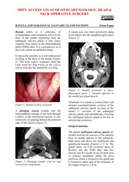
A ranula may also track posteriorly along<br />
tissue planes into the parapharyngeal space<br />
(Figure 3).<br />
Figure 3: <strong>Ranula</strong> extension to parapharyngeal<br />
space *, situated adjacent to<br />
the medial pterygoid muscle<br />
Treatment <strong>of</strong> a ranula is controversial, and<br />
includes marsupialization, excision <strong>of</strong> the<br />
sublingual salivary gland, excision <strong>of</strong> the<br />
ranula +/- excision <strong>of</strong> the sublingual<br />
salivary gland, and sclerotherapy. Excising<br />
the sublingual salivary gland is the key to<br />
minimizing recurrence.<br />
Surgical anatomy<br />
*<br />
The paired sublingual salivary glands are<br />
located beneath the mucosa <strong>of</strong> the anterior<br />
floor <strong>of</strong> mouth, anterior to the submandibular<br />
ducts and above the mylohyoid and<br />
geniohyoid muscles (Figures 4, 5, 6). The<br />
glands drain via 8-20 excretory ducts <strong>of</strong><br />
Rivinus into the SMG duct and also<br />
directly into the mouth on an elevated crest<br />
<strong>of</strong> mucous membrane called the plica<br />
fimbriata which is formed by the gland and<br />
is located to either side <strong>of</strong> the frenulum <strong>of</strong><br />
the tongue (Figures 5 & 6).
Lingual nerve<br />
Sublingual gland<br />
Submandibular duct<br />
Submandibular gland<br />
Mylohyoid muscle<br />
Geniohyoid muscle<br />
Figure 4: Superior, intraoral view <strong>of</strong> SMG,<br />
duct, lingual nerve and mylohyoid and<br />
geniohyoid muscles<br />
Lingual nerve<br />
Figure 5: Intraoral view <strong>of</strong> left sublingual<br />
gland with ducts <strong>of</strong> Rivinus, SMG and<br />
duct, lingual nerve and mylohyoid muscles<br />
Figure 6: View <strong>of</strong> right sublingual gland<br />
Submandibular duct<br />
Sublingual gland<br />
Submandibular gland<br />
Mylohyoid muscle<br />
Lingual nerve<br />
Intraoral SMG<br />
Floor <strong>of</strong> mouth<br />
Ducts <strong>of</strong> Rivinus<br />
Sublingual gland<br />
Mylohyoid<br />
Submental artery<br />
Cervical SMG<br />
SMGSubmandibular<br />
The lingual nerve crosses deep to the<br />
submandibular duct in the lateral floor <strong>of</strong><br />
mouth. In the anterior floor <strong>of</strong> mouth it is<br />
located posterior to the duct (Figures 4 &<br />
5).<br />
The submandibular duct is located<br />
immediately deep to the mucosa <strong>of</strong> the<br />
anterior and lateral floor <strong>of</strong> mouth, and<br />
opens into the oral cavity to either side <strong>of</strong><br />
the frenulum (Figures 4 & 5).<br />
Ranine veins are visible on the ventral<br />
surface <strong>of</strong> the tongue, and accompany the<br />
hypoglossal nerve (Figure 7).<br />
Figure 7: XIIn accompanied by ranine<br />
veins<br />
The detailed surgical anatomy <strong>of</strong> the<br />
SMG and submandibular triangle are<br />
dealt with in detail in the chapter on<br />
submandibular gland excision.<br />
Operative steps<br />
The following description applies to<br />
excision <strong>of</strong> a simple ranula, the sublingual<br />
salivary gland, and a plunging ranula.<br />
Anaesthesia<br />
A broad spectrum antibiotic is<br />
administered for 24 hours. With excision<br />
<strong>of</strong> a plunging ranula the anaesthetist should<br />
avoid muscle paralysis as it is useful to<br />
monitor the movement <strong>of</strong> the lower lip<br />
should the marginal mandibular nerve be<br />
surgically irritated.<br />
Positioning and draping<br />
The patient is placed in a supine position<br />
with neck extended. The skin <strong>of</strong> the<br />
anterior neck and lower face is sterilised.<br />
Draping is done in such a way that the<br />
2
mouth and upper neck are exposed. A selfretaining<br />
retractor is used to open the<br />
mouth. A silk traction suture is passed<br />
through the tip <strong>of</strong> the tongue to better<br />
expose the anterior floor <strong>of</strong> the mouth.<br />
Excision <strong>of</strong> a simple ranula (Figure 1)<br />
The mucosa is incised over the ranula<br />
taking care not to enter the sac. A<br />
submucosal dissection plane is established<br />
over the wall <strong>of</strong> the ranula. Using sharp<br />
and blunt dissection, the cyst is excised,<br />
taking care not to injure the submandibular<br />
duct or the lingual nerve. Should the<br />
submandibular duct be injured, then it is<br />
simply translocated to the lateral floor <strong>of</strong><br />
mouth by mobilising the duct, and passing<br />
it through a stab incision in the mucosa <strong>of</strong><br />
the lateral floor <strong>of</strong> mouth. It is secured to<br />
the oral mucosa with a suture passed<br />
through the side <strong>of</strong> the duct (Figure 8).<br />
Figure 8: Submandibular duct transposed<br />
to right lateral floor <strong>of</strong> mouth, and sutured<br />
to mucosa with a vicryl suture<br />
Excision <strong>of</strong> sublingual salivary gland<br />
The sublingual gland is surprisingly large,<br />
and is located immediately deep to the<br />
mucosa just anterior to the submandibular<br />
duct in the anterior floor <strong>of</strong> mouth. It may<br />
be excised by using electrocautery and<br />
blunt dissection (Figure 9). Structures at<br />
risk <strong>of</strong> injury are the submandibular duct<br />
and lingual nerve.<br />
Figure 9: The sublingual salivary gland<br />
and submandibular duct<br />
Excision <strong>of</strong> plunging ranula<br />
Sublingual gland<br />
Submandibular duct<br />
Readers are referred to the chapter on<br />
submandibular salivary gland excision<br />
for the surgical anatomy, as much <strong>of</strong> the<br />
dissection is similar. A horizontal incision,<br />
placed in a skin crease and at least 3cms<br />
below the mandible or at the level <strong>of</strong> the<br />
hyoid bone, and extending anteriorly from<br />
the anterior border <strong>of</strong> the sternocleidomastoid<br />
muscle, is made through skin,<br />
subcutaneous tissue and platysma. The<br />
common facial and anterior facial veins are<br />
identified posteriorly, and divided and<br />
ligated if required for access. The ranula is<br />
identified in the anterior part <strong>of</strong> the<br />
submandibular triangle (Figure 10).<br />
Figure 10: Plunging ranula in right neck<br />
3
The anterior belly <strong>of</strong> digastric is identified<br />
and retracted anteriorly. The mylohyoid<br />
muscle is identified deep to and behind the<br />
anterior belly <strong>of</strong> digastric (Figure 11).<br />
Area <strong>of</strong> Dehiscence<br />
Anterior belly digastric<br />
Mylohyoid<br />
Hyoglossus<br />
Figure 11: Muscles encountered with SMG<br />
excision, and area <strong>of</strong> dehiscence in<br />
mylohyoid through which plunging ranula<br />
typically passes into the neck<br />
The surgeon may have to mobilise and<br />
resect the SMG for better access (Figure<br />
12).<br />
Figure 12: SMG mobilised to better expose<br />
plunging ranula<br />
The ranula is mobilized with sharp and<br />
blunt dissection from the surrounding<br />
muscles and the SMG posteriorly. It is<br />
traced to where it generally passes through<br />
a dehiscence in the mylohyoid muscle, or<br />
less commonly behind the mylohyoid, into<br />
the floor <strong>of</strong> the mouth. The surgeon then<br />
completes the resection transorally,<br />
including resection <strong>of</strong> the sublingual<br />
salivary gland.<br />
Should the SMG have been preserved, then<br />
the status <strong>of</strong> the submandibular duct is<br />
checked to determine whether it needs to<br />
be translocated. The mucosal defect in the<br />
floor <strong>of</strong> mouth is then closed with<br />
absorbable sutures, and the neck is closed<br />
in layers over a suction drain.<br />
Author & Editor<br />
Johan Fagan MBChB, FCORL, MMed<br />
Pr<strong>of</strong>essor and Chairman<br />
Division <strong>of</strong> Otolaryngology<br />
<strong>University</strong> <strong>of</strong> <strong>Cape</strong> <strong>Town</strong><br />
<strong>Cape</strong> <strong>Town</strong><br />
South Africa<br />
johannes.fagan@uct.ac.za<br />
THE OPEN ACCESS ATLAS OF<br />
OTOLARYNGOLOGY, HEAD &<br />
NECK OPERATIVE SURGERY<br />
www.entdev.uct.ac.za<br />
The Open Access Atlas <strong>of</strong> Otolaryngology, Head &<br />
Neck Operative Surgery by Johan Fagan (Editor)<br />
johannes.fagan@uct.ac.za is licensed under a Creative<br />
Commons Attribution - Non-Commercial 3.0 Unported<br />
License<br />
4


