

open access atlas of otolaryngology, head & neck - Vula - University ...
open access atlas of otolaryngology, head & neck - Vula - University ...
open access atlas of otolaryngology, head & neck - Vula - University ...

Create successful ePaper yourself
Turn your PDF publications into a flip-book with our unique Google optimized e-Paper software.
OPEN ACCESS ATLAS OF OTOLARYNGOLOGY, HEAD &<br />
NECK OPERATIVE SURGERY<br />
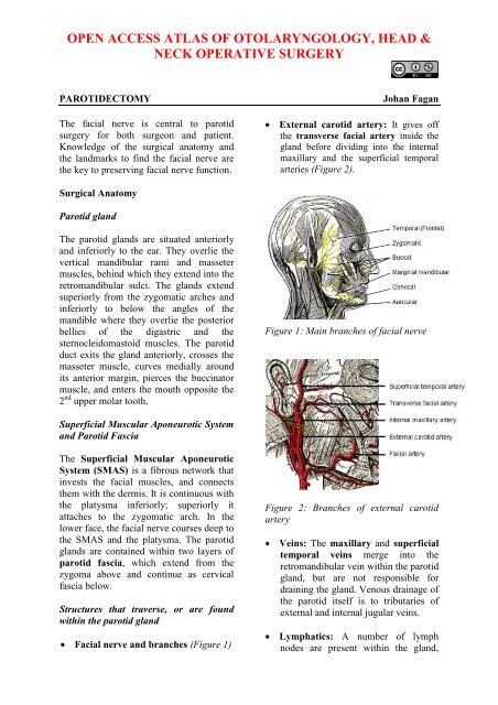
PAROTIDECTOMY Johan Fagan<br />
The facial nerve is central to parotid<br />
surgery for both surgeon and patient.<br />
Knowledge <strong>of</strong> the surgical anatomy and<br />
the landmarks to find the facial nerve are<br />
the key to preserving facial nerve function.<br />
Surgical Anatomy<br />
Parotid gland<br />
The parotid glands are situated anteriorly<br />
and inferiorly to the ear. They overlie the<br />
vertical mandibular rami and masseter<br />
muscles, behind which they extend into the<br />
retromandibular sulci. The glands extend<br />
superiorly from the zygomatic arches and<br />
inferiorly to below the angles <strong>of</strong> the<br />
mandible where they overlie the posterior<br />
bellies <strong>of</strong> the digastric and the<br />
sternocleidomastoid muscles. The parotid<br />
duct exits the gland anteriorly, crosses the<br />
masseter muscle, curves medially around<br />
its anterior margin, pierces the buccinator<br />
muscle, and enters the mouth opposite the<br />
2 nd upper molar tooth.<br />
Superficial Muscular Aponeurotic System<br />
and Parotid Fascia<br />
The Superficial Muscular Aponeurotic<br />
System (SMAS) is a fibrous network that<br />
invests the facial muscles, and connects<br />
them with the dermis. It is continuous with<br />
the platysma inferiorly; superiorly it<br />
attaches to the zygomatic arch. In the<br />
lower face, the facial nerve courses deep to<br />
the SMAS and the platysma. The parotid<br />
glands are contained within two layers <strong>of</strong><br />
parotid fascia, which extend from the<br />
zygoma above and continue as cervical<br />
fascia below.<br />
Structures that traverse, or are found<br />
within the parotid gland<br />
Facial nerve and branches (Figure 1)<br />
External carotid artery: It gives <strong>of</strong>f<br />
the transverse facial artery inside the<br />
gland before dividing into the internal<br />
maxillary and the superficial temporal<br />
arteries (Figure 2).<br />
Figure 1: Main branches <strong>of</strong> facial nerve<br />
Figure 2: Branches <strong>of</strong> external carotid<br />
artery<br />
Veins: The maxillary and superficial<br />
temporal veins merge into the<br />
retromandibular vein within the parotid<br />
gland, but are not responsible for<br />
draining the gland. Venous drainage <strong>of</strong><br />
the parotid itself is to tributaries <strong>of</strong><br />
external and internal jugular veins.<br />
Lymphatics: A number <strong>of</strong> lymph<br />
nodes are present within the gland,
principally in the superficial lobe, and<br />
drain to Level 2 <strong>of</strong> the <strong>neck</strong>.<br />
Relevant surgical relations<br />
Posterior: Cartilage <strong>of</strong> external auditory<br />
meatus; tympanic bone, mastoid process,<br />
sternocleidomastoid muscle<br />
Deep: Styloid process, stylomandibular<br />
tunnel, parapharyngeal space, posterior<br />
belly <strong>of</strong> digastric, sternocleidomastoid<br />
muscle<br />
Superior: Zygomatic arch, temporomandibular<br />
joint<br />
Facial nerve<br />
The facial nerve exits the stylomastoid<br />
foramen, and enters the parotid gland.<br />
Although the branching pattern does vary<br />
from patient to patient, the trunk generally<br />
divides at the pes anserinus into upper and<br />
lower divisions that subsequently branch<br />
into temporal (frontal), zygomatic, buccal,<br />
marginal mandibular and cervical branches<br />
that innervate the muscles <strong>of</strong> facial<br />
expression. Small branches to the posterior<br />
belly <strong>of</strong> digastric, stylohyoid, and auricular<br />
muscles also arise from the trunk (Figure<br />
3).<br />
Figure 3: The facial nerve trunk dividing<br />
into superior and inferior division at the<br />
pes anserinus<br />
The nerve traverses the parotid gland, with<br />
about 2/3 <strong>of</strong> the gland substance being<br />
superficial to the nerve. As parotid<br />
dissection generally is directed along the<br />
facial nerve, the nerve in effect divides the<br />
parotid from a surgical perspective into<br />
superficial and deep lobes, although there<br />
is no natural s<strong>of</strong>t tissue dissection plane<br />
that separates the two lobes.<br />
The midfacial nerve branches have<br />
multiple cross-innervations; however the<br />
frontal and marginal mandibular branches<br />
do not have cross-innervations and injury<br />
to these branches is followed by paralysis<br />
<strong>of</strong> the fore<strong>head</strong> and depressors <strong>of</strong> the lower<br />
lip (Figure 4). Therefore unlike the<br />
temporal and marginal mandibular nerves,<br />
selected midfacial branches may be<br />
sacrificed without loss <strong>of</strong> facial function.<br />
Figure 4: Midfacial branches (yellow)<br />
interconnect whereas temporal and<br />
marginal mandibular (black) do not<br />
Locating the Facial Nerve<br />
It is useful to know preoperatively whether<br />
a parotid tumour is situated deep or<br />
superficial to the facial nerve. This<br />
facilitates surgical planning and facilitates<br />
preoperative consent relating to the<br />
likelihood <strong>of</strong> a temporary postoperative<br />
facial nerve weakness.<br />
2
Surface markings<br />
Facial nerve trunk: The trunk exits the<br />
skull at the stylomastoid foramen. This is<br />
situated at the deep end <strong>of</strong> the<br />
tympanomastoid suture line, which can be<br />
located at the junction between the mastoid<br />
process and the tympanic ring <strong>of</strong> the<br />
external ear canal<br />
Temporal (frontal) branch <strong>of</strong> facial<br />
nerve: The nerve crosses the zygomatic<br />
arch; it runs within the SMAS and lies<br />
superficial to the deep temporalis fascia. It<br />
courses more or less along a line drawn<br />
between the attachment <strong>of</strong> the lobule <strong>of</strong> the<br />
ear to a point 1.5 cm above the lateral<br />
aspect <strong>of</strong> the eyebrow. To avoid injury to<br />
the temporal branch dissect either in a<br />
subcutaneous plane or deep to the SMAS<br />
(Figure 1).<br />
Radiology<br />
Radiological investigation is not routinely<br />
required with parotid tumours. It is<br />
recommended for surgical planning with<br />
tumours that are large, fixed, and are<br />
associated with facial nerve involvement,<br />
trismus, and parapharyngeal space<br />
involvement. MRI is a valuable<br />
investigation with recurrence <strong>of</strong><br />
pleomorphic adenoma as it is <strong>of</strong>ten<br />
multifocal.<br />
The extratemporal facial nerve is not<br />
visible with ultrasound, CT or MRI. The<br />
retromandibular vein is however intimately<br />
associated with the facial nerve. The vein<br />
courses through the parotid gland<br />
immediately deep to the facial nerve, but<br />
rarely runs immediately superficial to the<br />
nerve (Figures 5 & 6). Reliance is<br />
therefore placed on the juxtaposition <strong>of</strong> the<br />
retromandibular vein and the nerve to<br />
predict whether a tumour is likely to be<br />
deep or superficial to the nerve.<br />
Figure 5: Facial nerve running superficial<br />
to retromandibular vein<br />
Figure 6: Facial nerve running deep, but<br />
close, to retromandibular vein<br />
Figure 7: Red arrows indicate<br />
retromandibular veins, and yellow arrow<br />
the course <strong>of</strong> the facial nerve in a<br />
superficial lobe pleomorphic adenoma<br />
The retromandibular vein can be clearly<br />
visualized on a CT with contrast, or an<br />
MRI (Figures 7, 8).<br />
3
Figure 8: Red arrows indicate retromandibular<br />
veins, and yellow arrow the<br />
course <strong>of</strong> the facial nerve in a deep lobe<br />
pleomorphic adenoma<br />
Radiology may also alert the surgeon to<br />
extension <strong>of</strong> a deep lobe parotid tumour<br />
through the stylomandibular tunnel into the<br />
parapharyngeal space (Figure 9).<br />
Figure 9: Tumour passing through stylomandibular<br />
tunnel to parapharyngeal<br />
space (Arrow indicates styloid process)<br />
Intraoperative location <strong>of</strong> facial nerve<br />
The facial nerve is usually explored by<br />
prograde dissection i.e. by locating the<br />
nerve trunk where it exits from the<br />
stylomastoid foramen, and then dissecting<br />
anteriorly along the trunk, the pes<br />
anserinus and the divisions and nerve<br />
branches. Occasionally this is not possible<br />
e.g. with a large fixed mass centered at the<br />
stylomastoid foramen. In such cases a<br />
retrograde dissection may be required after<br />
locating the temporal branch where it<br />
crosses the zygoma, the buccal branches<br />
which lie parallel to the parotid duct<br />
(Figure 10), or the marginal mandibular<br />
branch where is crosses the facial artery<br />
and vein just below or at the inferior<br />
margin <strong>of</strong> the mandible, where it is just<br />
deep to platysma (Figure 11).<br />
Figure 10: Buccal branches adjacent to<br />
the parotid duct<br />
Figure 11: Marginal mandibular nerve<br />
crossing facial artery and vein<br />
Marginal mandibular<br />
nerve<br />
The surgical landmarks for finding the<br />
facial nerve trunk at the stylomastoid<br />
foramen are remarkably constant, and all<br />
the landmarks should be identified at every<br />
Facial artery and vein<br />
Submandibular salivary<br />
gland<br />
4
operation to facilitate finding the nerve<br />
(Figures 12, 13).<br />
Figure 12: Schematic surgical landmarks<br />
for the facial nerve trunk<br />
Figure 13: Intraoperative surgical<br />
landmarks for the facial nerve trunk<br />
Posterior belly <strong>of</strong> digastric muscle: The<br />
nerve runs at the same depth below the<br />
skin surface, and bisects the angle between<br />
the muscle and the styloid process<br />
Cartilage pointer: This refers to the<br />
medial-most, pointed end <strong>of</strong> the cartilage<br />
<strong>of</strong> the external auditory meatus. The nerve<br />
exits the foramen approximately 1cm deep<br />
and 1cm inferior to this point<br />
Tympanic ring, mastoid process and<br />
tympanomastoid suture line: The<br />
tympanomastoid suture line is the most<br />
precise landmark for the facial nerve as it<br />
leads medially, directly to the stylomastoid<br />
foramen<br />
Styloid process: The facial nerve crosses<br />
the styloid process. Palpating the styloid<br />
process is therefore a useful means to<br />
determine the depth and position <strong>of</strong> the<br />
facial nerve<br />
Branch <strong>of</strong> occipital artery: A small<br />
branch <strong>of</strong> the occipital artery is commonly<br />
encountered just lateral to the facial nerve<br />
close to the stylomastoid foramen. Brisk<br />
arterial bleeding should therefore alert the<br />
surgeon to the proximity <strong>of</strong> the facial<br />
nerve; it is easily controlled with bipolar<br />
cautery.<br />
Electrical stimulation and monitoring<br />
These need not be routinely employed, but<br />
may be useful adjuncts to a sound<br />
knowledge <strong>of</strong> facial nerve anatomy in<br />
selected cases such as revision surgery and<br />
with large tumours. It may however not<br />
record facial stimulation with faulty<br />
equipment, and nerve fatigue following<br />
excessive mechanical or electrical<br />
stimulation, and use <strong>of</strong> a muscle relaxant.<br />
Electrophysiological monitoring: An<br />
EMG monitor may be used to detect<br />
contraction <strong>of</strong> the facial muscles when the<br />
facial nerve is mechanically or<br />
electrically stimulated.<br />
Facial nerve electrical stimulation:<br />
Battery operated or more sophisticated<br />
nerve stimulators may be employed<br />
intraoperatively to assist with finding the<br />
nerve, or to differentiate between nerve<br />
and blood vessels. Stimulating the nerve<br />
produces visible contraction <strong>of</strong> the facial<br />
musculature or an EMG signal.<br />
Types <strong>of</strong> Parotidectomy<br />
Partial parotidectomy: Resection <strong>of</strong><br />
parotid pathology with a margin <strong>of</strong> normal<br />
parotid tissue. This is the standard<br />
5
operation for benign pathology and<br />
favourable malignancies<br />
Superficial parotidectomy: Resection <strong>of</strong><br />
the entire superficial lobe <strong>of</strong> parotid<br />
(Figure 3) and is generally used for<br />
metastases to parotid lymph nodes e.g.<br />
from skin cancers, and for high grade<br />
malignant parotid tumours.<br />
Total parotidectomy: This involves<br />
resection <strong>of</strong> the entire parotid gland,<br />
usually with preservation <strong>of</strong> the facial<br />
nerve<br />
Preoperative consent<br />
Scar: usually very good healing except<br />
over the mastoid where some scarring may<br />
be occur<br />
Anaesthesia in the greater auricular<br />
distribution: Skin <strong>of</strong> inferior part <strong>of</strong><br />
auricle, and overlying the angle <strong>of</strong> the<br />
mandible<br />
Facial nerve weakness: Temporary<br />
weakness common (
Divide the greater auricular nerve as it<br />
crosses sternocleidomastoid muscle,<br />
posteriorly to the external jugular vein<br />
Figure 15: Exposure <strong>of</strong> parotid mass or<br />
gland<br />
Identify and skeletonise the posterior<br />
belly <strong>of</strong> the digastric muscle. Do not<br />
dissect cephalad <strong>of</strong> the muscle as one<br />
may injure the facial nerve (Figure 16)<br />
Digastric muscle<br />
External jugular vein<br />
(ligated)<br />
Greater auricular nerve<br />
Sternomastoid muscle<br />
Figure 16: Expose the sternomastoid and<br />
posterior belly <strong>of</strong> digastric muscle<br />
Skeletonise the cartilage <strong>of</strong> the external<br />
auditory canal up to the tragal pointer.<br />
This can be done quite quickly with<br />
electrocautery dissection as the facial<br />
nerve exits the stylomastoid foramen<br />
1cm deep to the tragal pointer<br />
Skeletonise the mastoid tip to the depth<br />
<strong>of</strong> the tragal pointer<br />
Identify all the following landmarks for<br />
the facial nerve (Figures 12, 13 & 17)<br />
o Tragal pointer (nerve 1 cm deep and<br />
inferior)<br />
o Tympanic ring<br />
o Anterior aspect <strong>of</strong> mastoid bone<br />
o Tympanomastoid suture line (leads<br />
directly to stylomastoid foramen)<br />
o Posterior belly <strong>of</strong> digastric muscle<br />
(Facial nerve at same depth, just<br />
above muscle)<br />
o Palpate the styloid process (facial<br />
nerve in angle between styloid and<br />
digastric, and crosses styloid more<br />
anteriorly)<br />
Figure 17: Identify facial nerve landmarks<br />
Cartilage pointer<br />
Tympanic ring<br />
VIIn<br />
Digastric<br />
Sternomastoid<br />
Locate the facial nerve trunk by blunt<br />
dissection with a fine haemostat<br />
(Figure 18)<br />
Figure 18: Location <strong>of</strong> facial nerve trunk,<br />
and superior and inferior release <strong>of</strong><br />
capsule and parotid tissues (yellow<br />
arrows)<br />
Use fine curved blunt tipped scissors<br />
for the remainder <strong>of</strong> the nerve<br />
Tympanomastoid suture<br />
Mastoid process<br />
7<br />
VIIn
dissection. Tunnel and spread the<br />
tissues overlying the facial nerve and<br />
its branches, and divide the parotid<br />
tissue overlying the nerve. It is<br />
important to dissect directly on the<br />
nerve so as not to lose sight <strong>of</strong> it.<br />
Never divide parotid tissue beyond<br />
exposed facial nerve. Wearing loupes<br />
e.g. with 2.5x magnification assists<br />
with the dissection, and enables one to<br />
better distinguish between blood<br />
vessels and nerves. Employ bipolar<br />
diathermy and fine silk ties for<br />
haemostasis.<br />
Dissect along the trunk to the pes<br />
anserinus<br />
Dissect back towards the stylomastoid<br />
foramen to exclude early branching<br />
from the trunk<br />
Divide the parotid fascia and parotid<br />
tissue superiorly and inferiorly to<br />
release the parotid posteriorly and to<br />
permit anterior mobilisation <strong>of</strong> the<br />
gland/tumour (Figure 17)<br />
Dissect along, and strip the superficial<br />
lobe <strong>of</strong>f the branches <strong>of</strong> facial nerve.<br />
Unless a complete superficial<br />
parotidectomy is done, only the<br />
branches close the mass are dissected<br />
and exposed.<br />
Identify the retromandibular vein as it<br />
crosses the medial to the facial nerve<br />
(Figures 5 & 6)<br />
If removing the superior part <strong>of</strong> the<br />
gland, identify/ligate the superficial<br />
temporal artery superiorly, just anterior<br />
to auricle<br />
If dissecting to the anterior border <strong>of</strong><br />
the gland, identify and transect the<br />
parotid duct<br />
Remove the tumour with a cuff <strong>of</strong> the<br />
superficial parotid lobe (Figure 3)<br />
Parotid dissection for deep lobe tumours<br />
The principles <strong>of</strong> resecting deep lobe<br />
tumours are to:<br />
Identify, dissect and free up the facial<br />
nerve from the underlying deep lobe or<br />
tumour, to provide <strong>access</strong> to the deep<br />
lobe. This may involve either a<br />
superficial parotidectomy, or simply<br />
reflecting the superficial lobe<br />
anteriorly, keeping the parotid duct<br />
intact, and replacing it at the<br />
conclusion <strong>of</strong> surgery (Figure 19)<br />
Figure 19: Reflecting superficial lobe for<br />
<strong>access</strong> to facial nerve and to deep lobe<br />
tumour<br />
Deliver the tumour either between, or<br />
inferior to the facial nerve or its<br />
branches, identifying the branches <strong>of</strong><br />
the facial nerve branches around the<br />
tumour, and removing tumour between<br />
splayed facial nerve branches (Figure<br />
20)<br />
The deep lobe <strong>of</strong> the parotid/tumour is<br />
bordered medially by the fat <strong>of</strong> the<br />
parapharyngeal space, and can be<br />
delivered from the parapharyngeal<br />
space by blunt dissection<br />
Be prepared to divide the external<br />
carotid, deep transverse facial and<br />
superficial temporal arteries and the<br />
retromandibular and superficial<br />
temporal veins if and when they are<br />
encountered during dissection<br />
8
Figure 20: Tumour resected by removing<br />
tumour between splayed facial nerve<br />
branches<br />
Additional <strong>access</strong> may be provided to<br />
the deep aspect <strong>of</strong> a tumour by dividing<br />
the styloid process and/or via a<br />
transcervical approach (Figure 21)<br />
Figure 21: Access to parapharyngeal<br />
space tumour extension by reflecting the<br />
superficial lobe and division <strong>of</strong> styloid<br />
process<br />
Tumour spillage<br />
Great care should be taken to avoid rupture<br />
and spillage <strong>of</strong> pleomorphic adenoma<br />
tissue into the operative site as it may lead<br />
to multifocal tumour recurrence, <strong>of</strong>ten<br />
more than 20yrs following surgery (Figure<br />
22).<br />
Figure 22: Multifocal recurrence <strong>of</strong><br />
pleomorphic adenoma<br />
A minor controlled capsular rupture may<br />
be simply managed by copiously irrigating<br />
the wound. With more extensive ruptures,<br />
especially <strong>of</strong> pleomorphic adenoma in the<br />
parapharyngeal space, some would<br />
advocate postoperative radiation therapy.<br />
Due to the multifocal nature <strong>of</strong> the<br />
recurrence, MRI is an important<br />
preoperative investigation for recurrence.<br />
Having to operate in a previously dissected<br />
field, the facial nerve is at greater risk <strong>of</strong><br />
injury, and should be monitored during<br />
surgery.<br />
Wound closure<br />
Confirm nerve continuity: Carefully<br />
inspect the nerve. One may stimulate the<br />
nerve with a nerve stimulator. Neuropraxia<br />
due to mechanical trauma may however<br />
cause failure <strong>of</strong> muscle contraction.<br />
Obtain meticulous haemostasis. Use ties<br />
and bipolar diathermy. Employ a Valsalva<br />
manoeuvre to identify venous bleeding.<br />
Sealed suction drain: Until drainage<br />
Facial nerve repair<br />
Unlike with malignant tumours, the facial<br />
nerve and its branches can virtually always<br />
be dissected free from benign neoplasms.<br />
Isolated midfacial branches may be<br />
sacrificed without causing visible facial<br />
dysfunction. Transection <strong>of</strong> the temporal<br />
(frontal) and marginal mandibular nerves<br />
however results in disfiguring facial<br />
asymmetry; these nerves should be<br />
repaired with 8/0 nylon/proline epineural<br />
sutures. When primary nerve repair is not<br />
possible due to undue tension or nerve<br />
resection, then the nerve can be grafted<br />
with greater auricular nerve, or sural nerve.<br />
The greater auricular nerve is<br />
approximately the same diameter as the<br />
facial nerve trunk, and has a few branches<br />
that can be used to graft more than one<br />
facial nerve branch. The sural nerve<br />
provides greater length and more branches<br />
and is better suited to bridging longer<br />
defects and for grafting to more peripheral<br />
branches (Figure 22).<br />
Figure 22: Sural nerve graft<br />
When the proximal end <strong>of</strong> the facial nerve<br />
is not available, e.g. with extensive<br />
proximal perineural tumour extension, then<br />
a hypoglossal-facial nerve interposition<br />
graft can be used to restore facial tone and<br />
movement. The nerve graft is sutured endto-end<br />
to the distal facial nerve(s), and<br />
end-to-side to the hypoglossal nerve after<br />
cutting about 25% into the side <strong>of</strong> the<br />
hypoglossal nerve to expose the nerve<br />
axons (Figure 23).<br />
Figure 23: Hypoglossal/facial nerve graft<br />
Author & Editor<br />
Johan Fagan MBChB, FCORL, MMed<br />
Pr<strong>of</strong>essor and Chairman<br />
Division <strong>of</strong> Otolaryngology<br />
<strong>University</strong> <strong>of</strong> Cape Town<br />
Cape Town<br />
South Africa<br />
johannes.fagan@uct.ac.za<br />
THE OPEN ACCESS ATLAS OF<br />
OTOLARYNGOLOGY, HEAD &<br />
NECK OPERATIVE SURGERY<br />
www.entdev.uct.ac.za<br />
Anastomosis to<br />
VIIn trunk<br />
The Open Access Atlas <strong>of</strong> Otolaryngology, Head &<br />
Neck Operative Surgery by Johan Fagan (Editor)<br />
johannes.fagan@uct.ac.za is licensed under a Creative<br />
Commons Attribution - Non-Commercial 3.0 Unported<br />
License<br />
Greater auricular n<br />
interposition graft<br />
Anastomosis to XIIn<br />
10


