Anti-Phosphotyrosine, clone 4G10 - Millipore
Anti-Phosphotyrosine, clone 4G10 - Millipore
Anti-Phosphotyrosine, clone 4G10 - Millipore

Create successful ePaper yourself
Turn your PDF publications into a flip-book with our unique Google optimized e-Paper software.
<strong>Anti</strong>-<strong>Phosphotyrosine</strong>, <strong>clone</strong> <strong>4G10</strong> ®<br />
Monoclonal <strong>Anti</strong>body<br />
Cat. # 05-321X<br />
Lot # DAM1676163<br />
FOR RESEARCH USE ONLY<br />
NOT FOR USE IN HUMANS<br />
Background<br />
Some of the tyrosine residues can be tagged<br />
with a phosphate group (phosphorylated) by<br />
protein kinases. (In its phosphorylated state, it<br />
is referred to as phosphotyrosine.). Tyrosine<br />
phosphorylation is considered as one of the<br />
key steps in signal transduction and regulation<br />
of enzymatic activity.<br />
The advent of anti-phosphotyrosine antibodies<br />
is one of significant events in signal<br />
transduction research. Before the availability<br />
of anti-phosphotyrosine antibodies, tyrosyl<br />
phospyhorylation of proteins and enzymes<br />
was investigated through hazardous and timeconsuming<br />
radioactive experiments. <strong>Anti</strong>phosphotyrosine<br />
antibodies are commonly<br />
used in western blots after the targeted<br />
proteins have been immunoprecipitated to<br />
measure the tyrosyl phosphorylation of the<br />
proteins. <strong>Anti</strong>-phosphotyrosine antibodies are<br />
also directly used on cell lysate to examine the<br />
overall change of tyrosine phosphorylation<br />
level in reponse to various treatments.<br />
Presentation<br />
Purified mouse monoclonal IgG2bκ in buffer<br />
containing 0.1 M Tris-glycine, pH 7.4, 0.15 M<br />
NaCl, 0.05% sodium azide. Liquid at 2-8ºC.<br />
IgG2bκ mouse monoclonal antibody produced<br />
in vitro by mouse-mouse hybridoma <strong>4G10</strong> ®<br />
(FOX-NY [NS-1 derivative] myeloma x spleen<br />
cells).<br />
Concentration<br />
1 mg/mL<br />
Specificity<br />
Recognizes tyrosine-phosphorylated proteins<br />
from all species.<br />
Species Cross-reactivity<br />
All species<br />
Immunogen<br />
Phosphotyramine-KLH.<br />
Molecular Weight<br />
Dependent upon the molecular weight of the<br />
tyrosine phosphorylated protein being<br />
detected.<br />
Method of Purification<br />
Protein G-Sepharose chromatography<br />
Storage and Handling<br />
Stable for 1 year at 2-8°C from date of receipt.<br />
NOTE: DO NOT FREEZE. For maximum<br />
recovery of the product, centrifuge the original<br />
vial prior to removing the cap. If the product<br />
has accidentally been frozen and thawed, spin<br />
it at 13,000 x g for 10 minutes at 2-8ºC. Save<br />
the supernatant for application.<br />
Control<br />
pack size: 50 µg<br />
Untreated A-431 (negative control) and EGF<br />
treated A-431 (positive control) whole cell<br />
lysates.<br />
Included Positive <strong>Anti</strong>gen Control: Catalog #<br />
12-302, EGF-stimulated A431 cell lysate is<br />
provided as a free positive antigen control for<br />
western immunoblotting. Aliquot as desired,<br />
refreeze immediately, and store at -20°C. The<br />
lysate is stable for 6 months at -20°C. Before<br />
use, add 2.5 µL of 2-mercaptoethanol/100 µL<br />
of lysate and boil for 5 minutes to reduce the<br />
preparation. Load 20 µg of reduced lysate per<br />
lane for immunoblot analysis.<br />
Quality Control Testing<br />
Store at 2-8ºC<br />
DO NOT FREEZE<br />
Routinely evaluated on EGF-treated human<br />
A431 carcinoma cells.<br />
Certificate of Analysis<br />
Applications<br />
Species<br />
Cross-Reactivity<br />
<strong>Anti</strong>body<br />
Isotype<br />
Epitope/<br />
Region<br />
Host<br />
Species<br />
Molecular<br />
Weight<br />
Accession #<br />
WB, IP A IgG2bκ N/A M Varies N/A<br />
page 1 of 2<br />
References<br />
1. Kanakura, Y., et al. (1991). J. Biol. Chem. 266:<br />
490.<br />
2. Cohen, B., et al. (1990). Proc. Natl. Acad. Sci.<br />
USA. 87: 4458.<br />
3. Druker, B.J., et al (1989). New Eng. J. Med.<br />
321: 1383.<br />
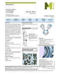
Western Blot Analysis: 0.5-2 µg/mL of this lot<br />
detected tyrosine-phosphorylated proteins in<br />
a modified RIPA lysate from EGF-treated<br />
human A431 carcinoma cells (Cohen, B.,<br />
1990; Druker, B. J.,1989; Kanakura, Y.,<br />
1991).<br />
Western Blot Analysis:<br />
Representative lot data.<br />
EGF-stimulated A431 cell lysate<br />
was resolved by electrophoresis,<br />
transferred to nitrocellulose and<br />
probed with antiphosphotyrosine<br />
(1 µg/mL).<br />
Proteins were visualized using a<br />
goat anti-mouse secondary<br />
antibody conjugated to HRP and<br />
a chemiluminescence detection<br />
system<br />
APPLICATION LEGEND: WB Western Blotting IP Immunoprecipitation IC Immunocytochemistry IF Immunofluorescence<br />
IH Immunohistochemistry (Tissue)<br />
SPECIES LEGEND: H Human M Mouse R Rat Rb Rabbit A All Species<br />
Please visit www.millipore.com for additional product information, test data and references.<br />
Submit your published journal article, and earn credit toward future <strong>Millipore</strong> purchases. Visit www.millipore.com/publication_rewards to learn more!<br />
Rev.A/2008-06-11/05-321XCA/JM
<strong>Anti</strong>-<strong>Phosphotyrosine</strong>, <strong>clone</strong> <strong>4G10</strong> ®<br />
Cat # 05-321X<br />
Lot # DAM1676163<br />
Additional Research Applications<br />
Immunoprecipitation: 2-4 µg of a previous lot immunoprecipitated quantitatively the phosphotyrosine containing proteins in the lysate of a<br />
confluent culture (10 cm dish) of cells expressing an activated tyrosine kinase. To preserve phosphotyrosine, add: 0.2 mM sodium<br />
orthovanadate to the lysis buffer.<br />
PROTOCOL<br />
Western Blot<br />
1. Perform SDS-polyacrylamide gel electrophoresis (SDS-PAGE) on a cell lysate sample (cell lysis buffer: 50 mM Tris-HCl, pH 7.4; 1% NP-40;<br />
0.25% sodium deoxycholate; 150 mM NaCl; 1 mM EDTA; 1 mM PMSF; 1 µg/mL aprotinin, leupeptin, pepstatin; 1 mM Na3VO4; 1 mM NaF)<br />
and transfer the proteins to nitrocellulose. Wash the blotted nitrocellulose twice with water.<br />
2. Block the blotted nitrocellulose in freshly prepared TBS containing 3% nonfat dry milk (Catalog # 20-200), (TBS-MLK) for 45-90 minutes at<br />
room temperature with constant agitation.<br />
3. Incubate the nitrocellulose with 0.5-2 µg/mL of anti-<strong>Phosphotyrosine</strong>, <strong>clone</strong> <strong>4G10</strong> ® , diluted in freshly prepared TBS-MLK overnight with<br />
agitation at 4°C.<br />
4. Wash the nitrocellulose twice with water.<br />
5. Incubate the nitrocellulose in the secondary reagent of choice (a goat anti-mouse HRP conjugated, Catalog # 12-349, 1:5000 dilution, was<br />
used) in TBS-MLK for 1.5 hours at room temperature with agitation.<br />
6. Wash the nitrocellulose with water twice.<br />
7. Wash the nitrocellulose in TBS-0.05% Tween 20 for 3-5 minutes.<br />
8. Rinse the nitrocellulose in 4-5 changes of water.<br />
9. Use detection method of choice (enhanced chemiluminescence with a 30 second exposure was used).<br />
Immunoprecipitation<br />
1. Add 2-4 µg of anti-<strong>Phosphotyrosine</strong>, <strong>clone</strong> <strong>4G10</strong> ® and 60 µL (30 µL packed beads) of washed Protein G agarose bead slurry (Catalog # 16-<br />
266) to 500 µL of TBS in a microcentrifuge tube.<br />
2. Gently rock the reaction mixture at 4°C for 1 ho ur.<br />
3. Collect the agarose beads by pulsing (5 seconds in the microcentrifuge at 14,000 x g), and drain off the supernatant. Wash the beads 3<br />
times with either ice-cold cell lysis buffer or TBS.<br />
4. Dilute the cell lysate to roughly 1 µg/µL total cell protein with TBS.<br />
5. Add 500 µg-1mg cell lysate to the reaction mixture.<br />
6. Gently rock the reaction mixture at 4°C for 1 ho ur.<br />
7. Collect the agarose beads by pulsing (5 seconds in the microcentrifuge at 14,000 x g), and drain off the supernatant. Wash the beads 3<br />
times with either ice-cold cell lysis buffer or TBS.<br />
8. Resuspend the agarose beads in 60 µL 2X Laemmli sample buffer.<br />
9. Store the beads frozen for future analysis or boil the beads for 5 minutes.<br />
10. Collect the beads after boiling using a microcentrifuge pulse.<br />
11. Perform SDS-PAGE and immunoblot analysis on a sample of the supernatant fraction<br />
RELATED PRODUCTS (specific) RELATED PRODUCTS (non-specific)<br />
cat # description cat # description<br />
16-104<br />
16-199<br />
12-110<br />
16-105<br />
12-349<br />
05-321<br />
05-321X<br />
16-204<br />
16-101<br />
16-184<br />
17-153<br />
05-777<br />
17-123<br />
06-427<br />
16-103<br />
12-256<br />
<strong>Anti</strong>-<strong>Phosphotyrosine</strong>, <strong>clone</strong> <strong>4G10</strong>® , FITC conjugate IPVH00010 Immobilon-P 26.5 cm x 3.75 m Roll PVDF 0.45 µm<br />
<strong>Anti</strong>-<strong>Phosphotyrosine</strong>, recombinant <strong>clone</strong> <strong>4G10</strong> ® , agarose<br />
conjugate<br />
IPFL00010<br />
<strong>Phosphotyrosine</strong> control (EGF-stim A431 cell lysate) IPVH07850<br />
<strong>Anti</strong>-<strong>Phosphotyrosine</strong>, <strong>clone</strong> <strong>4G10</strong>® , HRP conjugat ISEQ00010<br />
Goat <strong>Anti</strong>-Mouse IgG, HRP conjugate ISEQ07850<br />
<strong>Anti</strong>-<strong>Phosphotyrosine</strong>, <strong>clone</strong> <strong>4G10</strong> ®<br />
IPFL07810<br />
<strong>Anti</strong>-<strong>Phosphotyrosine</strong>, <strong>clone</strong> <strong>4G10</strong> ®<br />
<strong>Anti</strong>-<strong>Phosphotyrosine</strong>, recombinant <strong>clone</strong> <strong>4G10</strong> ® , biotin<br />
conjugate<br />
<strong>Anti</strong>-<strong>Phosphotyrosine</strong>, <strong>clone</strong> <strong>4G10</strong> ® , agarose conjugate<br />
<strong>Anti</strong>-<strong>Phosphotyrosine</strong>, recombinant <strong>clone</strong> <strong>4G10</strong> ® , HRP<br />
conjugate<br />
<strong>Anti</strong>-<strong>Phosphotyrosine</strong> Immunoblotting Kit (<strong>4G10</strong> ® ), ECL<br />
Detection<br />
<strong>Anti</strong>-<strong>Phosphotyrosine</strong>, recombinant <strong>clone</strong> <strong>4G10</strong> ®<br />
<strong>Anti</strong>-<strong>Phosphotyrosine</strong> Immunoblotting Kit (<strong>4G10</strong> ® , HRP<br />
conjugate)<br />
<strong>Anti</strong>-<strong>Phosphotyrosine</strong><br />
<strong>Anti</strong>-<strong>Phosphotyrosine</strong>, <strong>clone</strong> <strong>4G10</strong> ® , biotin conjugate<br />
<strong>Phosphotyrosine</strong> Molecular Weight Standards<br />
WBKLS0100<br />
Immobilon-FL 26.5 cm x 3.75 m Roll PVDF 0.45 µm<br />
Immobilon-P 7 x 8.4 cm PVDF 0.45 mm (sheet) 50/pk<br />
Immobilon-P SQ 26.5 cm x 3.75 m 1 roll PVDF 0.2 µm<br />
Immobilon-P 7 x 8.4 cm PVDF 0.2 mm (sheet) 50/pk<br />
Immobilon-FL 7 x 8.4 cm PVDF 0.45 mm (sheet) 10/pk<br />
Immobilon Western Chemilum HRP Substrate 100 mL<br />
antibodies Multiplex products biotools cell culture enzymes kits proteins/peptides siRNA/cDNA products<br />
Please visit www.millipore.com for additional product information, test data and references<br />
28820 Single Oak Drive • Temecula, CA 92590<br />
Technical Support: T: 1-800-MILLIPORE (1-800-645-5476) • F: 1-800-437-7502<br />
FOR RESEARCH USE ONLY. Not for use in diagnostic or therapeutic applications. Purchase of this Product does not include any right to resell or transfer, either as a stand-alone product or as<br />
a component of another product. Any use of this Product for purposes other than research is strictly prohibited without prior written authorization from an authorized officer of <strong>Millipore</strong> Corporation.<br />
Upstate®, Chemicon® and all other trademarks are owned by <strong>Millipore</strong> Corporation. Copyright ©2008 <strong>Millipore</strong> Corporation. All rights reserved.<br />
17-373<br />
2060<br />
2500<br />
B2080-<br />
175GM<br />
Spray & Glow ECL WB Detection System 1 ea<br />
Re-Blot Western Blot Recycling Kit<br />
Re-Blot Plus Western Blot Recycling Kit<br />
Blot Quick Blocker Membrane Blocking Agent 175G<br />
page 2 of 2