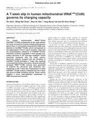
Xenopus PSM and is restricted in the caudal PSM. This Wnt/ β-catenin signaling activity is essential for Mespo expression. Increasing Wnt/β-catenin signaling activity by injecting pCS– Lef1–VP16 enhances Mespo expression in the PSM, and decreasing Wnt/β-catenin signaling activity by injecting pCS– Lef1–EnR downregulates Mespo expression (Fig. 4). We also found that Wnt/β-catenin signaling, acting upstream of Mespo, directly controls Mespo expression, based on the recruitment of Lef1 to the site in the Mespo 5′-regulatory region (Fig. 6B). Since Mespo expression is essential for segmental patterning, we concluded that Wnt/β-catenin signaling controls segmentation process by regulating Mespo expression. J. Wang et al. / Developmental Biology 304 (<strong>2007</strong>) 836–847 Fig. 8. Interaction between PI3-K/AKT and Wnt/β-catenin signaling in PSM. (A) Western blot of β-catenin in the nucleus and cytosol of the caudal PSM cells from embryos treated with either DMSO or LY294002 for 2 h. (B) Western blot of β-catenin, phosphorylated β-catenin, Akt and phosphorylated Akt in the caudal PSM cells treated with DMSO for 2.5 h, or LY294002 for 1.5 h or 2.5 h. (C–E) Four-cell-stage embryos were injected in VMZ of one cell with pCS–LacZ as control (C) or together with pCS–Lef1–VP16 (D, E). At the neurula stage, injected embryos were treated with DMSO (C, D) or LY294002 (E) for 2 h, then fixed, stained with X-gal to reveal the tracer (light blue) and analyzed by whole-mount in situ hybridization for Mespo. Arrow in panel D indicates the increase of Mespo expression in the injected side and arrowhead indicates the expression of Mespo in the uninjected side. Arrow in panel E indicates the increased Mespo expression in the injected side and arrowhead indicates the decrease of Mespo expression in the uninjected side. Regulation of Mespo expression in the PSM The bHLH family member Mespo is a key regulator of genes associated with proper segmentation and successful somitogenesis (Yoon and Wold, 2000). FGF and RA signaling were proposed to act upstream of Mespo. It has been shown that blocking FGF signaling by SU5402 or increasing RA signaling by treating with RA caused a decrease of Mespo expression (Moreno and Kintner, 2004). However, the molecular nature of how Mespo expression is regulated remains unclear. We show here that Wnt/β-catenin signaling is required for Mespo expression. Increasing Wnt/β-catenin signaling activity by overexpressing β-catenin or Lef1–VP16 increases the 845
846 J. Wang et al. / Developmental Biology 304 (<strong>2007</strong>) 836–847 expression of Mespo (Fig. 4B and Supplemental Fig. 2B), and interfering Wnt/β-catenin signaling by overexpressing Lef1– EnR resulted in the opposite effect (Fig. 4C and Supplemental Fig. 2D). Our results also show that the PI3-K/AKT pathway participates in regulating Mespo expression (Fig. 7E). Since it has been proposed that MKK1/ERK signaling is involved in regulating Mespo expression (Moreno and Kintner, 2004), it seems that FGF signaling contributes to the expression of Mespo through PI3-K/AKT and MKK1/ERK pathway together. Thus, multiple signaling pathways regulate expression of Mespo that is a key regulator of somitogenesis. Interaction of signals in somitogenesis Previous studies have shown that FGF, Wnt and RA signaling are all involved in somitogenesis (Aulehla et al., 2003; Dubrulle et al., 2001; Galceran et al., 2004; Hofmann et al., 2004; Vermot and Pourquie, 2005). However, whether or how these signals interact to precisely control somite formation is poorly understood. In this report, we have provided evidence that Wnt/β-catenin signaling is involved in controlling somitogenesis by interacting with FGF signaling. FGF signaling downstream effector PI3-K/AKT regulated the stability of β-catenin in the nucleus of the PSM cells (Fig. 8A). Several lines have also shown that RA and FGF signaling interact to regulate somitogenesis. RA signaling may inhibit the MAPK/ERK pathway by activating the expression of MKP3, and FGF signaling seems to regulate RA signaling activity by regulating the expression of enzymes for RA production and degradation (Moreno and Kintner, 2004). Further experiments are needed to explore whether Wnt and RA signaling interact in somitogenesis. Acknowledgments The authors are grateful to Drs. D. Sparrow, E. Joseph, C. Niehrs and J.C. Smith for sharing plasmids, Dr. Schneider (Max-Planck-Institut fur Entwicklungsbiologie) for β-catenin antibody. We thank Drs. W. Wu and Q. Tao for discussion of the project. This work was supported by the National Natural Science Foundation of China (90408005, 30270650) and the National Key Project for Basic Science Research of China (2001CB509901, 2005CB522704) to X.D. We thank Dr. Dangsheng Li for his professional polish of the MS. Appendix A. Supplementary data Supplementary data associated with this article can be found, in the online version, at doi:10.1016/j.ydbio.2006.12.034. References Aberle, H., Bauer, A., Stappert, J., Kispert, A., Kemler, R., 1997. beta-Catenin is a target for the ubiquitin–proteasome pathway. EMBO J. 16, 3797–3804. Aulehla, A., Wehrle, C., Brand-Saberi, B., Kemler, R., Gossler, A., Kanzler, B., Herrmann, B.G., 2003. Wnt3a plays a major role in the segmentation clock controlling somitogenesis. Dev. Cell 4, 395–406. Conlon, F.L., Smith, J.C., 1999. Interference with brachyury function inhibits convergent extension, causes apoptosis, and reveals separate requirements in the FGF and activin signalling pathways. Dev. Biol. 213, 85–100. Delfini, M.C., Dubrulle, J., Malapert, P., Chal, J., Pourquie, O., 2005. Control of the segmentation process by graded MAPK/ERK activation in the chick embryo. Proc. Natl. Acad. Sci. U. S. A. 102, 11343–11348. Ding, X., Hausen, P., Steinbeisser, H., 1998. Pre-MBT patterning of early gene regulation in Xenopus: the role of the cortical rotation and mesoderm induction. Mech. Dev. 70, 15–24. Dubrulle, J., Pourquie, O., 2004. fgf8 mRNA decay establishes a gradient that couples axial elongation to patterning in the vertebrate embryo. Nature 427, 419–422. Dubrulle, J., McGrew, M.J., Pourquie, O., 2001. FGF signaling controls somite boundary position and regulates segmentation clock control of spatiotemporal Hox gene activation. Cell 106, 219–232. Galceran, J., Farinas, I., Depew, M.J., Clevers, H., Grosschedl, R., 1999. Wnt3a−/−-like phenotype and limb deficiency in Lef1(−/−)Tcf1(−/−) mice. Genes Dev. 13, 709–717. Galceran, J., Sustmann, C., Hsu, S.C., Folberth, S., Grosschedl, R., 2004. LEF1mediated regulation of Delta-like1 links Wnt and Notch signaling in somitogenesis. Genes Dev. 18, 2718–2723. Geng, X., Xiao, L., Lin, G.F., Hu, R., Wang, J.H., Rupp, R.A., Ding, X., 2003. Lef/Tcf-dependent Wnt/beta-catenin signaling during Xenopus axis specification. FEBS Lett. 547, 1–6. Glinka, A., Wu, W., Delius, H., Monaghan, A.P., Blumenstock, C., Niehrs, C., 1998. Dickkopf-1 is a member of a new family of secreted proteins and functions in head induction. Nature 391, 357–362. Haq, S., Michael, A., Andreucci, M., Bhattacharya, K., Dotto, P., Walters, B., Woodgett, J., Kilter, H., Force, T., 2003. Stabilization of beta-catenin by a Wnt-independent mechanism regulates cardiomyocyte growth. Proc. Natl. Acad. Sci. U. S. A. 100, 4610–4615. Harland, R.M., 1991. In situ hybridization: an improved whole-mount method for Xenopus embryos. Methods Cell Biol. 36, 685–695. He, Z., Li, J., Zhen, C., Feng, L., Ding, X., 2005. Knockdown of p53 by RNAi in ES cells facilitates RA-induced differentiation into muscle cells. Biochem. Biophys. Res. Commun. 335, 676–683. Hofmann, M., Schuster-Gossler, K., Watabe-Rudolph, M., Aulehla, A., Herrmann, B.G., Gossler, A., 2004. WNT signaling, in synergy with T/ TBX6, controls Notch signaling by regulating Dll1 expression in the presomitic mesoderm of mouse embryos. Genes Dev. 18, 2712–2717. Ikeya, M., Takada, S., 2001. Wnt-3a is required for somite specification along the anteroposterior axis of the mouse embryo and for regulation of cdx-1 expression. Mech. Dev. 103, 27–33. Joseph, E.M., Cassetta, L.A., 1999. Mespo: a novel basic helix–loop–helix gene expressed in the presomitic mesoderm and posterior tailbud of Xenopus embryos. Mech. Dev. 82, 191–194. Kazanskaya, O., Glinka, A., del Barco Barrantes, I., Stannek, P., Niehrs, C., Wu, W., 2004. R-Spondin2 is a secreted activator of Wnt/beta-catenin signaling and is required for Xenopus myogenesis. Dev. Cell 7, 525–534. Kim, S.H., Jen, W.C., De Robertis, E.M., Kintner, C., 2000. The protocadherin PAPC establishes segmental boundaries during somitogenesis in Xenopus embryos. Curr. Biol. 10, 821–830. Kintner, C.R., Brockes, J.P., 1985. Monoclonal antibodies to the cells of a regenerating limb. J. Embryol. Exp. Morphol. 89, 37–55. Knight, Z.A., Gonzalez, B., Feldman, M.E., Zunder, E.R., Goldenberg, D.D., Williams, O., Loewith, R., Stokoe, D., Balla, A., Toth, B., Balla, T., Weiss, W. A., Williams, R.L., Shokat, K.M., 2006. A pharmacological map of the PI3-K family defines a role for p110alpha in insulin signaling. Cell 125, 733–747. Kroll, K.L., Amaya, E., 1996. Transgenic Xenopus embryos from sperm nuclear transplantations reveal FGF signaling requirements during gastrulation. Development 122, 3173–3183. Moreno, T.A., Kintner, C., 2004. Regulation of segmental patterning by retinoic acid signaling during Xenopus somitogenesis. Dev. Cell 6, 205–218. Nieuwkoop, P.D., Faber, J., 1967. Normal Table of Xenopus laevis. North Holland, Amsterdam. Polakis, P., 2001. More than one way to skin a catenin. Cell 105, 563–566. Pourquie, O., 2001. Vertebrate somitogenesis. Annu. Rev. Cell Dev. Biol. 17, 311–350.