2007-DevelopmentalBiology.pdf
2007-DevelopmentalBiology.pdf
2007-DevelopmentalBiology.pdf

You also want an ePaper? Increase the reach of your titles
YUMPU automatically turns print PDFs into web optimized ePapers that Google loves.
the increased Mespo expression induced by injecting pCS–<br />
Lef1–VP16, while Mespo expression is clearly reduced in the<br />
uninjected side (61%, N=90, Fig. 8E). Thus injecting pCS–<br />
Lef1–VP16 expressing the constitutively active Lef1–VP16<br />
minimizes the effect of blocking the PI3-K/AKT pathway.<br />
Taken together, these results indicate that the PI3-K/AKT<br />
pathway participates in regulating Wnt/β-catenin signaling<br />
mediated Mespo expression, through the regulation of βcatenin<br />
stability.<br />
J. Wang et al. / Developmental Biology 304 (<strong>2007</strong>) 836–847<br />
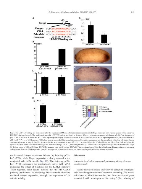
Fig. 5. The LEF/TCF binding site is responsible for the expression of Mespo. (A) Schematic representation of Mespo promoters from various species with a conserved<br />
LEF/TCF binding site (red). The position of potential LEF/TCF binding site (box) in Xenopus Mespo 5′-upstream sequence is indicated. (B–D) Fold induction of<br />
pCS–Lef1–VP16 in pGL3basic and p-4317Luc reporter plasmids (B), luciferase activities of p-4317Luc and p-4317mLuc reporter plasmids (C) or fold induction of<br />
pCS–Lef1–VP16 in p-4317Luc and p-4317mLuc reporter plasmids (D) in animal cap assays. Plasmids were injected into the animal pole at the four-cell stage. Animal<br />
caps were dissected at stage 8.5 and luciferase activities were measured at stage 12.5. RLU, relative light units. (E) Luciferase activities of the indicated plasmids<br />
injected into both VMZ cells at four-cell stage and measured at stage 19. RLU, relative light units. (F) Expression of endogenous Mespo mRNA at the tailbud stage.<br />
(G–I) Expression of GFP mRNA in p-4317GFP transgenic embryos (G) or in p-4317mGFP transgenic embryos (H) at the tailbud stage. The percentages of transgenic<br />
embryos that show the PSM expression (purple), non-specific expression (brown), and no detected signal (white) are shown in panel I.<br />
Discussion<br />
Mespo is involved in segmental patterning during Xenopus<br />
somitogenesis<br />
Mespo knock out mouse shows severe defects in somitogenesis,<br />
including perturbation of segmental patterning. The mutant<br />
mice have no identifiable somites, and the expression of genes<br />
associated with somitogenesis like Mesp2 (the ortholog of<br />
843