2007-DevelopmentalBiology.pdf
2007-DevelopmentalBiology.pdf
2007-DevelopmentalBiology.pdf

You also want an ePaper? Increase the reach of your titles
YUMPU automatically turns print PDFs into web optimized ePapers that Google loves.
844 J. Wang et al. / Developmental Biology 304 (<strong>2007</strong>) 836–847<br />
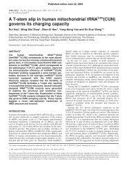
Fig. 6. Lef1 binds to site L in the Mespo promoter in vivo. (A) ChIP assay of<br />
embryos injected with pCS–Lef1–HA and p-511, or pCS–Lef1–HA and p-<br />
511m in both VMZ cells at four-cell stage to detect the binding of Lef1 to site L.<br />
(B) ChIP assay detecting the binding of Lef1 to endogenous Mespo regulatory<br />
sequence. pCS–Lef1–HA was injected into both VMZ cells of embryos at fourcell<br />
stage.<br />
Xenopus Thy), PAPC and Dll-3 (the ortholog of XDelta-1) is<br />
abolished in absence of Mespo (Yoon and Wold, 2000). This<br />
phenotype implies that Mespo is involved in establishing<br />
segmental patterning in somitogenesis. Here, we report that in<br />
Xenopus embryos depleting Mespo by Mespo Mo causes<br />
disorder of somite cell arrangement. During Xenopus somitogenesis,<br />
the rostral PSM cells compact to form cell aggregates<br />
named somitomeres, which later separate from the PSM to form<br />
somites. The nuclei of somite cells line up in the middle of each<br />
somite, representing the well-organized segmental pattern.<br />
There is no such distinct morphological feature in Mespodepleting<br />
Xenopus embryos. Instead, nuclei are randomly<br />
distributed, indicating the disorganization of somite cells (Fig.<br />
1H). The expression patterns of genes associated with<br />
segmental patterning are also perturbed (Fig. 2). Thus, our<br />
data indicate that Mespo functions in establishing the segmental<br />
pattern by means of regulating the genes involved in Xenopus<br />
somitogenesis.<br />
Wnt/β-catenin signaling controls gene expressions in<br />
somitogenesis<br />
Previous studies have demonstrated that Wnt3a is involved<br />
in somitogenesis (Aulehla et al., 2003; Galceran et al., 2004;<br />
Hofmann et al., 2004; Ikeya and Takada, 2001). In chick<br />
embryos, increasing Wnt/β-catenin signaling activity by<br />
implanting beads covered with Wnt3a expressing cells induced<br />
smaller somites (Aulehla et al., 2003). In mouse embryos, it<br />
has been proposed that Wnt3a is involved in somitogenesis via<br />
Axin2 (Aulehla et al., 2003) and Wnt/β-catenin signaling<br />
controlled segmentation by directly regulating Dll1 expression<br />
(Galceran et al., 2004; Hofmann et al., 2004). However, the<br />
effect of Wnt/β-catenin signaling in somitogenesis has not<br />
been fully understood. Our study provides new information on<br />
the role of Wnt/β-catenin signaling in somitogenesis. We<br />
checked XWnt3a and XWnt8 expression in Xenopus embryos<br />
and found that XWnt3a expression is restricted in neural crest<br />
cells, while XWnt8 expression is detected in the caudal PSM<br />
(Figs. 3A, B, E–H). Furthermore, the Wnt antagonist Dkk1 is<br />
expressed in somitomeres (Figs. 3C–F). These observations<br />
suggested that the activity of Wnt/β-catenin signaling exists in<br />
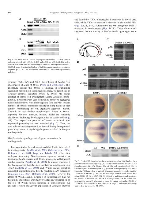
Fig. 7. PI3-K/AKT signaling regulates Mespo expression. (A) Hatched lines<br />
indicate the three caudal regions (C, R, and S) used for western blot of Akt and<br />
phosphorylated Akt. (B) Western blot of Akt and phosphorylated Akt in<br />
different regions indicated in panel A. (C) Western blot of phosphorylated Akt in<br />
the caudal PSM equivalent to region C (illustrated in panel A) treated with either<br />
LY294002 or DMSO. (D–G) The neurula stage embryos were treated with<br />
either DMSO (D, F), or LY294002 (E, G) for 2 h, and then stained for Mespo<br />
and Paraxis as indicated. (H) RT–PCR of Mespo mRNA in the caudal PSM<br />
equivalent to region C (illustrated in panel A) treated with either DMSO or<br />
LY294002. The caudal PSM were dissected at stage 23 and treated with drugs<br />
for 2 h, then harvested for RT–PCR.