

Buccinator myomucosal flap - Vula - University of Cape Town
Buccinator myomucosal flap - Vula - University of Cape Town
Buccinator myomucosal flap - Vula - University of Cape Town

Create successful ePaper yourself
Turn your PDF publications into a flip-book with our unique Google optimized e-Paper software.
OPEN ACCESS ATLAS OF OTOLARYNGOLOGY, HEAD &<br />
NECK OPERATIVE SURGERY<br />
BUCCINATOR MYOMUCOSAL FLAP Johan Fagan<br />
The <strong>Buccinator</strong> Myomucosal Flap is an<br />
axial <strong>flap</strong>, based on the facial and/or buccal<br />
arteries. It is a flexible and versatile <strong>flap</strong><br />
well suited to reconstructing s<strong>of</strong>t tissue<br />
defects <strong>of</strong> the oral cavity, oropharynx and<br />
nasal septum. Unlike most free <strong>flap</strong>s, it<br />
provides mucosal cover, as opposed to skin<br />
cover, and is sensate. The donor site can<br />
usually be closed primarily without<br />
causing deformity or scarring. The <strong>flap</strong> is<br />
about 5mm thick, and comprises buccal<br />
mucosa, submucosa and buccinator<br />
muscle, with the feeding vessels and<br />
vascular plexus.<br />
Relevant Anatomy<br />
<strong>Buccinator</strong> muscle<br />
The buccinator muscle is a thin,<br />
quadrilateral muscle in the cheek. It<br />
originates from the outer surfaces <strong>of</strong> the<br />
alveolar processes <strong>of</strong> the maxilla and<br />
mandible. Posteriorly it arises from the<br />
pterygomandibular raphe. Anteriorly it<br />
inserts into the orbicularis oris muscle.<br />
Laterally it is related to the ramus <strong>of</strong> the<br />
mandible, the masseter and medial<br />
pterygoid muscles, the buccal fat pad and<br />
the buccopharyngeal fascia. Medially, it is<br />
covered by the submucosa and mucosa <strong>of</strong><br />
the cheek. It is part <strong>of</strong> the pharyngealbuccal-orbicularis<br />
sphincter system and<br />
functions to facilitate whistling, sucking,<br />
propelling food during mastication and<br />
voiding the buccal cavity.<br />
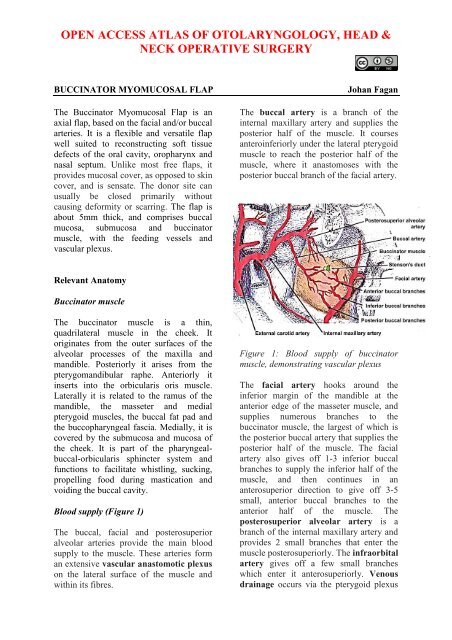
Blood supply (Figure 1)<br />
The buccal, facial and posterosuperior<br />
alveolar arteries provide the main blood<br />
supply to the muscle. These arteries form<br />
an extensive vascular anastomotic plexus<br />
on the lateral surface <strong>of</strong> the muscle and<br />
within its fibres.<br />
The buccal artery is a branch <strong>of</strong> the<br />
internal maxillary artery and supplies the<br />
posterior half <strong>of</strong> the muscle. It courses<br />
anteroinferiorly under the lateral pterygoid<br />
muscle to reach the posterior half <strong>of</strong> the<br />
muscle, where it anastomoses with the<br />
posterior buccal branch <strong>of</strong> the facial artery.<br />
Figure 1: Blood supply <strong>of</strong> buccinator<br />
muscle, demonstrating vascular plexus<br />
The facial artery hooks around the<br />
inferior margin <strong>of</strong> the mandible at the<br />
anterior edge <strong>of</strong> the masseter muscle, and<br />
supplies numerous branches to the<br />
buccinator muscle, the largest <strong>of</strong> which is<br />
the posterior buccal artery that supplies the<br />
posterior half <strong>of</strong> the muscle. The facial<br />
artery also gives <strong>of</strong>f 1-3 inferior buccal<br />
branches to supply the inferior half <strong>of</strong> the<br />
muscle, and then continues in an<br />
anterosuperior direction to give <strong>of</strong>f 3-5<br />
small, anterior buccal branches to the<br />
anterior half <strong>of</strong> the muscle. The<br />
posterosuperior alveolar artery is a<br />
branch <strong>of</strong> the internal maxillary artery and<br />
provides 2 small branches that enter the<br />
muscle posterosuperiorly. The infraorbital<br />
artery gives <strong>of</strong>f a few small branches<br />
which enter it anterosuperiorly. Venous<br />
drainage occurs via the pterygoid plexus
and internal maxillary vein. It lies<br />
posterior, superior and superficial to the<br />
buccinator muscle and drains into the<br />
buccal vein via the deep facial vein.<br />
Anteriorly, the deep facial vein drains into<br />
the facial vein proper.<br />
Innervation<br />
Mucosal sensory innervation is by the<br />
long buccal nerve, a branch <strong>of</strong> the<br />
maxillary division <strong>of</strong> the trigeminal nerve,<br />
which courses with the buccal branch <strong>of</strong><br />
the internal maxillary artery. Motor<br />
innervation <strong>of</strong> the buccinator muscle is<br />
via the temporal and cervical divisions <strong>of</strong><br />
the facial nerve laterally in the buccal fat<br />
pad.<br />
Parotid duct<br />
The duct pierces the buccinator muscle<br />
opposite the 2 nd upper molar, slightly<br />
above the center <strong>of</strong> the muscle, and should<br />
be identified and preserved when raising<br />
the <strong>flap</strong>.<br />
Elevation <strong>of</strong> <strong>flap</strong><br />
A <strong>flap</strong> as large as 7x5cm may be raised.<br />
The extent is limited by the parotid duct<br />
posterosuperiorly, the oral commissure<br />
anteriorly and the pterygomandibular raphe<br />
posteriorly. It may be based posteriorly<br />
(buccal artery), or anteriorly (facial<br />
artery) or superiorly (facial artery).<br />
Although not essential, a handheld Doppler<br />
can be used to map out the facial artery<br />
and the buccal artery before raising the<br />
<strong>flap</strong> (Figure 2). If a Doppler is not used,<br />
then care has to be taken to place the initial<br />
incisions so as not to separate the vessels<br />
from the <strong>flap</strong>.<br />
The buccal mucosa and the buccinator<br />
muscle are incised to the level <strong>of</strong> the<br />
buccopharyngeal fascia, and the <strong>flap</strong><br />
elevated, generally in an anterior to<br />
posterior direction. The <strong>flap</strong> is elevated in<br />
the loose areolar plane between the<br />
buccinator muscle and the<br />
buccopharyngeal fascia.<br />
Figure 2: Vessels mapped out with<br />
handheld Doppler probe<br />
The artery that the <strong>flap</strong> will be based on is<br />
identified, and dissection proceeds<br />
between the artery and the<br />
buccopharyngeal fascia, directed towards<br />
the origin <strong>of</strong> the vascular pedicle, keeping<br />
the vessel in full view, using dissecting<br />
scissors (Figures 3, 4). Preservation <strong>of</strong> the<br />
buccopharyngeal fascia prevents herniation<br />
<strong>of</strong> buccal fat and avoids injury to branches<br />
<strong>of</strong> the facial nerve. Minor bleeding vessels<br />
are coagulated with bipolar electrocautery.<br />
Figure 3: Flap elevation commences after<br />
identifying artery (Black arrow)<br />
2
Figure 4: Flap elevated keeping artery in<br />
view<br />
The <strong>flap</strong> is rotated to fill the s<strong>of</strong>t tissue<br />
defect. The mucosa and muscle are<br />
generally not divided where the <strong>flap</strong> is<br />
pedicled. The pedicle may however be<br />
isolated to facilitate rotation, and to create<br />
a ‘buccinator <strong>myomucosal</strong> neurovascular<br />
island pedicle <strong>flap</strong>’. If the<br />
pedicle has to cross the alveolus e.g. to<br />
reach the floor <strong>of</strong> mouth, then care has to<br />
be taken that the pedicle does not interpose<br />
between the molar teeth, as this may<br />
interfere with mastication and might injure<br />
the pedicle. In such cases some teeth may<br />
need to be extracted to create space for the<br />
pedicle, or an island <strong>flap</strong> has to be created;<br />
alternatively, the vascular pedicle may be<br />
divided after a delay <strong>of</strong> a few weeks. Due<br />
to the elasticity <strong>of</strong> the buccal mucosa, the<br />
donor site can generally be closed<br />
primarily with vicryl absorbable sutures.<br />
Posteriorly Based <strong>Buccinator</strong> Flap<br />
This <strong>flap</strong> can be rotated to reconstruct<br />
defects <strong>of</strong> the oropharynx (s<strong>of</strong>t palate, base<br />
<strong>of</strong> tongue, tonsil fossa), lateral floor <strong>of</strong><br />
mouth, and lateral tongue. It is based<br />
posteriorly on the buccal artery and<br />
buccal venous plexus (Figure 5).<br />
Once the course <strong>of</strong> the buccal artery has<br />
been identified with Doppler ultrasound,<br />
Figure 5: Blood supply <strong>of</strong> posteriorly<br />
based buccinator <strong>flap</strong><br />
the buccal mucosa and the buccinator<br />
muscle are incised to the level <strong>of</strong> the<br />
buccopharyngeal fascia, and the <strong>flap</strong><br />
elevated in an anterior-to-posterior<br />
direction up to the pterygomandibular<br />
raphe where the neurovascular bundle<br />
enters the <strong>flap</strong>. The mucosa at the posterior<br />
end <strong>of</strong> the <strong>flap</strong> may also be divided from<br />
the underlying muscle and freed <strong>of</strong> its<br />
insertion to the pterygomandibular raphe,<br />
and passed through a short tunnel under<br />
the pterygomandibular ligament.<br />
Anteriorly Based <strong>Buccinator</strong> Flap<br />
The anteriorly based <strong>flap</strong> is well suited to<br />
reconstructing s<strong>of</strong>t tissue defects <strong>of</strong> the<br />
inferior alveolus, lateral and anterior floor<br />
<strong>of</strong> mouth and oral tongue and lower lip<br />
(Figure 6). It is based anteroinferiorly on<br />
the inferior and posterior buccal branches<br />
<strong>of</strong> the facial artery (Figure 7). It is<br />
however essential that the facial artery<br />
be preserved when dissecting Level 1b <strong>of</strong><br />
the neck. This requires division and<br />
ligation <strong>of</strong> branches <strong>of</strong> the facial artery<br />
where it passes through or alongside the<br />
submandibular salivary gland (Figure 8).<br />
3
Figure 6: Flap for anterior floor <strong>of</strong> mouth<br />
Figure 7: Blood supply <strong>of</strong> anteriorly based<br />
buccinator <strong>flap</strong><br />
Figure 8: Preserved facial artery in Level<br />
1<br />
The mucosa and the buccinator muscle are<br />
incised just posterior (approx 1cm) to the<br />
commissure <strong>of</strong> the mouth, and the facial<br />
artery is identified in the cheek. The artery<br />
is ligated and divided superiorly. The<br />
dissection continues in a plane lateral to<br />
the vessels, as the <strong>flap</strong> is raised from frontto-back,<br />
and from superiorly-to-inferiorly.<br />
Superiorly Based <strong>Buccinator</strong> Flap<br />
This is a reversed-flow <strong>flap</strong> based on the<br />
facial artery and its anterior buccal<br />
branches (Figure 9).<br />
Figure 9: Blood supply <strong>of</strong> superiorly based<br />
buccinator <strong>flap</strong><br />
It can be used for s<strong>of</strong>t tissue defects <strong>of</strong> the<br />
hard palate and superior alveolus (Figure<br />
10a), including oroantral and oronasal<br />
fistulae, as well as for nasal nasal septal<br />
defects.<br />
Figure 10a: Defect into antrum following<br />
alveolectomy<br />
4
Dissection is commenced at the<br />
anterior/inferior margin <strong>of</strong> the <strong>flap</strong>, where<br />
the facial artery is identified and ligated;<br />
the <strong>flap</strong> is then elevated in a superior<br />
direction, and inset with vicryl sutures. If<br />
the donor defect cannot be completely<br />
closed, then it is left to open to mucosalise<br />
(Figures 10b-d).<br />
<strong>Buccinator</strong><br />
Facial art<br />
Figure 10b: Elevating <strong>flap</strong> after mapping<br />
facial artery with Doppler (purple line)<br />
Figure 10c: Superiorly based pedicle with<br />
facial artery ligated at level <strong>of</strong> mandible<br />
Figure 10d: Flap inset to cover antral<br />
palatal defect<br />
Benefits <strong>of</strong> buccinator <strong>flap</strong><br />
The buccinator <strong>flap</strong> is a versatile <strong>flap</strong> that<br />
can be used to reconstruct a variety <strong>of</strong><br />
defects. It is very reliable, simple and<br />
quick to raise, replaces mucosa with<br />
mucosa, is sensate, and has very little<br />
donor site morbidity. It is remarkably<br />
elastic and malleable, and can be stretched<br />
to conform to complexly shaped defects<br />
and is an excellent alternative to radial free<br />
forearm <strong>flap</strong>s for reconstruction <strong>of</strong> small<br />
and moderate sized defects <strong>of</strong> the oral<br />
cavity and oropharynx (Figures 11 a, b, c).<br />
Figure 11a: Defect following anterolateral<br />
floor <strong>of</strong> mouth resection and<br />
marginal mandibulectomy<br />
5
Figure 11 b, c: Inset <strong>of</strong> <strong>flap</strong><br />
Useful reference<br />
Van Lierop A, Fagan JJ. <strong>Buccinator</strong><br />
<strong>myomucosal</strong> <strong>flap</strong>: Clinical results and<br />
review <strong>of</strong> anatomy, surgical technique and<br />
applications. J Laryngol Otol, 2008; 122:<br />
181-7<br />
Author & Editor<br />
Johan Fagan MBChB, FCORL, MMed<br />
Pr<strong>of</strong>essor and Chairman<br />
Division <strong>of</strong> Otolaryngology<br />
<strong>University</strong> <strong>of</strong> <strong>Cape</strong> <strong>Town</strong><br />
<strong>Cape</strong> <strong>Town</strong>, South Africa<br />
johannes.fagan@uct.ac.za<br />
THE OPEN ACCESS ATLAS OF<br />
OTOLARYNGOLOGY, HEAD &<br />
NECK OPERATIVE SURGERY<br />
www.entdev.uct.ac.za<br />
The Open Access Atlas <strong>of</strong> Otolaryngology, Head &<br />
Neck Operative Surgery by Johan Fagan (Editor)<br />
johannes.fagan@uct.ac.za is licensed under a Creative<br />
Commons Attribution-NonCommercial 3.0 Unported<br />
License<br />
6


