Download - Akademi Sains Malaysia
Download - Akademi Sains Malaysia
Download - Akademi Sains Malaysia

You also want an ePaper? Increase the reach of your titles
YUMPU automatically turns print PDFs into web optimized ePapers that Google loves.
8<br />
JJournal of Science and Technology in the Tropics (2010) 6: 5-9<br />
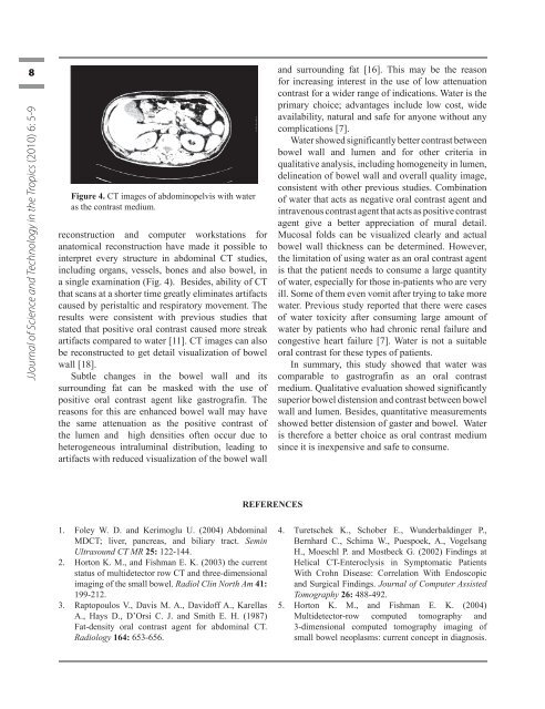
Figure 4. CT images of abdominopelvis with water<br />
as the contrast medium.<br />
reconstruction and computer workstations for<br />
anatomical reconstruction have made it possible to<br />
interpret every structure in abdominal CT studies,<br />
including organs, vessels, bones and also bowel, in<br />
a single examination (Fig. 4). Besides, ability of CT<br />
that scans at a shorter time greatly eliminates artifacts<br />
caused by peristaltic and respiratory movement. The<br />
results were consistent with previous studies that<br />
stated that positive oral contrast caused more streak<br />
artifacts compared to water [11]. CT images can also<br />
be reconstructed to get detail visualization of bowel<br />
wall [18].<br />
Subtle changes in the bowel wall and its<br />
surrounding fat can be masked with the use of<br />
positive oral contrast agent like gastrografin. The<br />
reasons for this are enhanced bowel wall may have<br />
the same attenuation as the positive contrast of<br />
the lumen and high densities often occur due to<br />
heterogeneous intraluminal distribution, leading to<br />
artifacts with reduced visualization of the bowel wall<br />
1. Foley W. D. and Kerimoglu U. (2004) Abdominal<br />
MDCT; liver, pancreas, and biliary tract. Semin<br />
Ultrasound CT MR 25: 122-144.<br />
2. Horton K. M., and Fishman E. K. (2003) the current<br />
status of multidetector row CT and three-dimensional<br />
imaging of the small bowel. Radiol Clin North Am 41:<br />
199-212.<br />
3. Raptopoulos V., Davis M. A., Davidoff A., Karellas<br />
A., Hays D., D’Orsi C. J. and Smith E. H. (1987)<br />
Fat-density oral contrast agent for abdominal CT.<br />
Radiology 164: 653-656.<br />
<br />
REFERENCES<br />
and surrounding fat [16]. This may be the reason<br />
for increasing interest in the use of low attenuation<br />
contrast for a wider range of indications. Water is the<br />
primary choice; advantages include low cost, wide<br />
availability, natural and safe for anyone without any<br />
complications [7].<br />
Water showed significantly better contrast between<br />
bowel wall and lumen and for other criteria in<br />
qualitative analysis, including homogeneity in lumen,<br />
delineation of bowel wall and overall quality image,<br />
consistent with other previous studies. Combination<br />
of water that acts as negative oral contrast agent and<br />
intravenous contrast agent that acts as positive contrast<br />
agent give a better appreciation of mural detail.<br />
Mucosal folds can be visualized clearly and actual<br />
bowel wall thickness can be determined. However,<br />
the limitation of using water as an oral contrast agent<br />
is that the patient needs to consume a large quantity<br />
of water, especially for those in-patients who are very<br />
ill. Some of them even vomit after trying to take more<br />
water. Previous study reported that there were cases<br />
of water toxicity after consuming large amount of<br />
water by patients who had chronic renal failure and<br />
congestive heart failure [7]. Water is not a suitable<br />
oral contrast for these types of patients.<br />
In summary, this study showed that water was<br />
comparable to gastrografin as an oral contrast<br />
medium. Qualitative evaluation showed significantly<br />
superior bowel distension and contrast between bowel<br />
wall and lumen. Besides, quantitative measurements<br />
showed better distension of gaster and bowel. Water<br />
is therefore a better choice as oral contrast medium<br />
since it is inexpensive and safe to consume.<br />
4. Turetschek K., Schober E., Wunderbaldinger P.,<br />
Bernhard C., Schima W., Puespoek, A., Vogelsang<br />
H., Moeschl P. and Mostbeck G. (2002) Findings at<br />
Helical CT-Enteroclysis in Symptomatic Patients<br />
With Crohn Disease: Correlation With Endoscopic<br />
and Surgical Findings. Journal of Computer Assisted<br />
Tomography 26: 488-492.<br />
5. Horton K. M., and Fishman E. K. (2004)<br />
Multidetector-row computed tomography and<br />
3-dimensional computed tomography imaging of<br />
small bowel neoplasms: current concept in diagnosis.<br />
Jostt vol 6.indd 8 7/22/10 10:08:32 PM