Anti-Human ICE (p20 subunit) Catalog # 06-503 Lot ... - Millipore
Anti-Human ICE (p20 subunit) Catalog # 06-503 Lot ... - Millipore
Anti-Human ICE (p20 subunit) Catalog # 06-503 Lot ... - Millipore
You also want an ePaper? Increase the reach of your titles
YUMPU automatically turns print PDFs into web optimized ePapers that Google loves.
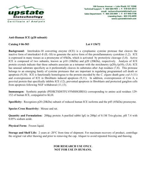
<strong>Anti</strong>-<strong>Human</strong> <strong>ICE</strong> (<strong>p20</strong> <strong>subunit</strong>)<br />
<strong>Catalog</strong> # <strong>06</strong>-<strong>503</strong> <strong>Lot</strong> # 15672<br />
Background: Interleukin-1ß converting enzyme (<strong>ICE</strong>) is a cytoplasmic cysteine protease that cleaves the<br />
inactive form of interleukin1-ß (IL-1ß) to generate the active form of this proinflammatory cyotokine (1,2). <strong>ICE</strong><br />
is expressed in many tissues as a proenzyme of 45kDa, which is activated by proteolytic cleavage (3,4). Active<br />
<strong>ICE</strong> is composed of two <strong>subunit</strong>s, known as p10 (10kDa) and <strong>p20</strong> (20kDa), respectively. Analysis of <strong>ICE</strong><br />
protein crystals indicate that these <strong>subunit</strong>s associate as a tetramer with the stoichmetry (<strong>p20</strong>)2/(p10)2 (5,6). <strong>ICE</strong><br />
has unusual substrate specificity as it preferentially cleaves its substrates after Asp residues (7,8). This protease<br />
belongs to an emerging family of cysteine proteases that are important in regulating programmed cell death or<br />
apoptosis (9,10). <strong>ICE</strong> is functionally homologous to the protein encoded by the C. elgans death gene ced-3 (11)<br />
and overexpression of <strong>ICE</strong> in fibroblasts induced apoptosis (9,11). In addition, overexpression of Crm A, a<br />
poxviral protein that specifically inhibits <strong>ICE</strong> (12), prevented apoptosis in fibroblasts and protected ganglion cells<br />
from apoptosis following NGF withdrawal (11,13).<br />
Immunogen: Synthetic peptide (PEHKTSDSTFLVFMSHGIREG) corresponding to amino acid residues 129-<br />
152 of human <strong>ICE</strong>, conjugated to KLH.<br />
Specificity: Recognizes <strong>p20</strong> (20kDa) <strong>subunit</strong> of reduced human <strong>ICE</strong> isoforms and the p45 (45kDa) proenzyme.<br />
Species Cross Reactivity: Mouse and rat.<br />
Quantity and Formulation: 200μg protein A-purified rabbit IgG in 200μl of 0.1M Tris-glycine, pH 7.4 with<br />
0.05% sodium azide.<br />
Physical Form: Frozen liquid.<br />
Storage and Shelf Life: 2 years at -20°C from time of shipment. For maximum recovery of product, centrifuge<br />
the original vial after thawing and prior to removing the cap. Aliquot to avoid repeated freezing and thawing.<br />
FOR RESEARCH USE ONLY.<br />
NOT FOR USE IN HUMANS.
Page Two of Three<br />
<strong>Catalog</strong> # <strong>06</strong>-<strong>503</strong><br />
<strong>Lot</strong> # 15672<br />
Background References:<br />
1. Black, R.A., et. al., FEBS Lett. 247: 386-390, (1989).<br />
2. Kostura, M.J., et. al., Proc. Natl. Acad. Sci. USA 86: 5227-5231 (1989).<br />
3. Cerreti, D.P., et. al., Science 256: 97-100, (1992).<br />
4. Thornberry, N.A., et. al., Nature 356: 768-774, (1992).<br />
5. Wilson, K.P., et. al., Nature 370: 270-275, (1994).<br />
6. Walker, N.P.C., et. al., Cell 78: 343-352, (1994).<br />
7. Sealth, P.R., et. al., J. Biol. Chem. 265: 14526-14528, (1990).<br />
8. Howard, A.D., et. al., J. Immunol. 147: 2964-2969, (1991).<br />
9. Kumar, S., et. al., Genes & Dev. 8: 1613-1626, (1994).<br />
10. Yuan, J., et. al., Cell 75: 641-652, (1993).<br />
11. Miura, M., et. al., Cell 75: 653-660, (1993).<br />
12. Ray, C., et. al., Cell 69: 597-604, (1992).<br />
13. Gagliardini, V., et. al., Science 263: 826-828, (1994)<br />
Additional References:<br />
Molineaux, et. al., PNAS 90: 1809-1813, (1993).<br />
Alnemvx, et. al., J. Biol. Chem. 270(9): 4312-4317, (1995).<br />
Quality Control Testing and Research Applications<br />
Western Immunoblot Analysis: Used at 0.5-2μg/ml, this lot of antibody detected the p45 proenzyme form of<br />
<strong>ICE</strong> in 20μg of cell lysate from human HL-60 cells and mouse ABE 8 1/2 cells, respectively. The<br />
immunoreactivity can be inhibited by the immunizing peptide.<br />
Immunoprecipitation: 5μg of this lot of antibody immunoprecipitated p45 proenzyme form of <strong>ICE</strong> from 500μg<br />
of human HL-60 cell lysates.<br />
Immunocytochemistry: At 10μg/ml, this lot of antibody gave positive immunostaining of HL60 cells.<br />
Western Immunoblot Protocol<br />
1. Perform SDS-polyacrylamide gel electrophoresis (SDS-PAGE) on a cell lysate sample (cell lysis buffer:<br />
50mM Tris-HCl, pH 7.4; 1% NP-40; 0.25% sodium deoxycholate; 150mM NaCl; 1mM EGTA; 1mM PMSF;<br />
1μg/ml aprotinin, leupeptin, pepstatin; 1mM Na3VO4; 1mM NaF) and transfer the proteins to nitrocellulose.<br />
Wash the blotted nitrocellulose twice with water.<br />
2. Block the blotted nitrocellulose in freshly prepared PBS containing 3% nonfat dry milk (MLK) for 20 minutes<br />
at 20-25°C with constant agitation.<br />
3. Incubate the nitrocellulose with 0.5-2mg/ml of a-<strong>Human</strong> <strong>ICE</strong> (<strong>p20</strong> <strong>subunit</strong>), diluted in freshly prepared<br />
PBS-MLK overnight with agitation at 4°C.<br />
4. Wash the nitrocellulose twice with water.<br />
5. Incubate the nitrocellulose in the secondary reagent of choice (a goat anti-rabbit IgG linked to horseradish<br />
peroxidase, 1:3000 dilution, was used) in PBS-MLK for 1.5 hours at room temperature with agitation.<br />
6. Wash the nitrocellulose with water twice.<br />
7. Wash the nitrocellulose in PBS-0.05% Tween 20 for 3-5 minutes.<br />
8. Rinse the nitrocellulose in 4-5 changes of water.<br />
9. Use detection method of choice (enhanced chemiluminescence was used).
Page Three of Three<br />
<strong>Catalog</strong> # <strong>06</strong>-<strong>503</strong><br />
<strong>Lot</strong> # 15672<br />
Immunoprecipitation Protocol<br />
1. Before beginning the immunoprecipitation, dilute the cell lysate to roughly 1μg/μl total cell protein in a<br />
microcentrifuge tube with PBS.<br />
2. Add 5mg of a-<strong>Human</strong> <strong>ICE</strong> (<strong>p20</strong> <strong>subunit</strong>), to 500μg-1mg cell lysate.<br />
3. Gently rock the reaction mixture at 4°C overnight.<br />
4. Capture the immunocomplex by adding 100μl of washed Protein G or A agarose bead slurry (50μl packed<br />
beads).<br />
5. Gently rock the reaction mixture at 4°C for 2 hours.<br />
6. Collect the agarose beads by pulsing (5 seconds in the microcentrifuge at 14,000 x g), and drain off the<br />
supernatant. Wash the beads 3 times with either ice-cold cell lysis buffer or PBS.<br />
7. Resuspend the agarose beads in 50μl 2X Laemmli sample buffer and boil for 5 minutes. Collect the beads by a<br />
microcentrifuge pulse. SDS-PAGE and subsequent immunoblot analysis can be performed on a sample of the<br />
supernatant, or the agarose beads can then be frozen for later use and reboiled for 5 minutes prior to SDS-<br />
PAGE.<br />
Immunocytochemistry<br />
1. Plate approximately 200μl of cell suspension into each well of a slide. Incubate 24 hr. in a 37°C CO2<br />
incubator.<br />
2. Wash the cells three times for 5 minutes with PBS. Do not shake cells.<br />
3. Add fix (ice-cold ethanol/acetic acid [95:5]) for 1 minute at room temperature.<br />
4. Wash the cells with PBS, twice, for 15 minutes. Do not shake.<br />
5. Cover the cells with of 8% albumin in PBS and incubate for 30 minutes at room temperature.<br />
6. Wash the cells two times with PBS, for 15 minutes per wash.<br />
7. Cover the cells with 10mg/ml a-<strong>Human</strong> <strong>ICE</strong> (<strong>p20</strong> <strong>subunit</strong>), in 1% albumin in PBS and incubate overnight at<br />
4°C in a humidified chamber.<br />
8. Wash the cells twice with PBS, for 15 minutes.<br />
9. Incubate the cells with a 1:100 dilution of goat anti-rabbit IgG fluorescein conjugated secondary antibody in<br />
PBS for 1 hour at room temperature.<br />
10. Wash the cells three times with PBS, for 5 minutes.<br />
11. Examine the cells under a fluorescent microscope.


