impaginato piccolo - Società Italiana di Parassitologia (SoIPa)
impaginato piccolo - Società Italiana di Parassitologia (SoIPa)
impaginato piccolo - Società Italiana di Parassitologia (SoIPa)

Create successful ePaper yourself
Turn your PDF publications into a flip-book with our unique Google optimized e-Paper software.
PARASSITOLOGIA<br />
A publication of the University of Rome La Sapienza<br />
Official Journal of the Italian Society of Parasitology<br />
EDITOR-IN-CHIEF M. Coluzzi<br />
ASSOCIATE/CORRESPONDING EDITORS<br />
General Parasitology L. Sacchi,<br />
Veterinary Parasitology G. Cringoli/D. Otranto<br />
Me<strong>di</strong>cal Parasitology F. Bruschi/E. Pozio<br />
Molecular Parasitology C. Ban<strong>di</strong><br />
Sanitary Parasitology A. della Torre<br />
EDITORIAL BOARD<br />
The Council (2004-2008) of the Italian Society<br />
of Parasitology: F. Bruschi, G. Garippa,<br />
C. Genchi, S. Giannetto, M.T. Manfre<strong>di</strong>,<br />
M. Pietrobelli, E. Pozio, L. Sacchi<br />
ADVISORY BOARD<br />
A. Aeschlimann, P. Ambroise-Thomas, V. Baimai,<br />
D.J. Bradley, R. Carter, A. Chabaud, C. Combes,<br />
C. Curtis, J. de Zulueta, K. Dietz, J.P. Dubey,<br />
T.H. Freyvogel, B.M. Greenwood, C. Louis,<br />
K. Marsh, S.A. Nadler, R.S. Nussenzweig, I.<br />
Paperna, J.M.E. Ribeiro, J.A. Rioux,<br />
D. Rollinson, R. Roncalli, M.W. Service,<br />
J.D. Smyth, Y.T. Touré, J. Vercruysse,<br />
D. Wakelin, D. Walliker, G.B. White<br />
EDITORIAL OFFICE<br />
Dipartimento <strong>di</strong> Scienze <strong>di</strong> Sanità Pubblica<br />
Sezione <strong>di</strong> <strong>Parassitologia</strong> “Ettore Biocca”<br />
Università <strong>di</strong> Roma “La Sapienza”<br />
Piazzale Aldo Moro 5, I-00185 Roma, Italy<br />
Tel ++39 06 4455780<br />
Fax ++39 06 49914653<br />
e-mail: mario.coluzzi@uniroma1.it<br />
PUBLISHER<br />
Lombardo E<strong>di</strong>tore, Divisione Perio<strong>di</strong>ci<br />
Production and Subscription Offices:<br />
Via Centrale 89 (Lama),<br />
I-06013 San Giustino (PG), Italy<br />
Tel ++39 075 8583860<br />
Fax ++39 075 8610415<br />
e-mail: infolombardo@lombardoe<strong>di</strong>tore.it<br />
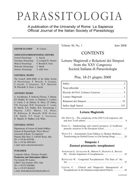
Volume 50, No. 1 June 2008<br />
CONTENTS<br />
Letture Magistrali e Relazioni dei Simposi<br />
from the XXV Congresso<br />
<strong>Società</strong> <strong>Italiana</strong> <strong>di</strong> <strong>Parassitologia</strong><br />
Pisa, 18-21 giugno 2008<br />
In<strong>di</strong>ce . . . . . . . . . . . . . . . . . . . . . . . . . . . . . . . . . . . . . . . I<br />
Nota e<strong>di</strong>toriale . . . . . . . . . . . . . . . . . . . . . . . . . . . . . . . . 3<br />
Ricordo del Prof. Lisimaco Casarosa . . . . . . . . . . . . . . . . . . 5<br />
Letture Magistrali . . . . . . . . . . . . . . . . . . . . . . . . . . . . . . 7<br />
Relazioni dei Simposi . . . . . . . . . . . . . . . . . . . . . . . . . . 33<br />
In<strong>di</strong>ce degli Autori. . . . . . . . . . . . . . . . . . . . . . . . . . . . 151<br />
Lettura Magistrale<br />
DEL PRETE G. - The complexity of the CD4 T-cell responses: old<br />
and new T-cell subsets . . . . . . . . . . . . . . . . . . . . . . . . . . . . . .<br />
POZIO E. - Epidemiology and control prospects of foodborne<br />
parasitic zoonoses in the European Union . . . . . . . . . . . . . . .<br />
WEINA P. J. - Artemisinins from Folklore to Modern Me<strong>di</strong>cine -<br />
Transforming an Herbal Extract to Life-Saving Drugs . . . . . .<br />
Simposio 1<br />
Zoonosi protozoarie: toxoplasmosi<br />
ANTONIAZZI E., GUAGLIANO R., MERONI V., PEZZOTTA S., BIANCHI<br />
P.E. - Ocular impaiment of toxoplasmosis . . . . . . . . . . . . . . .<br />
BUFFOLANO W. - Congenital Toxoplasmosis: The State of the<br />
Art . . . . . . . . . . . . . . . . . . . . . . . . . . . . . . . . . . . . . . . . . . . .<br />
CONTINI C. - Clinical and Diagnostic Management of<br />
Toxoplasmosis in the Immunocompromised Patient . . . . . . .<br />
9<br />
17<br />
25<br />
35<br />
37<br />
45<br />
(continued)
II<br />
P<br />
A R<br />
A SSIT<br />
O L<br />
O GIA<br />
Founded in 1959 by<br />
E. Biocca, A. Corradetti and O. Starkoff<br />
Contents<br />
MERONI V., GENCO F. - Toxoplasmosis in pregnancy: evaluation of<br />
<strong>di</strong>agnostic methods . . . . . . . . . . . . . . . . . . . . . . . . . . . . . . . . .<br />
PFAFF A.W., CANDOLFI E. - New insights in toxoplasmosis<br />
immunology during pregnancy. Perspective for vaccine prevention<br />
. . . . . . . . . . . . . . . . . . . . . . . . . . . . . . . . . . . . . . . . . . . . .<br />
RINALDI L., SCALA A. - Toxoplasmosis in livestock in Italy: an epidemiological<br />
update . . . . . . . . . . . . . . . . . . . . . . . . . . . . . . . .<br />
Simposio 2<br />
Il pianeta Malassezia<br />
CAFARCHIA C., OTRANTO D. - The Pathogenesis of Malassezia<br />
Yeasts . . . . . . . . . . . . . . . . . . . . . . . . . . . . . . . . . . . . . . . . . . .<br />
DIFONZO E.M., FAGGI E. - Skin <strong>di</strong>seases associated with<br />
Malassezia species in humans. Clinical features and <strong>di</strong>agnostic<br />
criteria . . . . . . . . . . . . . . . . . . . . . . . . . . . . . . . . . . . . . . . . . . .<br />
GALUPPI R., TAMPIERI M.P. - Epidemiology and variability of<br />
Malassezia spp. . . . . . . . . . . . . . . . . . . . . . . . . . . . . . . . . . . . .<br />
GUILLOT J., HADINA S., GUÉHO E. - The genus Malassezia: old<br />
facts and new concepts . . . . . . . . . . . . . . . . . . . . . . . . . . . . . .<br />
NARDONI S., CORAZZA M., MANCIANTI F. - Diagnostic and clinical<br />
features of animal malasseziosis . . . . . . . . . . . . . . . . . . . . . . .<br />
PEANO A., GALLO M.G. - Management of Malassezia-related<br />
<strong>di</strong>seases in the dog . . . . . . . . . . . . . . . . . . . . . . . . . . . . . . . . .<br />
PISSERI F., BERTOLI A., PISTELLI L.Essential oils in me<strong>di</strong>cine: principles<br />
of therapy . . . . . . . . . . . . . . . . . . . . . . . . . . . . . . . . . . .<br />
ROMANO C. - Uncommon cases of pityriasis versicolor . . . . . . .<br />
Simposio 3<br />
Aedes albopictus in Italia<br />
ANGELINI P., MACINI P., FINARELLI A.C., PO C., VENTURELLI C.,<br />
BELLINI R., DOTTORI M. - Chikungunya epidemic outbreak in<br />
Emilia-Romagna (Italy) during summer 2007 . . . . . . . . . . . . .<br />
BASEGGIO A. - Availability of insecticidal molecules to control<br />
Aedes albopictus (Skuse) . . . . . . . . . . . . . . . . . . . . . . . . . . . .<br />
VALERIO L., MARINI F., BONGIORNO G., FACCHINELLI L., POMBI M.,<br />
CAPUTO B., MAROLI M., DELLA TORRE A. - Blood-fee<strong>di</strong>ng preferences<br />
of Aedes albopictus (Diptera: Culicidae) in urban and<br />
rural settings within the Province of Rome, Italy . . . . . . . . . .<br />
CIGNINI B., DI DOMENICANTONIO R., CHIONNE M., SCIROCCHI A. -<br />
Decennial experience of the Municipality of Rome in the fight<br />
against Asian Tiger Mosquito . . . . . . . . . . . . . . . . . . . . . . . . .<br />
51<br />
55<br />
59<br />
65<br />
69<br />
73<br />
77<br />
81<br />
85<br />
89<br />
93<br />
97<br />
99<br />
103<br />
105<br />
(continued)
P<br />
A R<br />
A SSIT<br />
O L<br />
O GIA<br />
Founded in 1959 by<br />
E. Biocca, A. Corradetti and O. Starkoff<br />
Contents<br />
NICOLETTI L., CIUFOLINI M.G., FORTUNA C., MAGURANO F., FIO-<br />
RENTINI C., MARCHI A., BENEDETTI E., BUCCI P. - Arboviruses in<br />
Italy . . . . . . . . . . . . . . . . . . . . . . . . . . . . . . . . . . . . . . . . . . . . .<br />
PIETROBELLI M. - Importance of Aedes albopictus in Veterinary<br />
Me<strong>di</strong>cine . . . . . . . . . . . . . . . . . . . . . . . . . . . . . . . . . . . . . . . . .<br />
ROMI R., MAJORI G. - An overview of the lesson learned in almost<br />
20 years of fight against the “Tiger” mosquito . . . . . . . . . . . . .<br />
SEVERINI F., DI LUCA M., TOMA L., ROMI R. - Aedes albopictus in<br />
Rome: results and perspectives after 10 years of monitoring . . .<br />
TALBALAGHI A. - Tiger mosquito Control: new approaches to the<br />
issue in local context . . . . . . . . . . . . . . . . . . . . . . . . . . . . . . . .<br />
Tamburro A. - Control of the Asian tiger mosquito: technical and<br />
administrative aspects . . . . . . . . . . . . . . . . . . . . . . . . . . . . . . .<br />
VENTURELLI C., MASCALI ZEO S., MACINI P., ANGELINI P., BELLINI<br />
R., VERONESI R., MONTANARI M. - A Regional plan of the Emilia-romagna<br />
regional bureau for Aedes albopictus control - year<br />
2008 . . . . . . . . . . . . . . . . . . . . . . . . . . . . . . . . . . . . . . . . . . . .<br />
Simposio 4<br />
Il ruolo della ricerca nella lotta alla Malaria<br />
DELLA TORRE A., ARCA B., FAVIA G., PETRARCA V., COLUZZI M. -<br />
The role of research in molecular entomology in the fight<br />
against malaria vectors . . . . . . . . . . . . . . . . . . . . . . . . . . . . . . .<br />
ESPOSITO F., MAJORI G., COLUZZI M. - The role of research in the<br />
fight against malaria: the Italian contribution to malaria<br />
research in the frame of north-south cooperation in the last 25<br />
years . . . . . . . . . . . . . . . . . . . . . . . . . . . . . . . . . . . . . . . . . . . .<br />
SCHWARZER E., SKOROKHOD O.A., BARRERA V., ARESE P. -<br />
Hemozoin and the human monocyte-A brief review of their<br />
interactions . . . . . . . . . . . . . . . . . . . . . . . . . . . . . . . . . . . . . . .<br />
VERRA F., AVELLINO P., BANCONE G., MANGANO V., MODIANO D. -<br />
Genetic epidemiology of susceptibility to malaria: not only academic<br />
exercises . . . . . . . . . . . . . . . . . . . . . . . . . . . . . . . . . . . .<br />
III<br />
109<br />
113<br />
117<br />
121<br />
125<br />
127<br />
129<br />
BASILICO N., BOSISIO E., BUELLI F., CAMPIANI G., CASAGRANDE M.,<br />
CASTELLI F., COGHI P., CORBETT Y., CORTELEZZI L.,<br />
D’ALESSANDRO S., DELL’AGLI M., ESPOSITO F., FATTORUSSO C.,<br />
FATTORUSSO E., FINAURINI S., GALLI G.V., GEMMA S.,<br />
HABLUETZEL A., LUCANTONI L., MELATO S., MONTI D., OLLIARO<br />
P., OMODEO-SALÈ F., PARAPINI S., PERSICO M., RIZZI M., ROMEO<br />
S., ROSSI F., RUSCONI C., SPARATORE A., TAGLIALATELA SCAFATI<br />
O., VAN DEN BOGAART E., D. TARAMELLI, VAIANA N., YERBANGA<br />
S. - Old and new targets for innovative antimalarial compounds:<br />
the <strong>di</strong>fferent strategies of the Italian Malaria Network . . . . . . . 133<br />
137<br />
141<br />
143<br />
147
SOCIETÀ ITALIANA<br />
DI PARASSITOLOGIA<br />
SOCIETÀ ITALIANA<br />
DI PARASSITOLOGIA<br />
XXV Congresso Nazionale<br />
Letture Magistrali<br />
Relazioni dei Simposi<br />
REGIONE TOSCANA PROVINCIA DI PISA<br />
Comitato Organizzatore<br />
COMUNE DI PISA<br />
Fabrizio Bruschi, Presidente<br />
Francesca Mancianti, Vice-Presidente<br />
Massimo Masetti, Segretario<br />
Barbara Castagna,<br />
Lorena Chiumiento,<br />
Riccardo Forletta,<br />
Marta Magi,<br />
Fabio Macchioni,<br />
Simona Nardoni,<br />
Roberto Papini,<br />
Stefania Perrucci<br />
Azienda Ospedaliero<br />
Universitaria Pisana<br />
ORDINE DEI MEDICI CHIRURGHI<br />
E DEGLI ODONTOIATRI<br />
PROVINCIA DI PISA<br />
Comitato Scientifico<br />
Fabrizio Bruschi,<br />
insieme ai Chairpersons dei Simposi<br />
e delle Sessioni<br />
Pisa, 18-21 Giugno 2008<br />
E<strong>di</strong>tors<br />
F. Bruschi, F. Mancianti<br />
UNIVERSITÀ DI PISA<br />
ORDINE DEI VETERINARI<br />
PROVINCIA DI PISA
The in<strong>di</strong>vidual authors take responsability for linguistic quality of the articles and presentations.
<strong>Parassitologia</strong> 50, 2008<br />
Il Congresso Nazionale della <strong>Società</strong> <strong>Italiana</strong> <strong>di</strong> <strong>Parassitologia</strong> ritorna a Pisa dopo ventidue anni con la sua 25 a e<strong>di</strong>zione,<br />
organizzato dalle componenti me<strong>di</strong>che e veterinarie presenti nel nostro Ateneo.<br />
Esso giunge in un momento particolare della nostra <strong>Società</strong> perché se da un lato il livello della produzione scientifica<br />
è notevolmente aumentato, vedendo molti nostri colleghi firmare articoli sulle riviste internazionali piu’ prestigiose,<br />
non soltanto del settore parassitologico, la componente universitaria che costituisce la maggioranza degli appartenenti<br />
vive uno stato <strong>di</strong> profonda incertezza sul destino del proprio settore <strong>di</strong>sciplinare.<br />
Problematiche emergenti quali l’aumento dei flussi migratori nella nostra Nazione, la <strong>di</strong>ffusione <strong>di</strong> potenziali vettori<br />
<strong>di</strong> agenti virali (Chikungunia e dengue) infezioni opportuniste nei soggetti immunodepressi, soprattutto i trapiantati<br />
d’organo solido e <strong>di</strong> cellule staminali e l’emergere ed il ri-emegere <strong>di</strong> zoonosi parassitarie costituiscono delle sfide<br />
importanti per i Parassitologi italiani. Quest’ultimo problema <strong>di</strong> salute pubblica offre una formidabile opportunità <strong>di</strong><br />
stretta collaborazione tra la componente me<strong>di</strong>ca e quella veterinaria della nostra <strong>Società</strong>.<br />
Il Congresso si articola in quattro Simposi a cui hanno partecipato esperti italiani e stranieri del settore, de<strong>di</strong>cati<br />
a:<br />
1) Zoonosi protozoarie: Toxoplasmosi patrocinato dalla <strong>Società</strong> <strong>Italiana</strong> <strong>di</strong> Malattie Infettive e Tropicali<br />
(SIMIT),<br />
2) Il pianeta Malassezia: storia, epidemiologia, clinica, <strong>di</strong>agnostica e trattamento <strong>di</strong> una zoonosi emergente,<br />
patrocinato dalla Federazione <strong>Italiana</strong> <strong>di</strong> Micologia Umana ed Animale (FIMUA),<br />
3) Aedes albopictus in Italia: da insopportabile parassita a vettore <strong>di</strong> virus Chikungunya<br />
4) Il ruolo della ricerca nella lotta alla Malaria, sotto l’egida del’Italian malaria Network, da poco costituito.<br />
Le sessioni scientifiche, ben 14, comprendono 87 comunicazioni orali e 119 poster sui seguenti argomenti:<br />
Biologia molecolare, Parassiti e fauna acquatica, Immunologia, Patologia e Clinica, Parassiti e fauna selvatica,<br />
Entomologia me<strong>di</strong>ca e veterinaria, Protozoi <strong>di</strong> interesse sanitario, Epidemiologia e <strong>di</strong>agnosi delle malattie parassitarie,<br />
Micologia, Terapia e farmacoresistenza.<br />
In particolare quest’anno sono stati molto numerosi i contributi <strong>di</strong> argomento entomologico, sia in campo me<strong>di</strong>co<br />
che veterinario.<br />
Abbiamo comunque cercato <strong>di</strong> dare anche agli autori <strong>di</strong> poster la possibilità <strong>di</strong> intervenire brevemente, alla fine<br />
delle sessioni orali.<br />
Dalle proposte suggerite sono scaturiti un Workshop intitolato Pig Parasites in Italy from History to Epidemiology<br />
ed una sessione <strong>di</strong> case reports parassitologici, illustrati da infettivologi dei vari ospedali toscani.<br />
Questo fascicolo (Volume 50, No.1 ed il suo supplemmento) della rivista <strong>Parassitologia</strong>, giornale ufficiale della<br />
<strong>Società</strong> <strong>Italiana</strong> <strong>di</strong> <strong>Parassitologia</strong>, raccoglie le letture magistrali, le relazioni dei Simposi e gli abstracts delle comunicazioni<br />
scientifiche e dei poster.<br />
Il fascicolo inizia con un ricordo del Prof. Lisimaco Casarosa, un Maestro della <strong>Parassitologia</strong> italiana, che viene<br />
ricordato con affetto e profonda gratitu<strong>di</strong>ne da chi <strong>di</strong> noi ha qualche hanno <strong>di</strong> più.<br />
Mi è gra<strong>di</strong>to ringraziare tutti coloro che, in varia maniera, hanno contribuito all’organizzazione <strong>di</strong> questo importante<br />
evento e rivolgo un caloroso benvenuto nella città della Torre, con l’augurio <strong>di</strong> un proficuo lavoro.<br />
Pisa, Giugno 2008<br />
NOTA EDITORIALE<br />
Il Presidente del Comitato Organizzatore<br />
Prof. Fabrizio Bruschi
<strong>Parassitologia</strong> 50, 2008<br />
RICORDO DEL PROF. LISIMACO CASAROSA<br />
Con profonda e sincera commozione mi trovo oggi a commemorare il professor Lisimaco Casarosa,<br />
venuto a mancare il 27 agosto 2006 a Pisa.<br />
La prima volta che ho incontrato il professor Casarosa risale al 1975, quando ancora studentessa seguivo il corso<br />
<strong>di</strong> <strong>Parassitologia</strong> veterinaria da Lui tenuto. Mantengo tuttora vivido il ricordo dell’interesse che sapeva suscitare<br />
nello stu<strong>di</strong>o <strong>di</strong> una materia che altrimenti sarebbe potuta risultare arida e nozionistica. La nostra collaborazione<br />
scientifica ha avuto inizio dopo che, nel 1980, sono risultata vincitrice <strong>di</strong> un concorso per ricercatore per l’allora<br />
settore scientifico <strong>di</strong>sciplinare V32B <strong>di</strong> cui il professor Casarosa presiedeva la commissione. Da quel momento è<br />
cominciato un rapporto <strong>di</strong> profonda stima e <strong>di</strong> affetto reciproci che si è concluso solo con la Sua morte.<br />
Nato il 22 marzo 1920 e laureatosi in Me<strong>di</strong>cina Veterinaria a Pisa nel 1943, rivestì il ruolo <strong>di</strong> assistente incaricato presso<br />
l’Istituto <strong>di</strong> Anatomia nel 1945 e fu quin<strong>di</strong> nominato assistente or<strong>di</strong>nario presso la Cattedra <strong>di</strong> Patologia Generale<br />
ed Anatomia Patologica della nostra Facoltà. Dal 1948 al 1950 fu assistente incaricato e successivamente assistente<br />
or<strong>di</strong>nario nell’Istituto <strong>di</strong> Anatomia Patologica dell’Università <strong>di</strong> Pisa. Fu abilitato alla libera docenza in Patologia<br />
Generale ed Anatomia Patologica nel 1954. Dal 1948-49 fino al 1951-52 e 1952-53 fu incaricato degli insegnamenti<br />
<strong>di</strong> <strong>Parassitologia</strong> e <strong>di</strong> Ispezione degli Alimenti <strong>di</strong> Origine Animale, rispettivamente. Dopo una parentesi che lo volle a<br />
Messina richiamato dal suo maestro, il professor Bruno Romboli, e dove per alcuni anni tenne la <strong>di</strong>rezione dell’Istituto<br />
<strong>di</strong> Anatomia Patologica, fece ritorno a Pisa come professore or<strong>di</strong>nario. Direttore dell’Istituto <strong>di</strong> <strong>Parassitologia</strong> dalla sua<br />
costituzione fino al 1982, quando confluì nell’attuale Dipartimento <strong>di</strong> Patologia Animale, Profilassi ed Igiene degli<br />
Alimenti, fu insignito dell’Or<strong>di</strong>ne del Cherubino nel 1967. Le sue pubblicazioni hanno trattato argomenti inerenti l’anatomia<br />
e l’istologia patologica applicate alla parassitologia e quin<strong>di</strong> temi parassitologici in generale. La <strong>Parassitologia</strong><br />
è stato il suo interesse scientifico precipuo. Egli fu appassionato cultore <strong>di</strong> questa nuova <strong>di</strong>sciplina cui si de<strong>di</strong>cò con<br />
competenza ed entusiasmo. A partire dagli anni ‘90, in collaborazione con la sua equipe, ha promosso e coor<strong>di</strong>nato<br />
ricerche che riguardavano <strong>di</strong>versi aspetti della microascari<strong>di</strong>osi da larve <strong>di</strong> ascari<strong>di</strong> propri degli animali selvatici, svolte<br />
presso il Dipartimento <strong>di</strong> Patologia Animale, Profilassi e Igiene degli Alimenti dell’Università <strong>di</strong> Pisa. In particolare,<br />
l’attività <strong>di</strong> ricerca del Prof. Lisimaco Casarosa in questo periodo ha riguardato:<br />
- La descrizione morfologica <strong>di</strong> parassiti adulti e delle loro larve infestanti;<br />
- L’efficacia terapeutica sia nei confronti <strong>di</strong> parassiti adulti che <strong>di</strong> forme larvali;<br />
- La sensibilità <strong>di</strong> <strong>di</strong>verse specie animali alle microascari<strong>di</strong>osi;<br />
- Il pattern migratorio <strong>di</strong> larve ascari<strong>di</strong>che in seguito a <strong>di</strong>verse vie <strong>di</strong> somministrazione;<br />
- I quadri atomo-patologici macroscopici e microscopici indotti dalla migrazione <strong>di</strong> larve ascari<strong>di</strong>che;<br />
- La sintomatologia clinica osservabile in corso <strong>di</strong> tale migrazione;<br />
- La possibilità <strong>di</strong> trasmissione per via verticale della microascari<strong>di</strong>osi.<br />
I risultati <strong>di</strong> tali ricerche, basate in gran parte su modelli sperimentali, hanno trovato <strong>di</strong>ffusione su riviste scientifiche<br />
<strong>di</strong> carattere internazionale e hanno contribuito alla delucidazione del possibile ruolo che larve ascari<strong>di</strong>che<br />
<strong>di</strong>verse da quelle classicamente coinvolte, possono avere nella sindrome da larva migrante viscerale e oculare dell’uomo<br />
e degli animali, sia domestici che selvatici. Ricordo ancora la sua estrema precisione e l’approfon<strong>di</strong>mento<br />
accurato nella ricerca bibliografica e nell’archiviazione dei dati, che hanno portato alla stesura del libro<br />
“<strong>Parassitologia</strong> degli animali domestici”, testo tuttora fondamentale nella <strong>di</strong>dattica parassitologica e nella consultazione.<br />
Il professor Casarosa ha prestato particolare cura nell’espletamento dell’attività <strong>di</strong>dattica della<br />
<strong>Parassitologia</strong> ed è ancora ricordato da generazioni <strong>di</strong> veterinari per le sue spiegazioni chiare ed esaurienti, per le<br />
sue doti <strong>di</strong> umanità, per la sua <strong>di</strong>sponibilità nei confronti dei giovani ricercatori e dei suoi allievi, cui metteva a<br />
<strong>di</strong>sposizione i laboratori dell’Istituto e la sua vasta esperienza scientifica.<br />
Ma, cosa più importante, il Prof. Casarosa è stato per alcuni <strong>di</strong> noi un maestro e una figura paterna, a cui ci si<br />
poteva rivolgere per un consiglio, un parere, un aiuto, con la sicurezza <strong>di</strong> averlo sempre <strong>di</strong>sponibile, paziente,<br />
attento, partecipe.<br />
Il suo amore e il suo entusiasmo per il proprio lavoro <strong>di</strong> docente e stu<strong>di</strong>oso non sono mai venuti meno. Pur collocato<br />
a riposo dal 1996, continuava comunque la sua attività <strong>di</strong> ricerca e <strong>di</strong> consultazione in ambito parassitologico,<br />
con lo stesso entusiasmo e la stessa passione che lo hanno accompagnato in tutta la sua lunga e proficua carriera.<br />
Con la scomparsa del professor Lisimaco Casarosa si chiude una generazione <strong>di</strong> stu<strong>di</strong>osi che tanto ha contribuito<br />
alla nascita della scuola <strong>di</strong> parassitologia italiana, da cui hanno preso l’avvio le attuali linee <strong>di</strong> ricerca. La<br />
Sua profonda umanità, la Sua vasta cultura che spaziava in campi molto <strong>di</strong>versi e <strong>di</strong>stanti da quelli più strettamente<br />
professionali, la Sua onestà <strong>di</strong> uomo e <strong>di</strong> scienziato rimarranno sempre vive nel ricordo <strong>di</strong> quanti hanno avuto<br />
la fortuna <strong>di</strong> conoscerlo. Chi <strong>di</strong> noi ha strettamente collaborato con lui per molti anni, in particolare la dottoressa<br />
Marta Magi, il dottor Roberto Papini e la sottoscritta, suoi stretti collaboratori Lo ricorda con rimpianto.<br />
Francesca Mancianti
LETTURE<br />
MAGISTRALI
<strong>Parassitologia</strong> 50: 9-16, 2008<br />
The complexity of the CD4 T-cell responses: old and new<br />
T-cell subsets<br />
G. Del Prete<br />
Department of Internal Me<strong>di</strong>cine, University of Florence, Viale Morgagni 85 – 50134 Florence,Italy; phone +39-055-<br />
4378103, Fax 055-417289 e-mail: gdelprete@unifi.it<br />
Introduction<br />
Abbreviations used in the text: Antigen-presenting cell, APC; cytotoxic T lymphocyte antigen-4, CTLA-4;<br />
Dendritic cells, DCs; Helicobacter pylori Neutrophil Activating Protein, HP-NAP; inducible costimulator ligand,<br />
ICOS-L; Interferon, IFN; Interleukin, IL; Janus kinase, JAK; major histocompatibility complex, MHC;<br />
natural killer T cells, NKT; pathogen-associated molecular patterns, PAMPs; pattern recognition receptors,<br />
PRRs; programmed death-1 ligand, PD-L1; receptor, R; signal transducers and activators of transcription,<br />
STAT; T-cell receptor, TCR; Toll-like receptor, TLR; Transforming growth factor, TGF; Tumour necrosis factor,<br />
TNF.<br />
Abstract.The T-cell compartment of the immune system reacts to an enormous variety of antigens, inclu<strong>di</strong>ng<br />
self antigens, due to its a wide repertoire of T-cell clones. Self-reactive T cells undergo a negative selection<br />
process resulting in apoptosis of T cells with high affinity for self-peptides. Self-reactive T cells escaped<br />
to negative selection are then controlled by natural T regulatory (Treg) cells. Regulation also controls excessive<br />
effector T-cell responses. Three types of effector T cells are recognized: T helper 1 (Th1) cells, which<br />
protect against intracellular bacteria; Th2 cells, which play a role against parasites; Th17 cells, which would<br />
face extracellular bacteria, but also are involved in autoimmunity. Effector T-cell polarization is determined<br />
by the complex interaction of antigen-presenting cells with naïve T cells and involves a multitude of factors,<br />
inclu<strong>di</strong>ng the dominant cytokine environment, costimulatory molecules, type and load of antigen presented<br />
and signaling cascades. The decision for the immune response to go in a certain <strong>di</strong>rection is based not onto<br />
one signal alone, rather onto many <strong>di</strong>fferent elements acting synergistically, antagonistically and through<br />
feedback loops lea<strong>di</strong>ng to activation of Th1, Th2, or Th17 responses. Both Th1 and Th2 can be suppressed<br />
by adaptive Treg cells through contact-dependent mechanisms and/or cytokines.<br />
Key words: T-cell polarization, T-cell regulation, Th1, Th2, Th17, Treg<br />
Cells and products of the innate immunity, the B cellme<strong>di</strong>ated<br />
antibody responses and cytotoxic T lymphocytes<br />
are fundamental for protection from pathoges.<br />
However, T helper (Th) cells are the central elements of<br />
the effector branch of the immune system.<br />
Naive T helper (Th) cells are activated by recognition<br />
by their T-cell receptor (TCR) of a peptide antigen in<br />
the context of MHC class II molecules of antigen-presenting<br />
cells (APCs). After activation, Th cells begin to<br />
<strong>di</strong>vide, giving rise to clones of effector cells.<br />
In the last 20 years, CD4 + effector Th cells had been<br />
<strong>di</strong>vided into two main functional subsets, with <strong>di</strong>stinct<br />
cytokine-secretion profiles and unique functional characteristics<br />
for each type. In both mice and humans,<br />
these cells were referred to as Th1 or Th2 cells. A third<br />
subset with a mixed panel of Th1 and Th2 cytokine<br />
secretion and interme<strong>di</strong>ate functional properties was<br />
referred to as Th0 (Mosmann 1986, Del Prete 1991a,<br />
Correspondence: Gianfranco Del Prete<br />
Department of Internal Me<strong>di</strong>cine, University of Florence,<br />
Viale Morgagni 85 – 50134 Florence,Italy;<br />
Tel +39-055-4378103; Fax 055-417289,<br />
e-mail: gdelprete@unifi.it<br />
1991b). Th1 cells secrete interferon (IFN)-γ, and<br />
tumour necrosis factor (TNF)-α and TNF-β, which<br />
make these cells particularly effective in protection<br />
against intracellular pathogens, as well as in elimination<br />
of cancer cells (Kidd 2003). Th2 cells secrete interleukin<br />
(IL)-4, IL-5, IL-10 and IL-13, which up-regulate<br />
antibody production and target a number of parasites.<br />
Th2-derived IL-4 and IL-13 activate B cells to IgE production,<br />
IL-5-induces eosinophilia, and IL-3- and IL-4stimulate<br />
mast cell proliferation and degranulation.<br />
Th2-dominated responses against common environmental<br />
allergens are responsible for allergic <strong>di</strong>sorders<br />
(Romagnani 1997).<br />
Until recently the CD4 effector responses were defined<br />
accor<strong>di</strong>ng to the so-called “Th1/Th2 para<strong>di</strong>gm”.<br />
However, a third subset of effector CD4 cells, known as<br />
Th17 cells, was recently <strong>di</strong>scovered. Th17 cells secrete<br />
IL-17, IL-6, IL-22 and TNF-α and seem to play a role<br />
in tissue inflammation and activation of neutrophils to<br />
face extracellular bacteria.<br />
The existence of T suppressor (Ts) cells had been suggested<br />
in the past (Gershon 1972; Green 1983).<br />
However, since neither the cells nor their postulated<br />
soluble factors were ever characterized, the entire concept<br />
was underscored for many years. In the last<br />
decade, however, their existence has definitively been<br />
demonstrated and Ts cells have been re-named as T reg-
10<br />
ulatory (Treg) cells. Treg cells devoted to control<br />
immune responses to self-antigens were defined as<br />
“natural Treg cells” inclu<strong>di</strong>ng natural killer T (NKT)<br />
and CD4 + CD25 + Foxp3 + T cells. NKT cells represent a<br />
<strong>di</strong>stinct population of T cells showing properties of NK<br />
cells, but expressing α/β TCR, which specifically recognize<br />
glycolipids often expressed by pathogens and<br />
tumour cells (Bendelac 1997). NKT secrete large<br />
amounts of IL-4, IL-10, IFN-γ and transforming growth<br />
factor-β (TGF-β). It is generally accepted that Foxp3 is<br />
a master control gene for the development and function<br />
of natural CD4 + CD25 + Tregs, and there is no doubt<br />
that CD4 + CD25 + Foxp3 + T cells originate from the thymus<br />
as a <strong>di</strong>stinct T cell subset (Sakaguchi 2000, Holm<br />
2004), mainly devoted to control self-reactive T cells<br />
escaped to negative selection, thus ensuring peripheral<br />
tolerance to autoantigens and protecting from autoimmunity.<br />
However, the mechanism by which natural<br />
Tregs exert their suppressive activity is still elusive.<br />
Mechanisms of T-cell polarization<br />
Differentiation of naive Th cells into Th1, Th2 or Th17<br />
effector cells occurs upon <strong>di</strong>rect contact with APCs. A<br />
number of factors are involved in determining the<br />
nature of the effector phenotype that will develop,<br />
inclu<strong>di</strong>ng the nature and affinity of the antigen, the type<br />
of TCR signaling, the nature of the coreceptor signals,<br />
and, more importantly, the predominant cytokine environment.<br />
Th cells respond to the products of many signaling<br />
cascades from a wide range of membrane-bound<br />
receptors, inclu<strong>di</strong>ng cytokine receptors, and undergo<br />
four steps of development: (i) activation of particular<br />
cytokine genes; (ii) commitment to a certain effector<br />
phenotype (Th1, Th2, or Th17); (iii) inhibition of the<br />
opposing cytokine genes, and (iv) stabilization and<br />
potentiation of the phenotype (Grogan 2001). The<br />
developmental stages of effector T cells are me<strong>di</strong>ated<br />
by <strong>di</strong>fferent mechanisms, inclu<strong>di</strong>ng control of gene<br />
expression by intracellular signaling cascades from cellsurface<br />
receptors and chromatin remodelling.<br />
APCs initiate the first step in the development of adaptive<br />
immunity and tune the T-cell response accor<strong>di</strong>ng to<br />
the nature of the inva<strong>di</strong>ng pathogens. Pathogen-associated<br />
molecular patterns (PAMPs) of APCs through ligation<br />
of pattern recognition receptors (PRRs) start<br />
APC activation, in particular of DCs (Janeway 2002,<br />
Kapsenberg 2003). The most common receptors<br />
involved are the Toll-like receptors (TLRs), members of<br />
the IL-1R superfamily (Akira 2001), which <strong>di</strong>scriminate<br />
between <strong>di</strong>fferent types of pathogens. Receptors<br />
on DCs bind inflammation-associated tissue-specific<br />
factors, which are characteristic for the type of tissue<br />
and the pathogen-specific response pattern of that tissue.<br />
The bin<strong>di</strong>ng of tissue factors also activates DCs<br />
and constitutes an in<strong>di</strong>rect method of pathogeninduced<br />
maturation.Tissue factors include cytokines,<br />
chemokines, eicosanoids, heat-shock proteins and<br />
necrotic cell lipids (Beg 2002). Bone marrow DCs<br />
migrate as immature cells to sites of pathogen invasion,<br />
G. Del Prete - The complexity of the CD4 T-cell response<br />
where pathogens activate their maturation into<br />
immunostimulatory effector cells. Mature DCs migrate<br />
into lymphoid organs and provide naive T cells with<br />
antigen that stimulates specific TCRs, costimulatory<br />
molecules that prevent tolerance, and polarizing factors<br />
that determine which phenotype of effector T-cell will<br />
be produced. DC maturation results in the production<br />
of functionally <strong>di</strong>fferent effector DC subsets that<br />
release polarizing signals (the most important are<br />
cytokines), which selectively promote the development<br />
of Th1, Th2, or Th17 cells (Reis e Sousa 2001). Mature<br />
effector DCs, which are referred to as DC1, DC2 and<br />
regulatory DC, selectively express cytokines, coreceptors<br />
and other polarizing signals that promote the<br />
development of Th1, Th2 or regulatory T cells, respectively.<br />
The majority of TLRs me<strong>di</strong>ate the development of Th1promoting<br />
DCs (DC1), whereas most of the PRRs that<br />
me<strong>di</strong>ate Th2-cell-inducing DCs (DC2) remain undefined.<br />
However, it has recently been shown that the<br />
synthetic TLR2 ligand Pam3Cys can elicit a Th2-inducing<br />
phenotype in DCs (Redecke 2004). Therefore,<br />
accor<strong>di</strong>ng to the nature of the foreign antigen, DCs<br />
determine the intensity and the functional phenotype of<br />
the immune response generated. The next step in the<br />
development of a Th1 or a Th2 immune response is the<br />
physical interaction between the <strong>di</strong>fferent activated<br />
DCs and the naive Th cell. An immunological synapse<br />
is formed between T cells and APCs with a dynamic<br />
process occurring when a T-cell comes into physical<br />
contact with the DC (Boes 2002). The activated naive<br />
Th cell then matures into a cell able to perform Th1 or<br />
Th2 functions (Table 1). Whether a Th1 or a Th2<br />
response is induced is determined when TCRs recognize<br />
the specific antigen peptide and induce intracellular<br />
signals, such as protein kinase C (PKC), calcium<br />
ions, nuclear factor-κB, that help generate the appropriate<br />
immune response (Constant 1997, Rogers 1999<br />
, Noble 2000).<br />
Other signals are required for the activation of Th cells<br />
and these are dependent on cell interactions involving<br />
coreceptor molecules on the DC and the T-cell. For<br />
example, CD40, CD80, CD86, the programmed death-<br />
1 ligand (PD-L1), programmed death-2 ligand (PD-L2)<br />
and inducible costimulator ligand (ICOS-L) are<br />
expressed on APCs and their respective bin<strong>di</strong>ng partners<br />
CD40L, CD28/cytotoxic T lymphocyte antigen-4<br />
(CTLA-4), programmed death-receptor 1 (PD-1), and<br />
ICOS are expressed on T cells. In order to achieve<br />
proper <strong>di</strong>fferentiation of the activated naive T cells, Tcell<br />
polarizing cytokines are required. For secretion of<br />
these factors to occur at the optimal time, i.e. when the<br />
DC comes into contact with the T-cell, the DC must be<br />
restimulated by the bin<strong>di</strong>ng of CD40 to its ligand<br />
(CD40L) on the T-cell membrane (Cella 1996).<br />
Cytokines are the most influential factors that modulate<br />
the T-cell phenotype, and their action involves<br />
intracellular signals transmitted through cytokine<br />
receptors expressed on the T-cell surface. When<br />
cytokines bind to their receptors on naive Th cells, the
eceptor subunits move closer together and Janus<br />
kinase and signal transducers and activators of transcription<br />
(JAK-STAT) pathways are activated and<br />
induce tyrosine phosphorylation of the cytokine receptor.<br />
The STAT proteins bind to the phosphorylated<br />
tyrosines on the receptor and also become phosphorylated.<br />
Phosphorylated STAT proteins translocate to the<br />
nucleus where they act as DNA transcription factors.<br />
The development of Th1 cells is regulated by transcription<br />
factors, such as STAT-4 and T-bet, which are <strong>di</strong>fferent<br />
and antagonistic to those controlling the Th2<br />
development, which are STAT-6, GATA-3 and c-maf<br />
(Szabo 2003). STAT-4 and T-bet are activated when IL-<br />
12 is produced by DCs (Hsieh 1993), often in association<br />
with IFN-γ produced by NK cells, in response to<br />
the interaction of PAMPs with TLRs present on the surface<br />
of cells of the innate immunity (Iwasaki 2004). In<br />
contrast, Th2 transcription factors are activated when<br />
IL-4 production occurs (Le Gros 1990). The source of<br />
early IL-4 production remained unknown for many<br />
years. Two possibilities are now suggested: (a) IL-4 is<br />
produced by the naïve T cell following the interaction<br />
of its Notch receptors with the Jagged ligand on DCs<br />
(Amsen 2004); (b) at least in some parasitic infections,<br />
IL-4 is produced by a still-undefined cell type (non-<br />
T/non-B, c-kit + , FcεR1 - ) in response to stimulation by<br />
IL-25/IL-17E produced by macrophages or mast cells<br />
(Fallon 2006).<br />
Cytokine-induced Th1 polarizing signals<br />
Th1-cell development starts with the secretion of IL-12<br />
and type 1 IFNs (IFN-α and IFN-β) released by<br />
macrophages and DCs upon activation by intracellular<br />
pathogens (Farrar 2002). IL-12 acts in an autocrine<br />
manner to generate a positive feedback loop, producing<br />
further IL-12. The IL-12 production induces NK cells<br />
to release IFN-γ, which also reinforces the macrophage<br />
and DC production of IL-12 in another amplifying positive<br />
feedback loop. While IFN-γ, IL-12 and type-1<br />
G. Del Prete - The complexity of the CD4 T-cell response<br />
Table 1. Summary of the main activities of the <strong>di</strong>fferent CD4 T-cell subsets<br />
T-cell subset Protection against Possible mechanisms<br />
Th1 a Intracellular bacteria and some viruses Macrophage activation, cytotoxic activity,<br />
increased cytoxity of NK and CD8 cells<br />
Th2 b Helminth parasites Activity of IL-4, IL-5 & IL-13, me<strong>di</strong>ator release<br />
by activated mast cells and eosinophils<br />
Th17 c Extracellular bacteria, some fungi Granulocyte recruitment, chemokines<br />
Treg d Tolerance to self Control of excessive<br />
responses to non-self<br />
Inhibition of anti-self effector responses<br />
Th3, T1R, CD4+CD25+Foxp3+ through<br />
the release of IL-10 & TGF-β or other<br />
a Involved in organ-specific autoimmunity (Hashimoto’s thyroi<strong>di</strong>tis, atrophic gastritis, Crohn’s <strong>di</strong>sease, granulomatous <strong>di</strong>sorders.<br />
b Responsible for allergic <strong>di</strong>seases, mucus hyper-secretion induced by IL-9.<br />
c Involved in chronic inflammatory and autoimmune <strong>di</strong>seases (animal models), inflammatory bowel <strong>di</strong>sease, multiple sclerosis,<br />
rheumatoid and Lyme arthritis, chronic obstructive pulmonary <strong>di</strong>sease, other?<br />
d Compliant to autoimmunity when deficient or inactive; compliant to cancer when they inhibit tumor-specific effector T cells;<br />
responsible for immunodeficiency when hyper-active.<br />
IFNs <strong>di</strong>rectly induce T cells to <strong>di</strong>fferentiate into Th1<br />
cells, it is the IFN-γ from APCs and NK cells that also<br />
acts as an inhibitor of the Th2 pathway by preventing<br />
Th2 cell expansion (Murphy 2000). IFN-γ interaction<br />
with naive Th cells leads to the activation of STAT1,<br />
which then induces the expression of T-bet. T-bet production<br />
initiates the remodeling of the IFN-γ gene<br />
locus, the production of IFN-γ, the expression of the IL-<br />
12R and the stabilization of its own expression through<br />
the autocrine activity of IFN-γ (Mullen 2001). Once the<br />
IL-12R is expressed, this cytokine further reinforces<br />
the Th1 <strong>di</strong>fferentiation. IL-12 signalling activates<br />
STAT3, STAT4 and NF-κB to promote the production<br />
of cytokines associated with the Th1 phenotype and<br />
chromatin remodelling. The IFN-γ secreted by Th1 cells<br />
as they develop stimulates surroun<strong>di</strong>ng naive Th cells<br />
to polarize into Th1 cells, in a self-renewing paracrine<br />
loop ( Kidd 2003). IL-12 also up-regulates IL-18R<br />
expression, and DC-derived IL-18 potentiates the functions<br />
of IL-12 at a later stage in the development of Th1<br />
cells (Stoll 1998, Yoshimoto 1998). However, the role<br />
of IL-18 in promoting Th1 cell development is less crucial<br />
than that of IL-12 because partially redundant.<br />
Cytokine-induced Th2 polarizing signals<br />
The <strong>di</strong>fferentiation of Th2 effector cells primarily<br />
involves the action of IL-4, IL-6, IL-10 and IL-11. IL-4<br />
induces the production of STAT6 in naive T cells,<br />
which in turn activates the expression of the zinc finger<br />
transcription factor GATA-3 ( Kaplan 1996, Ouyang<br />
1998). GATA-3 and T-bet are mutually antagonistic.<br />
When IFN-γ, IL-12 and T-bet levels are high, GATA-3<br />
production is inhibited, whereas when IL-4 and GATA-<br />
3 levels increase, T-bet release is repressed. Both IL-4<br />
and TCR signaling are required to up-regulate GATA-3<br />
transcription, which induce remodeling of the Th2<br />
cytokine gene cluster (Zheng 1997), resulting in the<br />
release of IL-3, IL-4, IL-5, IL-9, IL-10 and IL-13 and in<br />
the inhibition of the expression of the IL-12R and<br />
11
12<br />
therefore of the Th1 development (Farrar 2002).<br />
Another transcription factor that is specific for Th2<br />
cells is c-MAF, which is also responsible for regulating<br />
IL-4 synthesis through the activation of the IL-4 promoter<br />
( Ho 1996). Once GATA-3 production reaches a<br />
certain threshold, its own gene expression is auto-activated,<br />
hence stabilizing the Th2 phenotype through an<br />
intrinsic positive-feedback loop (Farrar 2002). As Th2<br />
cells mature, they produce increasing levels of IL-4,<br />
which generates a paracrine loop and induces neighboring<br />
naive T cells to develop into Th2 cells.<br />
IL-6, another cytokine released by macrophages, mast<br />
cells and pulmonary DCs during the early stages of a<br />
Th2 response, induces the Th2 phenotype through the<br />
up-regulation of IL-4 and inhibition of STAT1 phosphorylation,<br />
thereby preventing IFN-γ gene expression<br />
(Dodge 2003, Detournay 2005). In humans, IL-11<br />
released by myeloid cells acts <strong>di</strong>rectly on T cells to stimulate<br />
IL-4 and IL-5 expression, while simultaneously<br />
inhibits IFN-γ production. IL-11 also suppresses IL-12<br />
secretion and therefore also contributes to Th2 <strong>di</strong>fferentiation<br />
through this in<strong>di</strong>rect mechanism ( Curti<br />
2001). Activated DC2 may induce Th2 <strong>di</strong>fferentiation<br />
in<strong>di</strong>rectly via the secretion of IL-10, which then inhibits<br />
IL-12 synthesis at mRNA level and thus the Th1 pathway<br />
(Koch 1996). IL-10 also down-regulates IL-12β2R<br />
expression (Romano 2005), suggesting that the development<br />
of the Th2 phenotype would be the default<br />
pathway, occurring spontaneously in the absence of IL-<br />
12, but this is a point of controversy (Langenkamp<br />
2000, Maldonado-López 2001). Whether DC2 secrete<br />
other soluble factors that promote Th2 cell development,<br />
remains unknown. Mast cell degranulation and<br />
me<strong>di</strong>ator release can reduce the capacity of DCs to<br />
induce Th1 cells and promote the development of IL-4secreting<br />
T cells (Mazzoni 2006) suggesting that mast<br />
cells may have a role in the development of the antigenspecific<br />
Th2 cells in mast cell-related <strong>di</strong>sorders, such as<br />
atopy. Several coreceptors are implicated in the activation<br />
and the reinforcement of the Th2 phenotype.<br />
Following Th cell activation, the costimulatory molecule<br />
ICOS is up-regulated and is retained on both effector<br />
and memory cells (Hutloff 1999). Its ligand (ICOS-<br />
L), is expressed on most APCs, inclu<strong>di</strong>ng DCs, B cells,<br />
activated monocytes, fibroblasts and endothelial cells<br />
(Wassink 2004). ICOS participates in the regulation of<br />
T-cell activation by supporting the release of many<br />
cytokines (Okamoto 2004). ICOS –/– T cells are selectively<br />
deficient in IL-4 production (Nurieva 2005), and<br />
inhibition of ICOS activity results in the arrest of Th2<br />
cell-me<strong>di</strong>ated allergic airway responses without change<br />
of Th1-me<strong>di</strong>ated IFN-γ secretion (Coyle 2000).<br />
Regulation of Th1/Th2 responses<br />
An impressive series of in vitro and in vivo data<br />
obtained in both experimental animals and humans,<br />
have shown that Th1 and Th2 responses are mutually<br />
regulated in a process known as re-<strong>di</strong>rection or immune<br />
deviation. IL-12, IL-18, IFN-γ and IFN-α not only<br />
G. Del Prete - The complexity of the CD4 T-cell response<br />
favour the development of Th1 cells, but also inhibit<br />
the development of Th2 cells. Likewise, a number of<br />
pathogen products or even synthetic adjuvants, which<br />
are agonists of TLRs present on DCs and/or NK cells<br />
and are able to induce the production of IL-12 and/or<br />
IFNs by cells of the innate immunity, promote the shifting<br />
of Th2 responses to the less polarized Th0, or even<br />
to the Th1 polarized profile and effector functions (Erb<br />
1998, Klinman 2004, Mohamadzadeh 2005, Revets<br />
2005, Amedei 2006, Filì 2006). For example, the<br />
Neutrophil Activating Protein (HP-NAP) of<br />
Helicobacter pylori, a TLR2 agonist, is able to re<strong>di</strong>rect<br />
to Th1 allergen-specific Th2 responses (Amedei 2006)<br />
(Figure 1). Even established human Th2 responses can<br />
be shifted, at least in vitro, to a Th1 profile by antigenstimulation<br />
in the presence of IL-12 (Annunziato<br />
2001) and this phenomenon seems to be due to the IL-<br />
12-induced long-term persistence of the β2 chain of the<br />
IL-12R, which is transiently expressed by Th2 cells<br />
after TCR stimulation alone (Smits 2001). Conversely,<br />
IL-4 inhibits Th1 cell development and shifts Th1<br />
responses to a less-polarized phenotype, even if established<br />
Th1 responses seem to be less susceptible to re<strong>di</strong>rection<br />
than Th2 responses (Ghoreschi 2003,<br />
Skapenko 2004).<br />
Fig. 1. Re-<strong>di</strong>rection to Th1 of mite allergen-specific CD4 Th2<br />
cells induced by H. pylori Neutrophil Activating Protein (HP-<br />
NAP). Mite allergen-induced T-cell lines were generated in<br />
the presence of me<strong>di</strong>um alone, HP-NAP or IL-12. T-cell<br />
blasts of each line were then cloned and allergen-specific T<br />
cell clones were stimulated for 48 h with me<strong>di</strong>um or the<br />
allergen in the presence of irra<strong>di</strong>ated autologous APC.<br />
Culture supernatants were then collected and assayed for<br />
their IFN-γ and IL-4 content. Clones able to produce IFN-γ,<br />
but not IL-4, were categorized as Th1, clones producing<br />
IL-4 but not IFN-γ were coded as Th2, whereas clones producing<br />
both IFN-γ and IL-4 were categorized as Th0. Results<br />
represent mean percent proportions (+ SD) of clones with<br />
the in<strong>di</strong>cated cytokine profile obtained from series of three<br />
T-cell lines for each con<strong>di</strong>tion.
Chemokines have several functions and exert chemotactic<br />
activity on several cell types, inclu<strong>di</strong>ng Th1 and<br />
Th2 cells (Zlotnik 2000). The selective recruitment of<br />
Th1 cells at the site of tissue inflammation usually<br />
excludes Th2 cells and vice versa. However, some<br />
chemokines can influence also the polarization of Th1<br />
or Th2 responses by <strong>di</strong>rectly interacting with these cells<br />
or their precursors and/or favouring the production of<br />
Th1- or Th2-promoting cytokines. It has been shown<br />
that IP-10/CXCL10 promotes the production of Th1and<br />
inhibits the production of Th2-cytokines, whereas<br />
PF-4/CXCL4 inhibits the production of Th1- and promotes<br />
the development of Th2-cytokines (Fliescher<br />
2002; Romagnani 2005).<br />
While cytokines or chemokines can re-<strong>di</strong>rect Th1/Th2<br />
responses to a less polarized or even to the opposite<br />
phenotype, both types of effector cells are regulated by<br />
a heterogenous family of cells referred to as adaptive<br />
Tregs. Adaptive Tregs include: i) Th3 cells that are<br />
induced by oral antigen administration and exert their<br />
suppressive activity via the production of TGF-β<br />
(Weiner 2001), ii) T regulatory Tr1 cells, which are<br />
induced in the presence of IL-10 and exert their suppressive<br />
activity through IL-10 production (Roncarolo<br />
2001), and (iii) a subset of CD4 + CD25 - cells that under<br />
certain con<strong>di</strong>tion can become CD4 + CD25 + and acquire<br />
the expression of Foxp3 (Chen 2003, Walker 2003).<br />
Neither Th3 nor Tr1 cells express Foxp3 and their suppressive<br />
effect is me<strong>di</strong>ated by their TGF-β and IL-10 on<br />
both Th1 and Th2 responses through a MHC- and antigen-unrestricted<br />
mechanism (Vieira 2004). The functional<br />
mechanisms and even the origin of adaptive<br />
CD4 + CD25 + Foxp3 + cells are still unclear. TGF-β1 can<br />
convert CD4 + CD25 - T cells into Tregs in vitro (Chen<br />
2003, Walker 2003, Fantini 2004, Zheng 2004).<br />
The Th17 lineage<br />
In recent years it has become evident that the <strong>di</strong>versity<br />
of CD4 effector T-cell responses has been underestimated.<br />
A lineage of IL-17-producing CD4 T helper<br />
(Th17) cells, which are <strong>di</strong>stinct from Th1 and Th2<br />
cells, was recently <strong>di</strong>scovered and shown to be crucial<br />
Table 2. Induction and effects of CD4 Th17 cells<br />
G. Del Prete - The complexity of the CD4 T-cell response<br />
in autoimmune <strong>di</strong>seases and defence against extracellular<br />
bacteria (Murphy 2003, Nakae 2003, Harrington<br />
2005, Langrish 2005, Park 2005). Th17 cells represent<br />
a <strong>di</strong>stinct subset of effector T cells induced as a consequence<br />
of IL-23 production by DCs (Aggarwal 2003).<br />
IL-23 is a hetero<strong>di</strong>mer that shares the p40 chain with<br />
IL-12, but <strong>di</strong>ffers in the presence of a p19 instead of the<br />
p35 chain. Similar subunits sharing occurs for the IL-<br />
12R and the IL-23R: the IL-12R is a hetero<strong>di</strong>mer composed<br />
of β1 and β2 chains, whereas the IL-23R contains<br />
the β1 chain but in combination with a specific<br />
receptor known as IL-23R (Kolls 2004). Th17 cells<br />
produce IL-17 or IL-17A and IL-17F, two of several<br />
members of the IL-17 family (Tato 2006), as well as IL-<br />
22 (a member of the IL-10 family) (Liang 2006). These<br />
cytokines take part in inflammation by stimulating<br />
fibroblasts, endothelial cells, epithelial cells and<br />
macrophages to produce chemokines, as well as granulocyte-<br />
and granulocyte-macrophage colony-stimulating<br />
factors (G-CSF, GM-CSF), with recruitment of polymorphonuclear<br />
leucocytes (Ye 2001) that may play an<br />
important role in the protection against extracellular<br />
bacteria (Cua 2003). In some con<strong>di</strong>tions, however, IL-<br />
17-induced inflammation is dominated by<br />
macrophages, with production of IL-1, IL-6, metalloproteinase<br />
(MMPP3) and inducible nitric oxide synthase<br />
(NOS2) (Park 2005) (Table 2). Th17 cells represent<br />
a <strong>di</strong>stinct subset which does not express T-bet,<br />
GATA-3, or Foxp3 that are characteristic of Th1, Th2<br />
and Treg cells, respectively, but the transcription factors<br />
involved in their development are still unknown<br />
(Dong 2006). Th17 cells are predominantly found in<br />
the lung and gut mucosa, suggesting a homeostatic role<br />
in these tissues ( Kryczek 2007). Both in vitro <strong>di</strong>fferentiation<br />
of Th17 cells and in vivo Th17-me<strong>di</strong>ated inflammation<br />
are dependent on the transcription factor<br />
retinoic acid receptor-related orphan receptor γ-t<br />
(RORγt) (Ivanov 2006). The generation of Th17 cells<br />
is inhibited by IL-4 and IFN-γ, possibly via down-regulation<br />
of the IL-23R (Iwakura 2006). Furthermore, IL-<br />
2, IL-25 and IL-27 play a role in abrogating Th17 cell<br />
development and in the suppression of inflammation in<br />
murine models of autoimmune <strong>di</strong>sease; however, their<br />
Inducing elements Target cells Main effects<br />
Klebsiella pneumoniae, Bacterioides fragilis,<br />
Borrelia burgdorferi, Bordetella pertussis,<br />
Can<strong>di</strong>da albicans<br />
Dendritic cells Production of IL-23, IL-6, TGF-β<br />
DCs + antigens + IL-23, IL-6, TGF-β Naïve T cell Differentiation into Th17 cells, production<br />
of IL-17A, IL-17F, IL-22, IL-6, TNF-α<br />
IL-17, IL-6, IL-22, TNF-α Endothelial cells, Fibroblasts,<br />
Epithelial cells, Macrophages<br />
Production of IL-1, IL-6, TNF-α, G-CSF,<br />
GM-CSF, MMPP3, NOS-2, CXCL1,<br />
CXCL2, CXCL5, CCL2, CCL5<br />
Cytokines & chemokines Granulocytes, Macrophages Granulocyte recruitment, chronic<br />
inflammation, autoimmunity<br />
13
14<br />
mechanisms of action remain undefined (Batten 2006,<br />
Kleinschek 2007). More recently, a functional antagonism<br />
between Th17 and Foxp3 + Treg cells has been<br />
reported (Bettelli 2006). Th17 seems to originate not<br />
only as a consequence of the production of IL-23 by<br />
DCs, but mainly because of the combined activity of<br />
IL-6 and TGF-β. Since TGF-β is also involved in the<br />
generation of Treg cells (Chen 2003, Walker 2003,<br />
Vieira 2004, Allan 2005), the fact that IL-6 inhibits<br />
their development suggests the existence of a <strong>di</strong>chotomy<br />
in the generation of pathogenic Th17 cells that can<br />
induce autoimmunity and that of Foxp3 + Treg cells that<br />
inhibit autoimmunity. Intrestingly, both IL-4 and IFN-γ<br />
inhibit the development of Th17 cells (Iwakura 2006)<br />
(Figure 2), whereas it is still unclear whether Th17 cells<br />
exert any inhibitory effect on the development of Th1<br />
and Th2 cells. Likewise, the precise effects of Tregs on<br />
Th17 cells are as yet unknown.<br />
In conclusion, the development of a polarized Th cell<br />
from a naïve T-cell is a complex process involving stimulation<br />
of TLRs, and activation and maturation of DCs,<br />
Fig. 2. The complexity of the effector and regulatory network<br />
of the CD4 T-cells. The availability of IL-4 in the absence of<br />
IL-12 or IFN-γ results in the preferential development of Th2<br />
effector cells, whereas the production of IL-12 by DCs<br />
favours the <strong>di</strong>fferentiation of antigen-activated naïve T cells<br />
into Th1 effectors. Th1 and Th2 cells <strong>di</strong>fferentiation tends to<br />
be mutually exclusive through the secretion of IFN-γ and IL-<br />
4, respectively. If a combination of IL-23, IL-6 and TGF-β is<br />
produced by DCs, then the Th17 development is favoured.<br />
However, both Th2 and Th1 cells can inhibit Th17 cells<br />
through their secretion of IL-4 and IFN-γ, respectively.<br />
Interestingly, TGF-β is important also for the development of<br />
Treg cells, but IL-6 is inhibitory on this T-cell subset. Dashed<br />
arrows in<strong>di</strong>cate inhibitory circuits.<br />
G. Del Prete - The complexity of the CD4 T-cell response<br />
which leads to TCR engagement and cytokine release<br />
that triggers <strong>di</strong>stinct signaling cascades. The phenotype<br />
of the T-cell that is generated is influenced by the tissue<br />
microenvironment, the dominant cytokines, costimulatory<br />
molecules and the nature and dose of antigen presented.<br />
In any case, it is becoming clear that the pathway<br />
of both effector and regulatory activities is much<br />
more complex and tightly regulated than so far<br />
thought.<br />
References<br />
Aggarwal S, Gurney AL. (2003) IL-17: prototype member of an<br />
emerging cytokine family. J Leukoc Biol 71:1–8.<br />
Akira S, Takeda K, Kaisho T. (2001) Toll-like receptors: critical proteins<br />
linking innate and acquired immunity. Nat Immunol<br />
2:675–80.<br />
Allan SE, Passerini L, Bacchetta R et al. (2005) The role of 2 Foxp3<br />
isoforms in the generation of human CD4+ Tregs. J Clin Invest<br />
115:3276–84.<br />
Amedei A, Cappon A, Codolo G et al. (2006) The neutrophil-activating<br />
protein of Helicobacter pylori promotes Th1 immune<br />
responses. J Clin Invest 116:1092–101.<br />
Amsen D, Blander JM, Lee GR, et al. (2004) Instruction of <strong>di</strong>stinct<br />
CD4 T helper cell fates by <strong>di</strong>fferent notch ligands on antigenpresenting<br />
cells. Cell 117:515–26.<br />
Annunziato F, Cosmi L, Manetti R et al. (2001) Reversal of human<br />
allergen-specific CRTH2+Th2 cells by IL-12 or the PS-DSP30<br />
oligodeoxynucleotide. J Allergy Clin Immunol 108:815–21.<br />
Batten M, Li J, Yi S et al. (2006) Interleukin 27 limits autoimmune<br />
encephalomyelitis by suppressing the development of interleukin<br />
17-producing T cells. Nat Immunol 7:929–36.<br />
Beg AA. (2002) Endogenous ligands of Toll-like receptors: implications<br />
for regulating inflammatory and immune responses.<br />
Trends Immunol 23:509–12.<br />
Bendelac A, Rivera MN, Park SH, Roark JH. (1997) Mouse CD1specific<br />
NK1+T cells: development, specificity, and function.<br />
Annu Rev Immunol 15:535–62.<br />
Bettelli E, Carrier Y, Gao W, et al. (2006) Reciprocal developmental<br />
pathways for the generation of pathogenic effector TH17 and<br />
regulatory T cells. Nature 444:235–38.<br />
Boes M, Cerny J, Massol R, et al. (2002) T-cell engagement of<br />
dendritic cells rapidly rearranges MHC class II transport. Nature<br />
418:983–8.<br />
Cella M, Scheidegger D, Palmer-Lehmann K, et al. (1996) Ligation<br />
of CD40 on dendritic cells triggers production of high levels of<br />
interleukin-12 and enhances T cell stimulatory capacity: T-T help<br />
via APC activation. J Exp Med 184:747–52.<br />
Chen W, Jin W, Hardegen N et al. (2003) Conversion of peripheral<br />
CD4+CD25- naïve T cells to CD4+CD25+ regulatory T cells<br />
by TGF-β induction of transcription factor Foxp3. J Exp Med<br />
198:1875–86.<br />
Constant SL, Bottomly K. (1997) Induction of Th1 and Th2 CD4+<br />
T cell responses: the alternative approaches. Annu Rev<br />
Immunol 15:297–322.<br />
Coyle AJ, Lehar S, Lloyd C et al. (2000) The CD28-related molecule<br />
ICOS is required for effective T cell-dependent immune<br />
responses. Immunity 13:95–105.<br />
Cua DJ, Sherlock J, Chen Y et al. (2003) Interleukin-23 rather than<br />
interleukin-12 is the critical cytokine for autoimmune inflamma-
tion of the brain. Nature 421:744–8.<br />
Curti A, Ratta M, Corinti S et al. (2001) Interleukin-11 induces Th2<br />
polarization of human CD4 T cells. Blood 97:2758–63.<br />
Del Prete G, De Carli M, Mastromauro C, et al. (1991a) Purified<br />
protein derivative of Mycobacterium tuberculosis and excretorysecretory<br />
antigen(s) of Toxocara canis expand in vitro human T<br />
cells with stable and opposite (type 1 T helper or type 2 T<br />
helper) profile of cytokine secretion. J Clin Invest 88:346-56<br />
Del Prete G, De Carli M, Ricci M, Romagnani S (1991b) Helper<br />
activity for immunoglobulin synthesis of T helper type 1 (Th1)<br />
and Th2 human T cell clones: the help of Th1 clones is limited<br />
by their cytolytic capacity. J Exp Med 174:809-813.<br />
Detournay O, Mazouz N, Goldman M, Toungouz M. (2005) IL-6<br />
produced by type I IFN DC controls IFN-gamma production by<br />
regulating the suppressive effect of CD4+ CD25+ regulatory T<br />
cells. Hum Immunol 66:460–8.<br />
Dodge IL, Carr MW, Cernadas M, Brenner MB. (2003) IL-6 production<br />
by pulmonary dendritic cells impedes Th1 immune<br />
responses. J Immunol 170:4457–64.<br />
Dong C. (2006) Diverisification of T-helper-cell lineages: fin<strong>di</strong>ng of<br />
the family root of IL-17-producing cells. Nat Rev Immunol<br />
6:329–33.<br />
Erb KJ, Holloway JW, Sobeck A, et al. (1998) Infection of mice<br />
with Mycobacterium bovis-Bacillus Calmette Guerin (BCG) suppresses<br />
allergen-induced airway eosinophilia. J Exp Med<br />
187:561–69.<br />
Fallon PG, Ballantyne SJ, Mangan NE et al. (2006) Identification of<br />
an interleukin (IL)-25-dependent cell population that provides IL-<br />
4, IL-5, and IL-13 at the onset of helminth expulsion. J Exp Med<br />
203:1105–16.<br />
Fantini MC, Becker C, Monteleone G, et al. (2004) TGF-β induces<br />
a regulatory phenotype on CD4+CD25- T cells through Foxp3<br />
induction and down-regulation of SMAD7. J Immunol<br />
172:149–53.<br />
Farrar JD, Asnagli H, Murphy KM. (2002) T helper subset development:<br />
roles of instruction, selection, and transcription. J Clin<br />
Invest 109:431–5.<br />
Filì L, Ferri S, Guarna F et al. (2006) Re<strong>di</strong>rection of allergen-specific<br />
T(H)2 responses by a mo<strong>di</strong>fied adenine through Toll-like<br />
receptor 7 interaction and IL-12/IFN release. J Allergy Clin<br />
Immunol 118:511–7.<br />
Fleischer J, Grage-Griebenow E, Kasper B et al. (2002) Platelet<br />
factor 4 inhibits proliferation and cytokine release of activated<br />
human T cells. J Immunol 169:770–7.<br />
Gershon RK, Cohen P, Hencin R, Liebhaber SA. (1972)<br />
Suppressor T cells. J Immunol 108:585–90.<br />
Ghoreschi K, Thomas P, Breit S et al. (2003) Interleukin-4 therapy<br />
of psoriasis induces Th2 responses and improves human<br />
autoimmune <strong>di</strong>sease. Nat Med 9:40–6.<br />
Green DR, Flood PM, Gershon RK. (1983) Immunoregulatory T cell<br />
pathways. Annu Rev Immunol 1:439–63.<br />
Grogan JL, Mohrs M, Harmon B, et al. (2001) Early transcription<br />
and silencing of cytokine genes underlie polarization of T helper<br />
cell subsets. Immunity 14:205–15.<br />
Harrington LE, Hatton RD, Mangan PR, et al. (2005) Interleukin<br />
17-producing CD4 + effector T cells develop via a lineage <strong>di</strong>stinct<br />
from the T helper type 1 and 2 lineages. Nat Immunol<br />
6:1123–32.<br />
Ho IC, Hodge MR, Rooney JW, Glimcher LH. (1996) The protooncogene<br />
c-maf is responsible for tissue-specific expression of<br />
G. Del Prete - The complexity of the CD4 T-cell response<br />
15<br />
interleukin-4. Cell 85:973–83.<br />
Holm TD, Nielsen J, Claesson MH.(2004) CD4 + CD25 + regulatory<br />
T cells: I. Phenotype and physiology. Acta Pathol Microbiol<br />
Immunol Scand 112:629–41.<br />
Hsieh CS, Macatonia SE, Tripp CS, et al. (1993) Development of<br />
TH1 CD4 + T cells through IL-12 produced by Listeria-induced<br />
macrophages. Science 260:547–9.<br />
Hutloff A, Dittrich AM, Beier KC, et al. (1999) ICOS is an inducible<br />
T-cell co-stimulator structurally and functionally related to CD28.<br />
Nature 397:263–6.<br />
Ivanov II, McKenzie BS, Zhou L, et al. (2006) The orphan nuclear<br />
receptor RORgammat <strong>di</strong>rects the <strong>di</strong>fferentiation program of<br />
proinflammatory IL-17T helper cells. Cell 126:1121–33.<br />
Iwakura Y, Ishigame H. (2006) The IL-23/Il-17 axis in inflammation.<br />
J Clin Invest 116:1218–22.<br />
Iwasaki A, Medzhitov R.(2004) Toll-like receptor control of the<br />
adaptive immune responses. Nat Immunol 5:987–95.<br />
Janeway CA, Medzhitov R. (2002) Innate immune recognition.<br />
Annu Rev Immunol 20:197–216.<br />
Kaplan MH, Schindler U, Smiley ST, Grusby MJ. (1996) Stat6 is<br />
required for me<strong>di</strong>ating responses to IL-4 and for the development<br />
of Th2 cells. Immunity 4:313–9.<br />
Kapsenberg ML. (2003) Dendritic-cell control of pathogen-driven<br />
T-cell polarization. Nat Rev Immunol 3:984–93.<br />
Kidd P. (2003) Th1/Th2 balance: the hypothesis, its limitations,<br />
and implications for health and <strong>di</strong>sease. Altern Med Rev<br />
8:223–46.<br />
Kleinschek MA, Owyang AM, Joyce-Shaikh B et al. (2007) IL-25<br />
regulates Th17 function in autoimmune inflammation. J Exp<br />
Med 204:161–70.<br />
Klinman DM. (2004) Immunotherapeutic uses of CpG<br />
oligodeoxynucleotides. Nat Rev Immunol 4:1–10.<br />
Kolls JK, Linden A. (2004) Interleukin-17 family members and<br />
inflammation. Immunity 21:467–74.<br />
Langenkamp A, Messi M, Lanzavecchia A, Sallusto F. (2000)<br />
Kinetics of dendritic cell activation: impact on priming of Th1,<br />
Th2 and nonpolarized T cells. Nat Immunol 1:311–6.<br />
Langrish CL, Chen Y, Blumenschein WM et al. (2005) IL-23 drives<br />
a pathogenic T cell population that induces autoimmune inflammation.<br />
J Exp Med 201:233–40<br />
Le Gros G, Ben-Sasson SZ, Seder R, et al. (1990) Generation of<br />
interleukin 4 (IL-4)-producing cells in vivo and in vitro: IL-2 and<br />
IL-4 are required for in vitro generation of IL-4-producing cells.<br />
J Exp Med 172:921–29.<br />
Liang SC, Tan XY, Luxenburg DP et al. (2006) Interleukin (IL)-22<br />
and IL-17 are co-expressed by Th17 cells and cooperatively<br />
enhance expression of antimicrobial peptides. J Exp Med<br />
203:2271–9.<br />
Maldonado-Lopez R, Moser M.(2001) Dendritic cell subsets and<br />
the regulation of Th1/Th2 responses. Semin Immunol<br />
13:275–82.<br />
Mazzoni A, Siraganian RP, Leifer CA, Segal DM. (2006) Dendritic<br />
cell modulation by mast cells controls the Th1/Th2 balance in<br />
respon<strong>di</strong>ng cells. J Immunol 177:3577–81.<br />
Mohamadzadeh M, Olson S, Kalina WV et al. (2005) Lactobacilli<br />
activate human dendritic cells that skew T cells toward T helper<br />
1 polarization. Proc Natl Acad Sci USA 102:2880–5.<br />
Mosmann TR, Chervinski H, Bond MW, et al. (1986) Two types of<br />
murine helper T cell clone. I. Definition accor<strong>di</strong>ng to profiles of<br />
lymphokine activities and secreted proteins. J. Immunol.
16<br />
136:2348-2357.<br />
Mullen AC, High FA, Hutchins AS et al. (2001) Role of T-bet in<br />
commitment of TH1 cells before IL-12-dependent selection.<br />
Science 292:1907–10.<br />
Murphy CA, Langrish CL, Chen Y et al. (2003) Divergent pro-and<br />
anti-inflammatory roles for IL-23 and IL-12 in joint autoimmune<br />
inflammation. J Exp Med 198:1951–57.<br />
Murphy KM, Ouyang W, Farrar JD, et al. (2000) Signaling and transcription<br />
in T helper development. Annu Rev Immunol<br />
18:451–94.<br />
Nakae S, Nambu A, Sudo K, Iwakura Y. (2003) Suppression of<br />
immune induction of collagen-induced arthritis in IL-17-deficient<br />
mice. J Immunol 171:6173–77.<br />
Noble A, Truman JP, Vyas B, et al. (2000) The balance of protein<br />
kinase C and calcium signaling <strong>di</strong>rects T cell subset development.<br />
J Immunol 164:1807–13.<br />
Nurieva RI. (2005) Regulation of immune and autoimmune<br />
responses by ICOS–B7h interaction. Clin Immunol 115:19–25.<br />
Okamoto N, Nukada Y, Tezuka K, et al. (2004) AILIM/ICOS signaling<br />
induces T-cell migration/polarization of memory/effector Tcells.<br />
Int Immunol 16:1515–22.<br />
Ouyang W, Ranganath SH, Weindel K, et al. (1998) Inhibition of<br />
Th1 development me<strong>di</strong>ated by GATA-3 through an IL-4-independent<br />
mechanism. Immunity 9:745–55.<br />
Park H, Li Z, Yang XO et al. (2005) A <strong>di</strong>stinct lineage of CD4 T<br />
cells regulates tissue inflammation by producing interleukin 17.<br />
Nat Immunol 11:1133–41.<br />
Redecke V, Hacker H, Datta SK, et al. (2004) Activation of Toll-like<br />
receptor 2 induces a Th2 immune response and promotes<br />
experimental asthma. J Immunol 172:2739–43.<br />
Reis e Sousa C, Edwards AD, Manickasingham SP, Schulz O.<br />
(2001) Con<strong>di</strong>tioning of dendritic cells by pathogen derived stimuli.<br />
Immunobiology 204:595–7.<br />
Revets H, Pynaert G, Grooten J, De Baetselier P. (2005)<br />
Lipoprotein I, a TLR2/4 ligand modulates Th2-driven allergic<br />
immune responses. J Immunol 174:1097–103.<br />
Rogers PR, Croft M. (1999) Peptide dose, affinity, and time of <strong>di</strong>fferentiation<br />
can contribute to the Th1/Th2 cytokine balance. J<br />
Immunol 163:1205–13.<br />
Romagnani P, Maggi L, Mazzinghi B et al. (2005) CXCR3-me<strong>di</strong>ated<br />
opposite effects of CXCL10 and CXCL4 on Th1 or Th2<br />
cytokine production. J Allergy Clin Immunol 116:1372–9.<br />
Romagnani S. (1997) The Th1/Th2 para<strong>di</strong>gm. Immunol Today<br />
18:263–6.<br />
Romano CC, Mendes-Giannini MJ, Duarte AJ, Benard G. (2005)<br />
The role of interleukin-10 in the <strong>di</strong>fferential expression of interleukin-12p70<br />
and its beta2 receptor on patients with active or<br />
treated paracocci<strong>di</strong>oidomycosis and healthy infected subjects.<br />
Clin Immunol 114:86–94.<br />
Roncarolo MG, Bacchetta R, Bor<strong>di</strong>gnon C, et al. (2001) Type 1 T<br />
G. Del Prete - The complexity of the CD4 T-cell response<br />
regulatory cells. Immunol. Rev 182:68–79.<br />
Sakaguchi S. (2000) Regulatory T cells: key controllers of immunologic<br />
self-tolerance. Cell 101:455–8<br />
Skapenko A, Niedobitek GU, Kalden JR, et al. (2004) IL-4 exerts<br />
a much more profound suppression of Th1 immunity in humans<br />
than in mice. J Immunol 172:6427–34.<br />
Smits HH, van Rietschoten JG, Hilkens CM et al. (2001) IL-12induced<br />
reversal of human Th2 cells is accompanied by full<br />
restoration of IL-12 responsiveness and loss of GATA-3 expression.<br />
Eur J Immunol 31:1055–65.<br />
Stoll S, Jonuleit H, Schmitt E, et al. (1998) Production of functional<br />
IL-18 by <strong>di</strong>fferent subtypes of murine and human dendritic<br />
cells (DC): DC-derived IL-18 enhances IL-12-dependent Th1<br />
development. Eur J Immunol 28:3231–9.<br />
Szabo SJ, Sullivan BM, Peng SL, Glimcher LH.(2003) Molecular<br />
mechanisms regulating Th1 immune responses. Annu Rev<br />
Immunol 21:713–58.<br />
Tato CM, O’Shea JJ. (2006) What does it mean to be just 17.<br />
Nature 441:166–8.<br />
Vieira PL, Christensen JR, Minaee S et al. (2004) IL-10-secreting<br />
regulatory T cells do not express Foxp3 but have comparable<br />
regulatory functions to naturally occurring CD4+CD25+ regulatory<br />
T cells. J Immunol 172:5986–93.<br />
Walker MR, Kasprowicz DJ, Gersuk VH et al. (2003) Induction of<br />
Foxp3 and acquisition of T regulatory activity by stimulated<br />
human CD4+CD25- T cells. J Clin Invest 112:1437–43.<br />
Wassink L, Vieira PL, Smits HH, et al. (2004) ICOS expression by<br />
activated human Th cells is enhanced by IL-12 and IL-23:<br />
increased ICOS expression enhances the effector function of<br />
both Th1 and Th2 cells. J Immunol 173:1779–86.<br />
Weiner HL. (2001) Induction and mechanism of action of transforming<br />
growth factor beta-secreting Th3 regulatory cells.<br />
Immunol Rev 182:207–14.<br />
Ye P, Rodriguez FH, Kanaly S et al. (2001) Requirement of IL-17<br />
receptor signalling for lung CXC chemokine and granulocyte<br />
colony-stimulating factor expression, neutrophil recruitment,<br />
and host defense. J Exp Med 194:519–27.<br />
Yoshimoto T, Takeda K, Tanaka T, et al. (1998) IL-12 up-regulates<br />
IL-18 receptor expression on T cells, Th1 cells, and B cells: synergism<br />
with IL-18 for IFN-gamma production. J Immunol<br />
161:3400–7.<br />
Zheng SG, Wang JH, Gray JD, et al. (2004) Natural and induced<br />
CD4+CD25+ educate CD4+CD25– cells to develop suppressive<br />
activity: the role of IL-2, TGF-b and IL-10. J Immunol<br />
172:5213–21.<br />
Zheng W, Flavell RA. (1997) The transcription factor GATA-3 is<br />
necessary and sufficient for Th2 cytokine gene expression in<br />
CD4 T cells. Cell 89:587–96.<br />
Zlotnik A, Yoshie O. (2000) Chemokines: a new classification system<br />
and their role in immunity. Immunity 12:121–7.
<strong>Parassitologia</strong> 50: 17-24, 2008<br />
Epidemiology and control prospects of foodborne parasitic<br />
zoonoses in the European Union<br />
E. Pozio<br />
Community Reference Laboratory for Parasites, Istituto Superiore <strong>di</strong> Sanità, viale Regina Elena 299, 00161 Rome, Italy;<br />
Introduction<br />
Abstract. In the 27 Member States of the European Union, zoonotic parasites transmitted by food are circulating<br />
with <strong>di</strong>fferent prevalence accor<strong>di</strong>ng to the country, the environmental con<strong>di</strong>tions, the human behaviour,<br />
and the socio-economic level. Foodborne parasites can be <strong>di</strong>vided in two main groups accor<strong>di</strong>ng to<br />
the way of transmission to humans. These foodborne parasites reach the human beings through the consumption<br />
of raw infected food such as muscle tissues of <strong>di</strong>fferent animal species (Toxoplasma gon<strong>di</strong>i,<br />
Sarcocystis hominis, Sarcocystis suishominis, Diphyllobotrium latum, Taenia solium, Taenia saginata,<br />
Opisthorchis felineus, Anisakis spp., Pseudoterranova spp., Trichinella spp.), or vegetables (Fasciola hepatica),<br />
and contaminated food and water resources (Giar<strong>di</strong>a duodenalis, Cryptospori<strong>di</strong>um spp., T. gon<strong>di</strong>i,<br />
Echinococcus granulosus sensu latu, Echinococcus multilocularis, T. solium, Taenia multiceps). As a general<br />
role, the control strategies should be based on the education of the consumers, farmers and shepherds,<br />
the improvement of farming con<strong>di</strong>tions, the improvement or the development of more sensitive methods<br />
to detect these parasites in slaughtered animals and in foodstuff, a control of sewage sludge on pastures<br />
and of drinking water resources, and the reduction of contacts between livestock and wild animals<br />
which frequently represent the most important reservoir of these pathogens.<br />
Keywords: Foodborne parasites; zoonoses; European Union; epidemiology; control.<br />
In the European Union, the Health and Consumer<br />
Protection Directorate General (DG SANCO) of the<br />
European Commission and the European Food Safety<br />
Authority (EFSA) have made special efforts (inclu<strong>di</strong>ng<br />
the designation of economic resources) to improve the<br />
microbiological and chemical quality of food in the<br />
European Union (European Community regulations EC<br />
178/2002, 852/2004, 853/2004, 854/2004, 882/2004,<br />
and 2075/2005). To this end, the DG SANCO has created<br />
a network of National Reference Laboratories<br />
(NRL), co-or<strong>di</strong>nated by Community Reference<br />
Laboratories (CRL), responsible for establishing EUwide<br />
standards for testing, routine procedures and reliable<br />
testing methods. On this purpose, 40 European<br />
laboratories have been appointed as CRLs (12 for biological<br />
risks, 13 for animal health, 13 for chemical<br />
risks, 1 for GMOs, and 1 for feed ad<strong>di</strong>tives). In 2006,<br />
the Division of Gastroenteric and Tissue Parasitic<br />
Diseases of the Istituto Superiore <strong>di</strong> Sanità has been<br />
designated as the CRL for foodborne parasites<br />
(European Community EC 776/2006). The coor<strong>di</strong>nation<br />
of NRLs for parasites has allowed to acquire information<br />
on what is known on the epidemiology of the<br />
most important foodborne parasitic zoonoses and to<br />
know the control prospects in the Member States.<br />
Correspondence: Edoardo Pozio,<br />
Community Reference Laboratory for Parasites, Istituto<br />
Superiore <strong>di</strong> Sanità, viale Regina Elena 299,<br />
00161 Rome, Italy<br />
Tel +39 06 49902304; Fax +39 06 49903561,<br />
e-mail: edoardo.pozio@iss.it<br />
In the 27 Member States, there is a wide panel of foodborne<br />
parasitic zoonoses circulating between animals<br />
and humans, yet the attention spent greatly varies by<br />
in<strong>di</strong>vidual country, depen<strong>di</strong>ng on the impact on animal<br />
husbandry, human health and related risk perception,<br />
and to available economical resources. Herein, the epidemiology<br />
of foodborne parasites in Europe is<br />
described, followed by general in<strong>di</strong>cations for the control<br />
of these parasites.<br />
Epidemiology of foodborne parasites<br />
Giar<strong>di</strong>a duodenalis<br />
Although the zoonotic transmission of G. duodenalis<br />
has been described (Traub et al., 2004; Savioli et al.,<br />
2006), its role as a zoonotic agent is still under debate<br />
(Cacciò et al., in press). Furthermore, there has been<br />
relatively little interest in this pathogen because the<br />
economic loss in terms of livestock seems to be limited.<br />
Cryptospori<strong>di</strong>um spp.<br />
The amount of information available in Europe on<br />
Cryptospori<strong>di</strong>um parasites in humans and animals<br />
greatly varies by specific country. Regar<strong>di</strong>ng human<br />
cryptospori<strong>di</strong>osis, in Denmark and other Nor<strong>di</strong>c countries,<br />
the annual incidence is estimated to be 3,340<br />
symptomatic cases per 100,000 population (Hörman et<br />
al., 2004). In France, among HIV-infected persons, the<br />
annual incidence of cryptospori<strong>di</strong>osis has been reduced<br />
to 12 per 10,000 population since the introduction of<br />
highly active retroviral therapy (HAART). Among children<br />
with <strong>di</strong>arrhoea in France, the prevalence ranges<br />
from 2.1% to 5.0% (Lacroix et al., 1987; Bretagne et<br />
al., 1990). In Germany, among children aged 1-14<br />
years, a prevalence of nearly 2.0% has been reported
18<br />
(Krause et al., 1995). In Italy, there is only limited<br />
information on the prevalence of cryptospori<strong>di</strong>osis<br />
(Giangaspero et al., 2007). In children, it has been<br />
reported to range from 1.9% to 7.2% across the country<br />
(Caprioli et al., 1989; Brandonisio et al., 1996).<br />
Since the introduction of HAART in 1995, the prevalence<br />
among HIV-positive people has decreased dramatically<br />
(Pozio and Gomez-Morales, 2005). In the<br />
Netherlands, case-control stu<strong>di</strong>es have reported a<br />
prevalence of 2.2 % among persons with gastroenteritis,<br />
whereas the prevalence was only 0.3% in controls<br />
(Mank et al., 1997, de Wit et al., 2001). In this country,<br />
there was a seasonal peak in the late summer. In<br />
Spain, accor<strong>di</strong>ng to the Microbiological Information<br />
System, which reports cases on a voluntary basis and<br />
covers about 25% of the Spanish population, from<br />
1995 to 2002 there were 823 cases of cryptospori<strong>di</strong>osis,<br />
with a yearly average of 103 cases.<br />
With regard to Cryptospori<strong>di</strong>um infection in livestock<br />
in Denmark, the herd prevalence has been reported to<br />
be age-specific; it is 16% among sows, 31% among<br />
piglets, and 100% among weaners; in cattle, it is 14%<br />
among cows, 96% among young calves, and 84% for<br />
older calves (Maddox-Hytell, et al., 2006). In France,<br />
the prevalence has been reported to be 43.4% among<br />
calves (Lefay et al., 2000); in goats, it is 16-58%<br />
among very young animals, decreasing to 2.5% in older<br />
animals (Castro-Harmida et al., 2005). In Germany, a<br />
prevalence as high as 50% has been reported in young<br />
calves and lambs with <strong>di</strong>arrhoea. In five German state<br />
veterinary laboratories, Cryptospori<strong>di</strong>um infection was<br />
<strong>di</strong>agnosed yearly in 19-36% of bovine faecal samples<br />
(Joachim et al., 2003); in pigs, horses, dogs and cats, the<br />
reported prevalence is 1-2% (Epe et al., 2004). In Italy,<br />
a high prevalence (up to 75%) has been detected in<br />
young calves, kids, and lambs, yet only on farms with a<br />
low level of hygiene such as the backyard livestock,<br />
whereas this parasite is practically non-existent on modern<br />
industrialised farms (Giangaspero et al., 2007). In<br />
the Netherlands, a one-year study performed at a dairy<br />
farm reported a prevalence of up to 39% in calves that<br />
were 1-3 weeks old; seasonal variation was also<br />
observed, with the lowest prevalence in June (2.4%)<br />
and the highest prevalence in December (22%)<br />
(Huetink et al., 2001). Moreover, in the Netherlands,<br />
stu<strong>di</strong>es in veal calves showed high prevalences on herd<br />
bases from 99% herd prevalence at a age of 1-6 weeks<br />
to 70% at an age of 35 weeks (Medema and Schijven,<br />
2001). In Spain, C. parvum oocysts were identified in<br />
59% of lambs aged from 1 day to 3 months and in 7.8%<br />
of ewes older than 1 year. In pigs, the prevalence was<br />
significantly higher in weaned 1-2-month-old piglets<br />
(59.2%) than in fattening 2-6-month-old pigs (34.3%)<br />
(Quilez et al., 1996). A prevalence of 7.4% was found<br />
in dogs (Causape et al., 1996). In Sweden, C. parvum<br />
was the second most common pathogen in <strong>di</strong>arrhoeic<br />
calves younger than 3 months (Björkman et al., 2003).<br />
Sarcocystis spp.<br />
Sarcocystis spp. are very common in domestic animals,<br />
and although two species are zoonotic (i.e., Sarcocystis<br />
E. Pozio - Foodborne parasitic zoonoses<br />
hominis, related to cattle, and Sarcocystis suishominis,<br />
related to pigs), the knowledge on the prevalence of<br />
these two protozoa in humans is very limited. Fewer<br />
than 10 cases of muscular sarcocystosis have been documented<br />
in Europe and fewer than 100 cases have been<br />
reported worldwide. Intestinal sarcocystosis was documented<br />
in 10.4% and 7.3% of faecal samples of<br />
asymptomatic children in Poland and Germany, respectively.<br />
In Germany, S. hominis has been detected in up<br />
to 63% of cattle, yet the identification of the parasite at<br />
the species level is questionable. In Germany and<br />
Austria, the prevalence of S. suihominis in pigs is lower<br />
than that of S. hominis in cattle (Fayer, 2004).<br />
Toxoplasma gon<strong>di</strong>i<br />
This zoonotic parasite is widespread in humans, though<br />
its prevalence greatly varies by country. In the UK, it is<br />
estimated that 16-40% of the general population is<br />
infected, whereas in continental Europe the estimates<br />
range from 50 to 80% (Dubey and Beattie, 1988). In<br />
some European countries (e.g., France and Austria),<br />
testing for T. gon<strong>di</strong>i infection is compulsory for all pregnant<br />
women, whereas in many other countries the costbenefit<br />
ratio of such mass screening is currently being<br />
debated (Remington et al., 1995). Because the epidemiological<br />
stu<strong>di</strong>es on the prevalence of T. gon<strong>di</strong>i antibo<strong>di</strong>es<br />
in humans have been carried out using <strong>di</strong>fferent tests,<br />
among <strong>di</strong>fferent groups, and at <strong>di</strong>fferent times, it is very<br />
<strong>di</strong>fficult to compare the results. In Italy, in the 1980s, the<br />
sero-prevalence was 17.9% for pae<strong>di</strong>atric ages, whereas<br />
it was as high as 48.5% in the general adult population.<br />
In pregnant women, the seroprevalence ranges from<br />
10.9% in Norway to 67.4% in France, with a prevalence<br />
of 21.1% in Sweden, 25.7% in Spain, 40.0% in Italy,<br />
and 50.0% in Belgium (Hall, et al., 2001).<br />
In livestock, the prevalence greatly varies depen<strong>di</strong>ng on<br />
the animal species, age, and type of bree<strong>di</strong>ng, with the<br />
highest prevalence in sheep and goats, in which the<br />
parasite can cause embryonic death and resorption,<br />
foetal death and mummification, abortion, stillbirth,<br />
and neonatal death. The <strong>di</strong>sease is more severe in goats<br />
than in sheep. Outbreaks of toxoplasmosis have been<br />
reported in pigs with mortality in piglets. In Poland, the<br />
seroprevalence has been found to be high in cattle<br />
(53.8%) and pigs (15%) (Sroka, 2001). In the<br />
Netherlands, the seroprevalence was very low in finishing<br />
pigs (1.8%) and in fattening calves (1.2%), whereas<br />
it was 30.9% among sows and 27.9% among dairy<br />
cattle (van Knapen et al., 1995). In Denmark, the seroprevalence<br />
was 0.5% in fattening pigs bred indoors,<br />
whereas it was higher among sows (29%) because they<br />
have outdoor access. In Portugal, the seroprevalence<br />
was 15.6% in fattening pigs with outdoor access and<br />
27.1% in free-range poultry. In Greece, the seroprevalence<br />
in cattle and sheep grazing in the same pasture<br />
was 39.7% and 26.2%, respectively (www.iss.it/crlp/).<br />
Diphyllobotrium latum<br />
There are three types of epidemiological situations in<br />
Europe: countries where human infection has been<br />
documented relatively frequently; countries where spo-
a<strong>di</strong>c or imported cases have been observed; and countries<br />
where this infection is unknown (Dupouy-Camet<br />
and Peduzzi, 2004). Specific <strong>di</strong>phyllobothriasis surveillance<br />
only exists in Estonia, Lithuania, and Poland. In<br />
Finland, at least 20 cases are reported each year. In<br />
Sweden, 10-50 cases are observed each year. In<br />
Estonia, between 1990 and 1997, the number of cases<br />
decreased from 715 to 440. In the region of the French,<br />
Italian, and Swiss Alps, more than 35 cases have been<br />
documented in humans. In Romania, although the historical<br />
foci of the Danube delta were subjected to massive<br />
treatment campaigns, cases continue to be reported.<br />
In Poland and Lithuania, a few cases are reported<br />
each year. Five cases were observed in Vienna between<br />
1991 and 2003. Two cases were reported in Spain, one<br />
of which was caused by salmon imported from an<br />
unidentified country. Three cases were reported in<br />
Greece. Cases, though rare, have also been reported in<br />
Norway and the Slovak Republic. No autochthonous<br />
human infection has been reported in the other EU<br />
countries. The species of fish that are most commonly<br />
infected with D. latum are perch, pike, burbot, big<br />
white fish, charr, lake trout, and rainbow trout. The<br />
reservoir hosts are fish-eating wild carnivorous mammals<br />
(e.g., bears, foxes, raccoon dogs, cats, and<br />
mustelids) and domestic and stray cats and dogs.<br />
Taenia solium<br />
Swine-related taenia has been era<strong>di</strong>cated from almost<br />
all EU countries; however, some foci continue to be<br />
reported in Bulgaria, Latvia, Lithuania, Poland,<br />
Portugal, and Romania. In these countries, this parasite<br />
is probably circulating among free-range and backyard<br />
pigs and wild boars, yet the only available data that I<br />
managed to obtain was on the incidence of cysticercosis<br />
in Romania, where about 15 cases are <strong>di</strong>agnosed<br />
annually, most of which are neurocysticercosis (C.<br />
Cretu, personal communication). Spora<strong>di</strong>c cases of<br />
neurocysticercosis have also been documented in other<br />
EU countries, yet it is not known if transmission<br />
occurred recently or in the past. In the past ten years,<br />
nobody has succeeded in obtaining cysticerci from pigs<br />
reared in the EU, which strongly suggests that this parasite<br />
is rare in the EU, if it exists at all.<br />
Taenia saginata<br />
Given that taeniosis is not subject to mandatory notification,<br />
its incidence is usually estimated based on the<br />
sale of drugs for treating it. In Europe, prevalence rates<br />
between 0.01% and 10% have been reported, with the<br />
highest rates in Slovakia (Cabaret et al., 2002).<br />
However, it is very <strong>di</strong>fficult to compare the prevalence<br />
rates because the stu<strong>di</strong>es conducted to estimate it have<br />
had very <strong>di</strong>fferent designs.<br />
The prevalence of bovine cysticercosis, which is usually<br />
estimated based on meat-inspection reports, ranges<br />
from 0.007 to 6.8%, with great variation among countries,<br />
regions and abattoirs (Cabaret et al., 2002).<br />
However, these data are grossly defective, since efforts<br />
on <strong>di</strong>agnostics are not identical from one site to another,<br />
and data are recorded over a long period of time.<br />
E. Pozio - Foodborne parasitic zoonoses<br />
19<br />
Bovine cysticercosis appears to be more common in<br />
Eastern Europe, compared to the rest of the continent.<br />
However, few EU countries report their data to the<br />
OIE, these data are rather fragmentary and reliable<br />
conclusions are <strong>di</strong>fficult to make. Moreover, very few<br />
stu<strong>di</strong>es report the age or bree<strong>di</strong>ng type of infected animals.<br />
Dorny et al. (2000) has demonstrated that the<br />
seroprevalence of bovine cysticercosis is positively correlated<br />
with age, which can be explained by the fact<br />
that infection is accidental and that the risk of exposure<br />
increases with the age of the animals. The simultaneous<br />
investigation of larval and adult stages in cattle and<br />
humans, respectively, has been investigated and a good<br />
relationship between the infections in the interme<strong>di</strong>ate<br />
and final hosts has been detected (Spearman coefficient<br />
= 0.81, p < 0.05), suggesting that attention must be<br />
paid to cattle infection and human faeces <strong>di</strong>spersion in<br />
the environment (Dorny and Praet, 2007).<br />
Taenia multiceps<br />
In the EU, the number of documented infections in<br />
humans is a few dozen, yet under<strong>di</strong>agnosis exists. In<br />
Europe, T. multiceps coenuri have been detected in<br />
sheep in Bulgaria, France, Great Britain, Ireland, Italy,<br />
and Romania, yet it is very likely that infection is<br />
underreported in other countries, given that adult<br />
worms have been detected in canides (domestic dog<br />
and/or wolves) of Estonia, Italy, and Spain (Lloyd,<br />
1998; Scala et al., 2007).<br />
Echinococcus granulosus sensu latu<br />
This is one of the most prevalent foodborne parasites in<br />
the EU, with <strong>di</strong>fferent species/genotypes in <strong>di</strong>fferent<br />
geographical regions of Europe. In countries of the<br />
Me<strong>di</strong>terranean basin (i.e., Bulgaria, France, Italy,<br />
Portugal, Spain, and Romania) and in Ireland and the<br />
United Kingdom, where sheep farming prevails, the<br />
genotypes G1, G2, and G3, which are considered as E.<br />
granulosus s.s., are prevalent. The presence of these<br />
genotypes also coincides with the high prevalence of<br />
hydatidosis in humans (Romig et al., 2006). For example,<br />
in Bulgaria, there is an average incidence of 6.3<br />
cases per 100,000 population, with a 0.8% mortality<br />
rate, yet the incidence can reach 27.5 cases per<br />
100,000 population in endemic areas (i.e., in Gypsy villages)<br />
(www.iss.it/crlp/). In Italy, the national level<br />
incidence is 1.3 cases per 100,000 population, yet the<br />
incidence reaches 4-8 cases per 100,000 population on<br />
the island of Sar<strong>di</strong>nia. Echinococcus equinus (G4 genotype)<br />
is known from parts of Great Britain, Ireland,<br />
Belgium, Switzerland, Italy and Spain (Eckert and<br />
Thompson, 1988), and epidemiological evidence from<br />
Great Britain suggests that it may not be infective to<br />
humans (Thompson, 1995). Echinococcus ortleppi (G5<br />
genotype) is adapted to transmission by cattle. The previous<br />
cattle-based lifecycles in central Europe are attributed<br />
to this species. Frequent records from slaughtered<br />
animals occurred until as late as the 1980s (Eckert and<br />
Thompson, 1988), yet in many regions the taxon is now<br />
considered to be extinct; in Switzerland, the<br />
Netherlands and Italy, occurrence is spora<strong>di</strong>c (Casulli et
20<br />
al., 2008). A single patient in the Netherlands has been<br />
the only proven case of human cystic echinococcosis<br />
caused by this species. The pig genotype G7, considered<br />
also as Echinococcus canadensis with the genetically<br />
close related genotype G10 (European cervid strain),<br />
has been detected in domestic pigs from Spain, Poland,<br />
Lithuania, Slovakia, Romania, Italy, and the Ukraine<br />
(Bart et al., 2004; Romig et al., 2006; Varcasia et al.,<br />
2006). The presence of G7 is expected in most countries<br />
in Eastern and Southeastern Europe (Eckert and<br />
Thompson, 1988), where pigs are often kept by small<br />
holders and where home slaughter is a common practise.<br />
The presence of cystic echinococcosis in pigs is in<br />
itself not <strong>di</strong>agnostic, since other genotypes can develop<br />
in this interme<strong>di</strong>ate host. Apart from domestic pigs, the<br />
wild boar (Sus scrofa) is involved in transmission of G7<br />
in Spain and Ukraine (Romig et al., 2006) and probably<br />
elsewhere. Even where G7 is frequently found in pigs<br />
and dogs, human cases are rare, suggesting a low infectivity<br />
of this taxon for humans (Pawlowski et al., 1993).<br />
A variant of the pig strain (known as “G9”) has only<br />
been found in humans of Poland (Scott et al., 1997),<br />
and the identity and host range of this genotype are still<br />
under debate. The presence of the newly described<br />
European cervid strain (G10) has been confirmed in<br />
northeastern Finland and in Sweden (Lavikainen et al.,<br />
2003). Transmission currently seems to be based on<br />
wolves (Canis lupus) and semi-domesticated reindeer<br />
(Rangifer tarandus). It is suspected that the wild forest<br />
reindeer and moose (Alces alces) populations are also<br />
involved in transmission, since they are the main prey of<br />
wolves in this region. Before domestic sleigh dogs were<br />
replaced by snowmobiles, the typical transmission pattern<br />
in this area was between dogs and reindeer. G10<br />
appears also to have low infectivity for humans. In the<br />
past (when dogs were the main definitive hosts), spora<strong>di</strong>c<br />
cases were reported, yet currently no human<br />
patient is on record from the endemic area.<br />
Echinococcus multilocularis<br />
In Europe, this parasite is mainly transmitted in a<br />
wildlife cycle involving the red fox (Vulpes vulpes) and<br />
the raccoon dog (Nyctereutes procyonoides), as final<br />
hosts, and rodents (e.g., Microtus arvalis, Arvicola terrestris,<br />
Ondatra zibethicus), as interme<strong>di</strong>ate hosts.<br />
Most of the parasites biomass occurs in these hosts,<br />
although domestic dogs, in particular, may play a key<br />
role in transmission to humans due to close contact<br />
(Gottstein et al., 2001). Whether or not the geographical<br />
range of E. multilocularis has expanded in recent<br />
years cannot be proven because of the lack of adequate<br />
retrospective stu<strong>di</strong>es in newly recognized endemic<br />
areas (Romig et al., 2006). Today the parasite is spread<br />
with <strong>di</strong>fferent prevalence rates in Denmark, Belgium,<br />
France, Germany, Hungary, northern Italy, Lithuania,<br />
Poland, Slovakia, and the Netherlands (Romig et al.,<br />
2006; Casulli et al., 2005). Drastic increases of fox and<br />
raccoon dog population densities resulted in an<br />
increase of the parasite biomass. Alveolar echinococcosis<br />
in humans shows a very low prevalence level<br />
(Eckert et al., 2001). The long asymptomatic period,<br />
E. Pozio - Foodborne parasitic zoonoses<br />
which varies among patients, also makes it <strong>di</strong>fficult to<br />
determine the time and place of infection. The prevalence<br />
in highly endemic areas ranges from 2 to 40 per<br />
100,000 population, with peaks of 152/100,000 in<br />
recognised risk groups of France (Eckert et al., 2001).<br />
Accor<strong>di</strong>ng to a review of 210 cases from central<br />
Europe, 61.4% of patients were engaged in professional<br />
or part-time farming, gardening, or other outdoor<br />
activities, whereas 70.5% owned dogs (Kern et al.,<br />
2003). The adaptation of foxes to urban environments<br />
is known for several towns and cities with infection<br />
rates of 44% in Zurich, 43% in Geneva, 17% in<br />
Stuttgart, and 1% in Vienna. Since the prevalence of<br />
human alveolar echinococcosis can be high where people<br />
live in close contact with infected definitive hosts,<br />
the increasingly close association between fox and man<br />
in urban areas is cause for concern.<br />
Opisthorchis felineus<br />
This trematode has been spora<strong>di</strong>cally documented in<br />
humans in Germany (Bernhard, 1985; Sanger et al.,<br />
1991) and in red foxes and cats in Germany and Poland<br />
(Schuster et al., 2003). The only other EU country in<br />
which this pathogen has been reported is Italy, where it<br />
has been detected in cats and dogs of Pisa (Rivolta,<br />
1884), and in cats of Turin (Perroncito, 1901).<br />
Afterwards, this parasite sank into oblivion until 2003,<br />
when two cases were documented in humans for the<br />
consumption of carpaccio of a tench (Tinca tinca) from<br />
the Trasimeno lake (Crotti et al., 2007). In 2006 and<br />
2007, three outbreaks involving 30 persons occurred<br />
for the consumption of carpaccio of tenches from the<br />
Trasimeno and Bolsena lakes (Central Italy) (Crotti et<br />
al., 2007; Armignacco et al., 2008). Epidemiological<br />
investigations have shown that 30-80% of tenches<br />
from the Bolsena, Bracciano, Trasimeno, and Vico<br />
lakes are infected (Bossu et al., 2008). No other fish<br />
species from these lakes harboured O. felineus metacercariae.<br />
Moreover, this parasite has been detected in<br />
faecal samples of stray cats from the Trasimeno and<br />
Bolsena lakes (Crotti and Crotti, 2007; Bossu et al.,<br />
2008).<br />
Fasciola hepatica<br />
Because fascioliasis is not subject to mandatory notification,<br />
the prevalence in humans can only be estimated<br />
based on the retrospective analysis carried out at the<br />
laboratory of hospitals or health centres. In Europe,<br />
France is considered to be an important endemic area,<br />
with more than 9,000 cases between 1950 and 1983<br />
(Anonymous, 1988; Danis et al., 1985; Gaillet et al.,<br />
1983). Most cases were documented in the areas of<br />
Lyon, Bretagne Nord, and South West. The <strong>di</strong>sease is<br />
also documented in Portugal, where more than 1,600<br />
cases were <strong>di</strong>agnosed in the northern part of the country<br />
between 1970 and 1992 (Sampaio-Silva et al.,<br />
1996). In Spain, human fascioliasis appears to be<br />
underestimated and mainly <strong>di</strong>stributed in northern<br />
regions (autonomous communities of the Pais Vasco,<br />
Castilla-Leòn, Cantabria, Navarra and Rioja) (Sorribes<br />
et al., 1990). Spora<strong>di</strong>c cases are <strong>di</strong>agnosed in many
other EU countries, yet the impact of this pathogen on<br />
human health in Europe is unknown. In animals, which<br />
are the only reservoir of this parasite in Europe, the<br />
infection is widespread in cattle, sheep and goats, yet<br />
the prevalence greatly varies by type of bree<strong>di</strong>ng, characteristics<br />
of the habitat, and climate.<br />
Anisakidae<br />
In Europe, anisakiasis mainly occurs in Western countries,<br />
where there is a higher consumption of sea fish.<br />
Approximately 2,000 cases have been documented in<br />
Europe, mostly in France, the Netherlands, and Spain<br />
(Bouree et al., 1995; Smith, 1999; Au<strong>di</strong>cata et al.,<br />
2002), although dozens of cases have been also documented<br />
in Belgium, Italy, and the United Kingdom<br />
(Mattiucci et al., 2007). In the last years, there has<br />
been a marked increase in prevalence, probably<br />
because of the use of new <strong>di</strong>agnostic techniques, in particular,<br />
endoscopy. However, the increase in prevalence<br />
is probably also related to two other factors: i) the<br />
growing preference for raw or lightly cooked seafood;<br />
and ii) the increasing population size of potential definitive<br />
hosts, although this second factor should be<br />
proved with more convincing data, since the published<br />
results are contrasting. A very large number of fish and<br />
cephalopod species act as hosts for Anisakis spp. (200<br />
fish and 25 cephalopod species) and Pseudoterranova<br />
(75 fish species in the North Atlantic only), and the<br />
global market easily allows that infected fish originating<br />
from far sea are consumed in EU few hours after<br />
fishing.<br />
Trichinella<br />
In Europe, the source of human trichinellosis varies by<br />
country. In the original 15 EU countries, in the past 30<br />
years only in France, Germany, Italy and Spain, human<br />
infections for the consumption of autochthonous<br />
domestic and/or wild animals have been reported<br />
(Pozio, 2007). In the new Member States, outbreaks of<br />
trichinellosis for the consumption of local animals have<br />
occurred in Bulgaria, Estonia, Latvia, Lithuania,<br />
Poland, Romania, and the Slovak Republic (Pozio,<br />
2007). Today in Western Europe, there are increasingly<br />
reports of trichinellosis among immigrants from<br />
Eastern countries, who acquire the infection in the<br />
country of origin. The most prevalent species is<br />
Trichinella britovi, which has been detected in all countries<br />
but Cyprus, Denmark, Ireland, Luxembourg,<br />
Malta, and the UK (Pozio, 2007). The second most<br />
prevalent species is T. spiralis, which circulates prevalently<br />
among wild boars and domestic pigs. T. britovi is<br />
more widespread than T. spiralis in sylvatic carnivores<br />
(89% vs. 11%), whereas T. spiralis is more widespread<br />
than T. britovi in both sylvatic swine (62% vs. 38%)<br />
and domestic swine (82% vs. 18%) and in rodents<br />
(75% vs. 25%). Trichinella nativa is restricted to carnivorous<br />
mammals of countries of the Scan<strong>di</strong>navian<br />
peninsula and Estonia, and seldom it has been documented<br />
in Latvia and Lithuania (Pozio, 2007).<br />
Trichinella pseudospiralis shows a spora<strong>di</strong>c <strong>di</strong>stribution<br />
in sylvatic animals of Bulgaria, Denmark, France,<br />
E. Pozio - Foodborne parasitic zoonoses<br />
Finland, Germany, Hungary, Italy, and Sweden, though<br />
infections have also been documented in domestic pigs<br />
and synanthropic rats of the Slovak Republic (Pozio and<br />
Murrell, 2006; www.iss.it/site/Trichinella/index.asp).<br />
Control prospects<br />
21<br />
This brief review shows the broad spectrum of foodborne<br />
parasitic zoonoses circulating in EU countries<br />
and the prevalence of these zoonoses in humans.<br />
However, from a practical standpoint, the control<br />
strategies that have been developed in Europe are limited<br />
and only concern a few of the pathogens, whereas<br />
most of them are not subject to any form or control or<br />
there are only control regulations in some Member<br />
States. Furthermore, most of the <strong>di</strong>seases induced by<br />
these pathogens are not subject to mandatory notification<br />
or are subject to mandatory notification in some<br />
countries only.<br />
In general, very little attention has been placed on the<br />
epidemiology of foodborne parasitic zoonoses caused<br />
by protozoa. One of the main causes of this in<strong>di</strong>fference<br />
is probably the lack of an inexpensive, rapid, and<br />
simple method for detecting animals infected with<br />
these pathogens which can be used at the slaughterhouse.<br />
None of the zoonotic protozoa is subject to controls in<br />
livestock, at the farm or slaughterhouse, or in derived<br />
food products during their processing. Only in some<br />
EU countries (e.g., Denmark, Germany, the<br />
Netherlands, Sweden, and the UK), the detection of<br />
Cryptospori<strong>di</strong>um sp. in humans or in water resources<br />
should be notified to regional health authorities, yet<br />
never if the parasite is detected in animals. However,<br />
there has been increasing interest in T. gon<strong>di</strong>i, because<br />
of its pathogenicity to both humans and animals and<br />
for the economic loss resulting from abortions in sheep,<br />
goats and pigs. To this regard, initial attempts have<br />
been made to produce Toxoplasma-free pigs (e.g., in<br />
Denmark and the Netherlands), although the type of<br />
pigsty that should be built to avoid contamination of<br />
the pigsty environment with T. gon<strong>di</strong>i oocysts shed by<br />
cats entails bree<strong>di</strong>ng the pigs indoors for the entire<br />
bree<strong>di</strong>ng period, which is in contrast with the new regulations<br />
on animal welfare, which require that pigs<br />
have outdoor access.<br />
The control of zoonotic parasitic infections in both saltwater<br />
and freshwater fish only consists of visually<br />
examining few fish from each stock at the fish market<br />
by the veterinary services. Only in few cases, the muscle<br />
tissues of fish are tested by candling. Evidently, this<br />
control approach cannot prevent infected fish from<br />
reaching the consumer. Thus preventive measures can<br />
only be based on consumer education and to appropriately<br />
frozen fish which should be consumed raw (at<br />
least -20 in the core of the fish product for at least 52<br />
hours); smoking and marination are insufficient. Dry<br />
salting can be successfully used, provi<strong>di</strong>ng that the salt<br />
reaches all of the e<strong>di</strong>ble parts of the fish in concentrated<br />
form, but it is <strong>di</strong>fficult to establish a protocol.<br />
In livestock, most of the zoonotic parasites (e.g.,
22<br />
Taenia spp., Echinococcus granulosus, Fasciola hepatica)<br />
are detected only by veterinary visual inspection at<br />
the slaughterhouse. The only pathogens that are<br />
searched for using a specific test (<strong>di</strong>gestion) are nematodes<br />
belonging to the genus Trichinella, for which<br />
there exists a well-defined regulation for all susceptible<br />
animals used for human consumption (EC regulation<br />
2075/2005). Unfortunately, the <strong>di</strong>gestion test is one of<br />
the few <strong>di</strong>agnostic test which does not use positive and<br />
negative control samples; therefore, it is very important<br />
the validation of the test before its use in the routine.<br />
Taenia solium has been successfully controlled in<br />
Europe through consumer education, improved pig<br />
farming, and the appropriate treatment of wastewater,<br />
and we can currently affirm that the era<strong>di</strong>cation of this<br />
zoonosis is at hand. By contrast, the same success was<br />
not obtained for T. saginata as above reported. In most<br />
Member States, the consumption of beef carpaccio is<br />
very common, and the use of improperly treated wastewater<br />
for agriculture purposes is probably one of the<br />
main causes of its circulation, together with poor<br />
human hygiene, favouring the transmission of this cestode.<br />
The low sensitivity of serological tests for detecting<br />
cysticerchi in cattle prevent the control of this<br />
zoonosis. In fact, these tests can detect only infections<br />
with more than 50 cysticerchi (Dorny et al., 2000),<br />
which are quite unusual in low endemic regions, such<br />
as those in Europe.<br />
The circulation of Taenia multiceps and Echinococcus<br />
gralulosus is almost exclusively due to the low socioeconomic<br />
level of shepherds, who continue to illegally<br />
slaughter animals in the field. As long as the rearing of<br />
sheep and goats will be a prerogative of people with a<br />
low socio-economic level, all efforts to control these<br />
parasites will fail. The best control strategies will be the<br />
education of shepherds and the need for authorization<br />
to rear animals based on objective evaluations.<br />
In most EU countries, improvements in pig-bree<strong>di</strong>ng<br />
practices have resulted in the era<strong>di</strong>cation of Trichinella<br />
spp. infections in pigs reared on industrial farms.<br />
However, these parasites continue to circulate among<br />
backyard and free-ranging pigs reared in poor sanitary<br />
con<strong>di</strong>tions, which survive in areas where there is a low<br />
socio-economic level. Moreover, given that all<br />
Trichinella species have a natural reservoir among<br />
wildlife, the era<strong>di</strong>cation of these nematodes is impossible.<br />
It is extremely important to educate hunters, whose<br />
common habit of leaving animal carcasses in the field<br />
after skinning or removing and <strong>di</strong>scar<strong>di</strong>ng the entrails<br />
increases the probability of transmission to new hosts<br />
(Pozio and Murrell, 2006).<br />
References<br />
Anonymous (1988). Parasitic <strong>di</strong>seases: hepatic <strong>di</strong>stomiasis<br />
caused by Fasciola hepatica. Wkly Epidemiol Rec (WHO)<br />
63:109-111.<br />
Armignacco O, Caterini L, Marucci G, Bossu T, Ferri F, Bernar<strong>di</strong>ni<br />
G, Ludovisi A, Gomez Morales M.A, Pozio E (2008). Emerging<br />
human opisthorchiasis in Central Italy. <strong>Parassitologia</strong> 50:<br />
(medesimo fascicolo).<br />
E. Pozio - Foodborne parasitic zoonoses<br />
Au<strong>di</strong>cata MT, Ansotegui IJ, de Corres LF, Kennedy MW (2002).<br />
Anisakis simplex: dangerous dead or alive? Trends Parasitol<br />
67:71-75.<br />
Bart JM, Morariu S, Knapp J, Pitulescu M, Toch L, Gottstein B<br />
(2004). Echinococcus granulosus strain typing: first molecular<br />
characterization of sheep (G1) and pig (G7) strains in Romania.<br />
Int Arch Hydatid 35:181.<br />
Bernhard K (1985). Nachweis besonderer Helminthenarten bei<br />
DDR-Bürgern. Angew Parasitol 26:223-224.<br />
Björkman C, Svensson C, Christensson B, de Ver<strong>di</strong>er K (2003).<br />
Cryptospori<strong>di</strong>um parvum and Giar<strong>di</strong>a intestinalis in calf <strong>di</strong>arrhoea<br />
in Sweden. Acta Vet Scand 44:145-152.<br />
Bossù T, Scaramozzino P, Martini E, Lorenzetti R, Brozzi A, De<br />
Liberato C (2008). Survey on the presence of Opisthorchis<br />
felineus (Platyhelminthes, Digenea) metacercariae in fish from<br />
Bolsena and Bracciano lakes (Central Italy). <strong>Parassitologia</strong> 50:<br />
(medesimo fascicolo).<br />
Bouree P, Paugam A, Petithory JC (1995). Anisakidosis: report of<br />
25 cases and review of the literature. Comp Immunol Microbiol<br />
Infect Dis 18:75-84.<br />
Brandonisio O, Marangi A, Panaro MA, Marzio R, Natalicchio MI,<br />
Zizzadoro P, De Santis U (1996). Prevalence of Cryptospori<strong>di</strong>um<br />
in children with enteritis in southern Italy. Eur J Epidemiol<br />
12:187-190.<br />
Bretagne S, Jacovella J, Breuil J, Guillot F, Liance M, Houin R<br />
(1990). Cryptospori<strong>di</strong>osis in children: epidemics and spora<strong>di</strong>c<br />
cases. Ann Pe<strong>di</strong>atr (Paris) 37:381-386 (in French).<br />
Cabaret J, Geerts S, Madeline M, Ballandonne C, Barbier D<br />
(2002). The use of urban sewage sludge on pastures: the cysticercosis<br />
threat. Vet Res 33:575-597.<br />
Cacciò SM, Beck R, Lalle M, Marinculic A, Pozio E (2008).<br />
Multilocus genotyping of Giar<strong>di</strong>a duodenalis reveals striking <strong>di</strong>fferences<br />
between assemblages A and B. Int J Parasitol (in<br />
press).<br />
Caprioli A, Gentile G, Baldassarri L, Bisicchia R, Romoli E, Donelli<br />
G (1989). Cryptospori<strong>di</strong>um as a common cause of childhood<br />
<strong>di</strong>arrhoea in Italy. Epidemiol Infect 102:537-540.<br />
Castro-Hermida JA, Delafosse A, Pors I, Ares-Mazás E, Chartier C<br />
(2005). Giar<strong>di</strong>a duodenalis and Cryptospori<strong>di</strong>um parvum infections<br />
in adult goats and their implications for neonatal kids. Vet<br />
Rec 157:623-627.<br />
Casulli A, Manfre<strong>di</strong> MT, La Rosa G, Di Cerbo AR, Genchi C, Pozio<br />
E (2008). Echinococcus ortleppi and E. granulosus G1, G2 and<br />
G3 genotypes in Italian bovines. Vet Parasitol (in press).<br />
Casulli A, Manfre<strong>di</strong> MT, La Rosa G, Di Cerbo AR, Dinkel A, Romig<br />
T, Deplazes P, Genchi C, Pozio E (2005). Echinococcus multilocularis<br />
in red foxes (Vulpes vulpes) of the Italian Alpine region: is<br />
there a focus of autochthonous transmission? Int J Parasitol<br />
35:1079-1083.<br />
Causape AC, Quilez J, Sanchez-Acedo C, del Cacho E (1996).<br />
Prevalence of intestinal parasites, inclu<strong>di</strong>ng Cryptospori<strong>di</strong>um<br />
parvum, in dogs in Zaragoza city, Spain. Vet Parasitol 67:161-<br />
167.<br />
Crotti D, Crotti S (2007). Opisthorchis felineus in deiezioni fecali<br />
della popolazione felina dell’isola Maggiore del Trasimeno (PG).<br />
Progresso Veterinario 6:272-274.<br />
Crotti D, D’Annibale ML, Crotti S (2007). Opistorchiasi autoctona<br />
al lago Trasimeno (Perugia): descrizione <strong>di</strong> due episo<strong>di</strong> epidemici<br />
da Opistorchis felineus e problematiche <strong>di</strong>agnostiche <strong>di</strong>fferenziali.<br />
Microbiologia Me<strong>di</strong>ca 22:36-41.<br />
Danis M, Nozais JP, Chandenier J (1985). La <strong>di</strong>stomatose à
Fasciola hepatica. II: La fasciolose humaine en France. Action<br />
Vet. 907, VII-VIII.<br />
de Wit MA, Koopmans MP, Kortbeek LM, van Leeuwen NJ,<br />
Bartelds AI, van Duynhoven YT (2001). Gastroenteritis in sentinel<br />
general practices, The Netherlands. Emerg Infect Dis 7:82-<br />
91.<br />
Dorny P, Praet N (2007). Taenia saginata in Europe. Vet Parasitol<br />
149:22-24.<br />
Dorny P, Vercammen F, Brandt J, Vansteenkiste W, Berkvens D,<br />
Geerts S (2000). Sero-epidemiological study of Taenia saginata<br />
cysticercosis in Belgian cattle. Vet Parasitol 88:43-49.<br />
Dubey JP, Beattie CP (1988). Toxoplasmosis of Animals and Man.<br />
Boca Raton, FL: CRC Press.<br />
Dupouy-Camet J, Peduzzi R (2004). Current situation of human<br />
<strong>di</strong>phyllobothriasis in Europe. Euro Surveill 9:31-55.<br />
Eckert J, Thompson RCA (1988). Echinococcus strains in Europe:<br />
a review. Trop Med Parasitol 39:1– 8.<br />
Eckert J, Schantz PM, Gasser RB, Torgerson PR, Bessonov AS,<br />
Movsessian SO. Geographic <strong>di</strong>stribution and prevalence. In:<br />
WHO/OIE manual on echinococcosis in humans and animals: a<br />
public health problem of global concern, e<strong>di</strong>ted by Eckert J,<br />
Gemmell MA, Meslin FX, Pawlowski ZS. Paris Office<br />
International des Epizooties, France, 2001, pp 100–142.<br />
Epe C, Coati N, Schnieder, T (2004). Results of parasitological<br />
examinations of faecal samples from horses, ruminants, pigs,<br />
dogs, cats, hedgehogs and rabbits between 1998 and 2002.<br />
Deutsche Tierärztliche Wochenschrift 111:43-47 (in German).<br />
European Community (2002). Regulation EC 178/2002 of 28<br />
January 2002 laying down the general principles and requirements<br />
of food law, establishing the European Food Safety<br />
Authority and laying down procedures in matters of food safety.<br />
Off J EC L 31:1-24.<br />
European Community (2004). Regulation EC 852/2004 of 29 April<br />
2004 on the hygiene of foodstuffs. Off J EC L 139:1-54.<br />
European Community (2004). Regulation EC 853/2004 of 29 April<br />
2004 laying down specific rules for the organisation of official<br />
controls on products of animal origin intended for human consumption.<br />
Off J EC L 226:83-127.<br />
European Community (2004). Regulation EC No 854/2004 of 29<br />
April 2004 laying down specific rules for the organisation of official<br />
controls on products of animal origin intended for human<br />
consumption. Off J EC L 139:206-319.<br />
European Community (2004). Regulation EC No 882/2004 of 29<br />
April 2004 on official controls performed to ensure the verification<br />
of compliance with feed and food law, animal health and<br />
animal welfare rules. Off J EC L 165:1-141.<br />
European Community (2005). Regulation EC No 2075/2005 of the<br />
European Parliament and of the Council of 5 December 2005<br />
laying down specific rules on official controls for Trichinella in<br />
meat. Off J EC L 338:60-82.<br />
European Community (2005). Regulation EC No 776/2006 of 23<br />
May 2006 amen<strong>di</strong>ng Annex VII to Regulation (EC) No 882/2004<br />
of the European Parliament and of the Council as regards<br />
Community reference laboratories. Off J EC L 136:3-8.<br />
Fayer R (2004). Sarcocystis spp. in human infections. Clin<br />
Microbiol Rew 17:894-902.<br />
Gaillet P, Liance M, Rivollet D, Houin R (1983). Situation de la<br />
fasciolose humaine en France, enquête retrospective portant<br />
sur les 30 derniéres années. Bull Soc Fr Parasitol 1:79-82.<br />
Giangaspero A, Berilli F, Brandonisio O (2007). Giar<strong>di</strong>a and<br />
Cryptospori<strong>di</strong>um and public health: the epidemiology scenario<br />
E. Pozio - Foodborne parasitic zoonoses<br />
23<br />
from the Italian perspective. Parasitol Res 101:1169-1182.<br />
Gottstein B, Saucy F, Deplazes P, Reichen J, Demierre G, Busato<br />
A, Zuercher C, Pugin P (2001). Is high prevalence of<br />
Echinococcus multilocularis in wild and domestic animals associated<br />
with <strong>di</strong>sease in humans? Emerg Infect Dis 7:408–412.<br />
Hall S, Ryan M, Buxton D (2001). The epidemiology of toxoplasma<br />
infection. In: Toxoplasmosis, a comprehensive clinical guide<br />
e<strong>di</strong>ted by D.H.M. Joynson, T.G. Wreightt. Cambridge University<br />
Press, Cambridge, UK, pp 58-124.<br />
Hörman A, Korpela H, Sutinen J, Wedel H, Hänninen ML (2004).<br />
Meta-analysis in assessment of the prevalence and annual incidence<br />
of Giar<strong>di</strong>a spp. and Cryptospori<strong>di</strong>um spp. infections in<br />
humans in the Nor<strong>di</strong>c countries. Int J Parasitol 34:1337-1346.<br />
Huetink RE, van der Giessen JW, Noordhuizen JP, Ploeger HW<br />
(2001). Epidemiology of Cryptospori<strong>di</strong>um spp. and Giar<strong>di</strong>a duodenalis<br />
on a dairy farm. Vet Parasitol 102:53-67.<br />
Joachim A, Krull T, Schwarzkopf J, Daugschies A (2003).<br />
Prevalence and control of bovine cryptospori<strong>di</strong>osis in German<br />
dairy herds. Vet Parasitol 112:277-288.<br />
Kern P, Bardonnet K, Renner E, Auer H, Pawlowski Z, Ammann<br />
RW, Vuitton DA, Kern P (2003). European echinococcosis registry:<br />
human alveolar echinococcosis, Europe, 1982-2000.<br />
Emerg Infect Dis 9:343-349.<br />
Krause W, Abraham A, Lehmann D (1995). Evidence of<br />
Cryptospori<strong>di</strong>um in children with symptomatic enteritis from the<br />
Leipzig administrative area 1987-1992. Appl Parasitol 36:66-71<br />
(in German).<br />
Lacroix C, Berthier M, Agius G, Bonneau D, Pallu B, Jacquemin JL<br />
(1987). Cryptospori<strong>di</strong>um oocysts in immunocompetent children:<br />
epidemiologic investigations in the day-care centers of<br />
Poitiers, France. Eur J Epidemiol 3:381-385.<br />
Lavikainen A, Lehtinen MJ, Meri T, Hirvela¨-Koski V, Meri S (2003).<br />
Molecular genetic characterization of the Fennoscan<strong>di</strong>an cervid<br />
strain, a new genotypic group (G10) of Echinococcus granulosus.<br />
Parasitology 127:207–215.<br />
Lefay D, Naciri M, Poirier P, Chermette R (2000). Prevalence of<br />
Cryptospori<strong>di</strong>um infection in calves in France. Vet Parasitol<br />
89:1-9.<br />
Lloyd S (1998). Other cestode infections hymenolepiosis, <strong>di</strong>phyllobotriosis,<br />
coenurosis, and other adult and larval cestodes. In:<br />
Zoonoses, biology, clinical practice, and Public Health control<br />
e<strong>di</strong>ted by SR Palmer, Lord Soulsby, DIH Simpson. Oxford<br />
University Press, Oxford, UK, pp 651-663.<br />
Maddox-Hyttel C, Langkjaer RB, Enemark HL, Vigre H (2006).<br />
Cryptospori<strong>di</strong>um and Giar<strong>di</strong>a in <strong>di</strong>fferent age groups of Danish<br />
cattle and pigs - occurrence and management associated risk<br />
factors. Vet Parasitol 141:48-59.<br />
Mank TG, Polderman AM, Zaat JOM, Roggeveen C, van Eijk<br />
JThM, Deelder AM. Persistent <strong>di</strong>arrhea in a general practice<br />
population in the Netherlands: prevalence of protozoal and<br />
other intestinal infections. In: Thesis intestinal protozoa and <strong>di</strong>arrhea<br />
in general practice. Jos Mathôt BV, Haarlem, the<br />
Netherlands, 1997, pp 47-64.<br />
Mattiucci S, Paoletti M, De Angelis M, Sereno S, Cancrini G<br />
(2007). Human anisakidosis in Italy: molecular and histological<br />
identification of two new cases. <strong>Parassitologia</strong> 49:226.<br />
Medema GJ, Schijven JF (2001). Modelling the sewage <strong>di</strong>scharge<br />
and <strong>di</strong>spersion of Cryptospori<strong>di</strong>um and Giar<strong>di</strong>a in surface water.<br />
Water Res 35:4307-4316.<br />
Pawlowski Z, Mrozewicz B, Stefaniak J, Schantz P, Wilson M,<br />
Jacquier P (1993). Echinococcus granulosus pig strain from
24<br />
Poland has a low infectivity to humans. Am J Trop Med Hyg<br />
Suppl 49:342–343.<br />
Perroncito E (1901). I parassiti dell’uomo e degli animali utili e le più<br />
comuni malattie da essi prodotte. Dottor Francesco Vallar<strong>di</strong><br />
E<strong>di</strong>tore, Milan, Italy, pp 1-400.<br />
Pozio E (2007). World <strong>di</strong>stribution of Trichinella spp. infections in<br />
animals and humans. Vet Parasitol 149:3-21.<br />
Pozio E, Morales MA (2005). The impact of HIV-protease inhibitors<br />
on opportunistic parasites. Trends Parasitol 21:58-63.<br />
Pozio E, Marucci G (2003). Trichinella-infected pork products: a<br />
dangerous gift. Trends Parasitol 19:338.<br />
Pozio E, Murrell KD (2006). Systematics and epidemiology of<br />
Trichinella. Adv Parasitol 63:367-439.<br />
Remington JS, McLeod R, Thulliez P, Desmonts G.<br />
Toxoplasmosis. In: Infectious Disease of the Fetus and<br />
Newborn e<strong>di</strong>ted by JS Remington, JO Klein. 5th ed.<br />
Philadelphia: W.B. Saunders Co., USA, 2001, pp 205–346.<br />
Rivolta S (1884). Sopra una specie <strong>di</strong> Distoma nel gatto e nel cane.<br />
Giornale Anatomia Fisiologica Patologia Animali 16:20-28.<br />
Romig T, Dinkel A, Mackenstedt U (2006). The present situation of<br />
echinococcosis in Europe. Parasitol Int 55:S187-191.<br />
Quilez J, Sanchez-Acedo C, Clavel A, del Cacho E, Lopez-Bernad<br />
F (1996). Prevalence of Cryptospori<strong>di</strong>um infections in pigs in<br />
Aragon (northeastern Spain). Vet Parasitol 67:83-88.<br />
Sampaio-Silva ML, Da Costa JM, Da Costa AM, Pires MA, Lopes<br />
SA, Castro AM, Monjour L (1996). Antigenic components of<br />
excretory-secretory products of adult Fasciola hepatica recognized<br />
in human infections. Am J Trop Med Hyg 54:146-148.<br />
Sanger R, Möller FW, Lafrenz M, Ziegler K (1991). An endemic<br />
focus of Opisthorchis felineus in Mecklenburg-<br />
Pommerania/Germany. Trop Med Parasitol 42:463.<br />
Savioli L, Smith H, Thompson A (2006). Giar<strong>di</strong>a and<br />
Cryptospori<strong>di</strong>um join the ‘Neglected Diseases Initiative’. Trends<br />
Parasitol 22:203-208.<br />
Scala A, Cancedda GM, Varcasia A, Ligios C, Garippa G, Genchi<br />
E. Pozio - Foodborne parasitic zoonoses<br />
C (2007). A survey of Taenia multiceps coenurosis in Sar<strong>di</strong>nian<br />
sheep. Vet Parasitol 143:294-298.<br />
Schuster R, Gregor B, Heidrich J, Nöckler K, Kyule M, Wittstatt U<br />
(2003). A sero-epidemiological survey on the occurrence of<br />
opisthorchiid liver flukes in red foxes (Vulpes vulpes) in Berlin,<br />
Germany. Parasitol Res 90:400-404.<br />
Scott JC, Stefaniak J, Pawlowski ZS, McManus DP (1997).<br />
Molecular genetic analysis of human cystic hydatid cases from<br />
Poland: identification of a new genotypic group (G9) of<br />
Echinococcus granulosus. Parasitology 114:37–43.<br />
Smith JW (1999). Ascaridoid nematodes and pathology of the alimentary<br />
tract and its associated organs in vertebrates, inclu<strong>di</strong>ng<br />
man: a literature review. Helminthol Abst 68:49-96.<br />
Sorribes J, Aubary M.J., Bargues M.D., Mas Comas S. (1990).<br />
Revisión de los casos humanos de Fascioliasis en España entre<br />
los años 1970 y 1989. In: Congreso de Zoonosis. 1°<br />
Interautonómico. Reunión de la International Zoonoses<br />
Association (IZA), Valencia, Spain, Resúmenes, 1986.<br />
Sroka J (2001). Seroepidemiology of toxoplasmosis in the Lublin<br />
region. Ann Agric Environ Med 8:25-31.<br />
Thompson RCA. Biology and systematics of Echinococcus. In:<br />
Echinococcus and hydatid <strong>di</strong>sease e<strong>di</strong>ted by RCA Thompson,<br />
AJ Lymbery. Wallingford CAB International, Oxfordshire, UK,<br />
1995, pp 1 – 50.<br />
Traub RJ, Monis PT, Robertson I, Irwin P, Mencke N, Thompson<br />
RC (2004). Epidemiological and molecular evidence supports<br />
the zoonotic transmission of Giar<strong>di</strong>a among humans and dogs<br />
living in the same community. Parasitology 128:253-262.<br />
van Knapen F, Kremers AF, Franchimont JH, Narucka U (1995).<br />
Prevalence of antibo<strong>di</strong>es to Toxoplasma gon<strong>di</strong>i in cattle and<br />
swine in The Netherlands: towards an integrated control of livestock<br />
production. Vet Q 17:87-91.<br />
Varcasia A, Canu S, Lightowlers MW, Scala A, Garippa G (2006).<br />
Molecular characterization of Echinococcus granulosus strains<br />
in Sar<strong>di</strong>nia. Parasitol Res 98:273-277.
<strong>Parassitologia</strong> 50: 25-29, 2008<br />
Artemisinins from Folklore to Modern Me<strong>di</strong>cine - Transforming<br />
an Herbal Extract to Life-Saving Drugs<br />
P.J. Weina<br />
Walter Reed Army Institute of Research, 503 Robert Grant Avenue, Silver Spring, Maryland 20910<br />
Abstract. The history of the artemisinins from Ge Hong in China during the 4 th century, to the re-<strong>di</strong>scovery<br />
of the qing hao derivatives in the 1970s, to the explosion of artemisinin derivatives and combinations<br />
throughout the world today is a fascinating story. The central and underappreciated role of the United<br />
States Army’s ‘drug company’ known as the Division of Experimental Therapeutics at the Walter Reed Army<br />
Institute of Research is a story worth relating. From being the first group outside China to extract the active<br />
component of qing hao, to lea<strong>di</strong>ng the work on neurotoxicity of the class in animals, to bringing a Good<br />
Manufacturing Practices intravenous formulation to the worldwide market is traced.<br />
Keywords: Malaria, Artemisinin, Artesunate, Qing hao.<br />
The Walter Reed Army Institute of Research (WRAIR)<br />
has a group within it called the Division of<br />
Experimental Therapeutics. This small of group of<br />
physicians and scientists comprise one of the most<br />
influential, successful, but relatively unknown antimalarial<br />
drug programs in the world. Since World War<br />
II, this group has filed over sixty Investigational New<br />
Drug Applications with the U.S. Food and Drug<br />
Administration for antimalarials and have had a hand<br />
in virtually every antimalarial licensed in the developed<br />
world. And yet, with all of this history, most of the<br />
older researchers and military officers at the WRAIR<br />
can still remember the first time they heard about<br />
artemisinin. At the time, few people knew it by this<br />
name though. Artemisinin was the name used by only a<br />
very few scientists and the name used by most<br />
researchers “Qing hao su”, had a certain exotic and<br />
lyrical ring to it that caught everyone’s attention. Not<br />
only its name, but also the remarkable promise of the<br />
activity of this new compound captured people’s attention<br />
like few other compounds had done in many, many<br />
years. The WRAIR always had teams looking for new<br />
leads in the field and the WRAIR teams quickly learned<br />
of the evolving miracle from the Far East. Dr. Wilbur<br />
Milhous, considered by many at the WRAIR as the “village<br />
elder” for the Division of Experimental<br />
Therapeutics, recalls, “It was 1982 and I was a fellow<br />
at Burroughs Wellcome. We received a report from the<br />
WHO (World Health Organization) malaria steering<br />
group in Geneva saying there was a promising new<br />
Chinese plant called qing hao.” Rumors of a miraculous<br />
new Chinese cure for malaria had been circulating<br />
for years in the very small circle of ‘science geeks’ who<br />
specialized in the search for new chemotherapeutic<br />
Correspondence: Peter J. Weina<br />
PhD, MD, FACP Colonel, Me<strong>di</strong>cal Corps, USA<br />
Walter Reed Army Institute of Research<br />
503 Robert Grant Avenue<br />
Silver Spring, Maryland 20910<br />
Tel: 301-319-9956; fax: 301-319-7360,<br />
e-mail: peter.weina@amedd.army.mil<br />
drugs for malaria at the time. The Chinese had first<br />
published their fin<strong>di</strong>ngs in the Chinese Me<strong>di</strong>cal Journal<br />
in 1979, but being in Chinese and having limited <strong>di</strong>stribution<br />
before the days of the internet explosion, only a<br />
few people outside of China knew of their work. When<br />
<strong>di</strong>scovered by scientists at the WHO, Chinese scientists<br />
were approached for samples of the plant so they could<br />
conduct their own assays, but they were rebuffed.<br />
Clearly in retrospect, it can appreciated that as this was<br />
just after the Nixon era and Mao Tse-tung was still in<br />
power, the Chinese were very skeptical about sharing<br />
information with the outside world. Their greatest fear<br />
was that it would be utilized by large western commercial<br />
pharmaceutical companies for monetary gain.<br />
Ironically, qing hao had been known to Chinese herbalists<br />
for more than 2,000 years. Although there is some<br />
confusion regar<strong>di</strong>ng the <strong>di</strong>fference between Artemisia<br />
annua L. and A. apaicea, known in the 1 st century BC<br />
as cao hao (or herbaceous hao) and in the 2 nd century<br />
BC as qing hao (or blue-green hao), both were in common<br />
use in Chinese herbal me<strong>di</strong>cine and both contain<br />
the antimalarial substance artemisinin. Elisabeth Hsu<br />
in the Transactions of the Royal Society of Tropical<br />
Me<strong>di</strong>cine and Hygiene describes and <strong>di</strong>ssects the sometimes<br />
confusing <strong>di</strong>fferentiation between the two plants<br />
and their use in the Chinese materia me<strong>di</strong>ca. This relatively<br />
common weed found in many parts of the world<br />
is better known to gardeners by its common name,<br />
sweet wormwood. Like cinchona, and many other<br />
members of the Compositae family to which it is related<br />
(sagebrush, tarragon, absinth), sweet wormwood is<br />
particularly noted and prized for its aromatic bitterness.<br />
For as long as this herb has been known and used<br />
though, its soon to be central role in antimalarial usage<br />
was only truly appreciated at the height of the Cultural<br />
Revolution when Mao ordered his scientists to solve<br />
the problem of malaria that was sprea<strong>di</strong>ng through<br />
China’s southern provinces and ravishing the<br />
Vietnamese military. Even though well known to people<br />
like Ge Hong, who in the 4 th century was the first to<br />
write about recommen<strong>di</strong>ng the drug qing hao for the
26<br />
treatment of ‘intermittent fevers’, it is Mao and the<br />
malaria problem the Vietnamese were having that is<br />
cre<strong>di</strong>ted with provi<strong>di</strong>ng the primary push needed for<br />
qing hao to be re<strong>di</strong>scovered by the Chinese scientific<br />
community. Working <strong>di</strong>ligently on the issue, by 1972<br />
the Chinese succeeded in crystallizing the active principle<br />
– qinghaosu, or artemisinin – and Mao’s scientists<br />
quickly moved to begin testing it on humans. Seven<br />
years later they reported their fin<strong>di</strong>ngs to the world.<br />
Accor<strong>di</strong>ng to the Chinese, artemisinin cured falciparum<br />
malaria more rapidly and with less toxicity than either<br />
chloroquine or quinine, and it was even effective<br />
against strains that were chloroquine-resistant. Much<br />
of this work has been extensively documented in the literature<br />
of late and can be found in various well written<br />
and engaging writings. Only a portion of this fascinating<br />
story will be recounted below.<br />
What is little known though is the role of the U.S.<br />
Army’s Division of Experimental Therapeutics in the<br />
development of the artemisinins. The reports of<br />
artemisinin’s success came as somewhat of a surprise to<br />
the U.S. military drug development community.<br />
Current drug developers within the WRAIR doubted<br />
the Chinese claims about artemisinin and resisted the<br />
idea that a natural product worked so well. Their skepticism<br />
was reminiscent in many ways of classic<br />
European physicians who knew all about the treatment<br />
of malaria but were resistant and reluctant to accept<br />
the claims of the “Jesuit bark” 300 years before. The<br />
Chinese, many of these skeptics pointed out, had made<br />
similar claims in the past, most conspicuously when<br />
they said they could cure malaria with acupuncture,<br />
only to be proven wrong later when this was clinically<br />
tested. Many believed that these stu<strong>di</strong>es, some dating<br />
back nearly ten years, offered proof that the Chinese<br />
could not be trusted in this case either.<br />
What the skeptics in the U.S. Army <strong>di</strong>d not know at the<br />
time was that the <strong>di</strong>scovery of qing hao came as a <strong>di</strong>rect<br />
result of the Vietnam War. Ho Chi Min asked Mao Tsetung<br />
to assist Vietnam in its war with the Americans by<br />
provi<strong>di</strong>ng new treatments for malaria. Unknown to<br />
most of the world, malaria was responsible for excee<strong>di</strong>ngly<br />
high mortality in Vietnamese troops. By May<br />
1969, the China Science Institute set up “Office 523”<br />
where the initial re-<strong>di</strong>scovery of artemisinin was first<br />
made. It is reported that it was Zhenxing Wei, a professor<br />
of Chinese Tra<strong>di</strong>tional Me<strong>di</strong>cine at the Research<br />
Institute of Shandon Province in China, who laid claim<br />
to having made this re-<strong>di</strong>scovery. Apparently, it first<br />
appeared in a Chinese recipe book dated 168 BC as a<br />
treatment for hemorrhoids. Later it appears in The<br />
Handbook for Emergency Treatments, a book written<br />
by Ge Hong in 340 AD, where he recommends using it<br />
as a treatment for ‘intermittent fevers’, although some<br />
claim the translation is a recommendation specifically<br />
for chills and fevers. Regardless, Ge Hong in his writing<br />
instructs fever-sufferers to soak qing hao in one<br />
sheng (about 1 liter) of tea, squeeze out the juice and<br />
P. J. Weina - History of Artemisinins<br />
drink the remaining liquid. Following Hang’s recipe,<br />
scientists prepared the concoction described and fed it<br />
to mice infected with Plasmo<strong>di</strong>um berghei, a lethal<br />
rodent malaria parasite. Incre<strong>di</strong>bly, they found that<br />
qing hao was as good as chloroquine and quinine at<br />
clearing the parasite and also cured chloroquine-resistant<br />
strains of the rodent malaria. Wei, with his knowledge<br />
of general tra<strong>di</strong>tional me<strong>di</strong>cine and his previous<br />
knowledge of Hong’s book describing anti-fever prescriptions,<br />
most of which contained artemisina leaves,<br />
had an advantage in fin<strong>di</strong>ng the active compound. He<br />
had made two prior attempts during his career at trying<br />
to find these active compounds from these preparations,<br />
in 1958, and again in 1963, but failed in both of<br />
these attempts. Finally, working in Office 523, he succeeded<br />
in October of 1970 in obtaining 30mg of a pure<br />
crystalline product that was not toxic. There are detractors<br />
from the Wei claim that assert the re-<strong>di</strong>scovery of<br />
the properties of qing hao is properly attributed to<br />
Professor Tu Youyou working at the same Office 523 in<br />
the early 1970s. They contend that it was Youyou’s<br />
group that showed the efficacy of this product in mice<br />
had a cure rate in the 95-100% range. What is known<br />
though in spite of the initial re-<strong>di</strong>scovery of Ge Hong’s<br />
work is that in August of 1972, this extract was eventually<br />
shown to have remarkable efficacy in 21 patients<br />
suffering with malaria in Beijing.<br />
In another part of the world, back on the campus of the<br />
WRAIR, it was Dr. Dan Klayman, Chief of the<br />
Department of Me<strong>di</strong>cinal Chemistry in WRAIR’s<br />
Division of Experimental Therapeutics, who decided to<br />
take a closer look at what was known of the Chinese<br />
study. The more he looked at the available data, the<br />
more his interest grew. Artemisinin was an endoperoxide,<br />
a molecule consisting of two oxygen atoms, but in<br />
a form he had never seen before in a drug. On exposure<br />
to air, endoperoxides normally become unstable and<br />
fall apart, yet the Chinese were claiming that<br />
artemisinin could be crystallized into a compound that<br />
would persist in the body long enough to destroy the<br />
parasite. China’s refusal to share their techniques left<br />
others with nothing to compare to or to even attempt<br />
to reproduce their work. The Chinese refusal to assist<br />
even the WHO was understandable in retrospect given<br />
the presence of multiple WRAIR representatives on the<br />
WHO chemotherapy and malaria steering groups.<br />
Rightly or wrongly, the Chinese feared that if they<br />
handed over the formula for artemisinin, WRAIR<br />
would patent it for the benefit of “capitalist” pharmaceutical<br />
firms. Under these circumstances, Klayman<br />
really had no choice but to gather his own samples of<br />
the plant if he wished to pursue this promising lead.<br />
Klayman recruited botanists from Experimental<br />
Therapeutics and the Smithsonian Institute in<br />
Washington to determine whether A. annua existed in<br />
North America. Astonishingly, they found it growing<br />
right in the back yard of the Nation’s Capital, near a little<br />
town steeped in Civil War history called Harper’s
Ferry along the Potomac River. The site was virtually<br />
‘just down the road’ from the WRAIR headquarters<br />
within commuting <strong>di</strong>stant from the main research lab.<br />
Klayman solicited the help of his colleague, Dr. Willis<br />
Reid, Chief of the Department of Parasitology in<br />
Experimental Therapeutics, who enlisted boy scouts<br />
from his Scout troop to harvest the plant. Klayman<br />
then set about the <strong>di</strong>fficult process of trying to duplicate<br />
the Chinese extraction process without the benefit<br />
of any roadmap of the proper processes to use. It took<br />
Klayman almost two years, but in 1984 he cracked it,<br />
and was featured on the May cover of one of the most<br />
prestigious U.S. journals, Science, with his announcement<br />
that artemisinin was a poorly-water-soluble crystalline<br />
compound. He revealed in his manuscript in<br />
Science that these two oxygen atoms were like a small<br />
nuclear weapon waiting to explode if given the right<br />
trigger. This trigger turned out to be free iron, and<br />
malaria-infected red blood cells are full of this free<br />
iron. Malaria pigment, or haem, that the parasite produces<br />
from the breakdown of hemoglobin served as the<br />
perfect trigger for this bomb. When artemisinin enters<br />
the free iron-rich haem in these red blood cells, the two<br />
oxygen atom molecule falls apart violently and triggers<br />
a cascade of free ra<strong>di</strong>cals that are toxic to the parasite.<br />
The more free iron in a parasitized red blood cell, the<br />
more artemisinin enters the cell, and the greater the<br />
killing effect of the drug. This reaction is so rapid, and<br />
artemisinin is so explosive, Klayman speculated the<br />
parasite would have little time to recognize its structure<br />
and develop resistance.<br />
With Klayman’s <strong>di</strong>scovery, work began rapidly on the<br />
various extracts of the parent artemisinin structure.<br />
Klayman’s work on the lipid soluble forms of<br />
artemisinin, artemether and arteether, was rapidly followed<br />
by work within the Parasitology and<br />
Pharmacology groups at Experimental Therapeutics.<br />
This work started to verify some of the Chinese claims<br />
of efficacy of this remarkable group of drugs. Arteether<br />
was selected by the U.S. Army and the WHO for development<br />
as an intramuscular sesame oil solution principally<br />
for the emergency treatment of severe malaria.<br />
Unfortunately, the promising work and optimism about<br />
this drug class was about to change. Dr. Thomas<br />
Brewer was the principal investigator working on the<br />
toxicology of this class and whose teams started to see<br />
some <strong>di</strong>sturbing results in work with rats and dogs<br />
treated with both arteether and artemether. All animals<br />
given high doses of these compounds developed a progressive<br />
neurologic defect, with eventual car<strong>di</strong>orespiratory<br />
collapse and death in five of six animals stu<strong>di</strong>ed.<br />
These neurologic fin<strong>di</strong>ngs included gait <strong>di</strong>sturbances,<br />
loss of spinal and pain response reflexes, and prominent<br />
loss of brain stem and eye reflexes. Pathologic<br />
examination of rat brain sections showed a dose-related,<br />
region-specific pattern of injury. The microscopic<br />
examination even showed complete loss of some critical<br />
brain cell bo<strong>di</strong>es in these rats, but worse for the<br />
progress in developing these drugs, the changes were<br />
P. J. Weina - History of Artemisinins<br />
27<br />
also seen in a second animal model, dogs. The publication<br />
of this work in 1994 effectively silenced the work<br />
on the artemisinin drug development program for the<br />
U.S. Army.<br />
Development of the artemisinins continued in many<br />
countries and along many fronts throughout the world<br />
despite the red flag of neurotoxicity raised by the U.S<br />
Army’s program. And despite this setback, even the<br />
U.S. Army’s drug development program had believers<br />
who refused to give up on this very promising group of<br />
antimalarial drugs. Chemists like Dr. A.J. Lin in<br />
Me<strong>di</strong>cinal Chemistry at Experimental Therapeutics<br />
worked tirelessly to try to find other artemisinin derivatives<br />
that would not have this toxicity. It was his<br />
team’s work that found water soluble extracts of<br />
artemisinin that preserved the active site endoperoxidase<br />
bridge but with some very <strong>di</strong>fferent properties<br />
than the lipid soluble forms. The lion’s share of the<br />
work with the pharmacodynamics and pharmacokinetics<br />
with virtually all of the artemisinin derivatives was<br />
done either personally or under the <strong>di</strong>rection of Dr.<br />
Qigui Li. It was Dr. Li’s <strong>di</strong>ligent and steadfast work<br />
with the precise and exhaustive preclinical animal work<br />
for most of the artemisinin derivatives that laid the<br />
groundwork for future successful efforts in making<br />
these important compounds available to those who<br />
needed these drugs the most. It was quickly <strong>di</strong>scovered<br />
that the water soluble forms, while relatively unstable<br />
compared to the lipid soluble forms, had little of the<br />
toxicity noted in arteether and artemether.<br />
The faith of those visionaries throughout the world<br />
who <strong>di</strong>d not give up on the artemisinins has since been<br />
justified many times over. Today artemisinin drugs are<br />
the main line of defense against drug-resistant malaria<br />
virtually everywhere in the world. The most rapidly acting<br />
of all is artesunate, a water-soluble derivative of<br />
artemisinin originally isolated by the Chinese. But<br />
although oral and intravenous forms of artesunate are<br />
manufactured in China and Vietnam, no Western pharmaceutical<br />
company were willing to make it. The reasons<br />
are all too pre<strong>di</strong>ctable. “Because the Chinese isolated<br />
it first, artesunate is not patentable,” says<br />
Milhous. “And without a patent no pharmaceutical<br />
firm is willing to pick up the co-development costs.”<br />
Much of this has changed recently with the predominance<br />
of public-private partnerships and large grants of<br />
money from donors such as the Bill and Melinda Gates<br />
Foundation. This change has made these products<br />
available in many parts of the world, but licensure in<br />
the Western world, inclu<strong>di</strong>ng the U.S. and the<br />
European Union in general, has been lagging severely<br />
behind the rest of the world. WRAIR took up the cause<br />
and concentrated first on two other derivatives: artemotil<br />
and artelinic acid, both of which were patentable.<br />
In March 2000, the Dutch company ARTECEF registered<br />
artemotil in Holland with work done by the<br />
WRAIR on this compound. The significance of the<br />
artemisinin drugs to parts of the world most heavily
28<br />
affected by malaria cannot be overstated or ignored.<br />
The WHO even published its guidelines in December<br />
of 2005, for the first time stating that artemisinins are<br />
first line therapy for much of malaria. These recommendations<br />
<strong>di</strong>d not take long to catch on in most of the<br />
world most severely affected by malaria. Table 1 shows<br />
the governmentally accepted first line artemisinin therapy<br />
recommendations as of 2007.<br />
Team,” there was a momentous decision to be made,<br />
and that was that quini<strong>di</strong>ne must be replaced. To<br />
understand the importance of this decision, one must<br />
first understand a little about quini<strong>di</strong>ne. While this<br />
drug has saved the lives of people with severe malaria<br />
in the United States over the time it has been available,<br />
it has toxicities that require admission to an intensive<br />
care unit to safely give the drug. Like its cousin quinine,<br />
quini<strong>di</strong>ne has several side effects associated with<br />
its use. Quini<strong>di</strong>ne happens to be “better” for the treatment<br />
of malaria, as far as how rapidly it works, but it<br />
is also much more toxic than its cousin quinine. Among<br />
the numerous side effects common to both drugs is<br />
prolongation of the “Q-T” interval. This action of the<br />
drug is why quini<strong>di</strong>ne was originally licensed in the<br />
United States. It was a pro-arrhythmic drug used to<br />
cause changes in the heart’s electrical impulses and correct<br />
unstable electrical rhythms in car<strong>di</strong>ology patients.<br />
The fact that quini<strong>di</strong>ne was already licensed for car<strong>di</strong>ac<br />
reasons in the U.S., and the fact that it was more effective<br />
for treating malaria than even quinine, made it a<br />
perfect can<strong>di</strong>date for being the drug of choice for<br />
severe malaria. But this ease in licensing came at a<br />
price, and this price was more <strong>di</strong>fficult and costly treatment<br />
of severe malaria in intensive care units.<br />
P. J. Weina - History of Artemisinins<br />
Table 1. Governmental Policy for 1 st Line Artemisinin Recommendations as of 2007<br />
The U.S. military’s most recent saga in the development<br />
of artemisinin compounds started in late 2000 when<br />
the drug development group at the WRAIR decided<br />
that it was time to replace the only currently licensed<br />
drug to treat severe and complicated malaria in the<br />
United States. In a committee first known as the<br />
“Severe and Complicated Malaria Working Group” and<br />
later as the “Intravenous Artesunate Integrated Product<br />
AS + AQ AS + SP AS + MQ AL DHA/Pip<br />
Africa Burun<strong>di</strong>, Cameroon,<br />
Congo, Côte d’Ivoire,<br />
Democratic Republic of Congo,<br />
Equatorial Guinea, Gabon,<br />
Ghana, Guinea, Liberia,<br />
Madagascar, Malawi, Mauritania,<br />
Senegal,<br />
Sao Tomé & Principe,<br />
Sierra Leone, Sudan (S),<br />
Tanzania (Zanzibar)<br />
Mozambique, Djibouti,<br />
Somalia, South Africa<br />
(Mpumalanga),<br />
Sudan (N)<br />
Asia Indonesia Afghanistan, In<strong>di</strong>a (5<br />
Provinces), Iran,<br />
Tajikistan, Yemen<br />
South<br />
America<br />
Cambo<strong>di</strong>a,<br />
Malaysia,<br />
Myanmar,<br />
Thailand<br />
Ecuador, Peru Bolivia,<br />
Peru,<br />
Venezuela<br />
Angola, Benin, Burkina<br />
Faso, Central African<br />
Republic, Chad, Comoros,<br />
Ethiopia, Gambia,<br />
Guinea Bissau, Kenya, Mali,<br />
Namibia, Niger, Nigeria,<br />
Rwanda, Uganda, South<br />
Africa (Kwa Zulu Natal),<br />
Tanzania (Mainland), Togo,<br />
Zambia, Zimbabwe<br />
Bangladesh, Bhutan, Laos,<br />
Sau<strong>di</strong> Arabia<br />
Brazil, Guyana, Suriname<br />
AS – artesunate; AQ – amo<strong>di</strong>aquine; SP – sulfadoxine/pryimethamine; MQ – mefloquine; AL – artemether/lumefantrine;<br />
DHA/Pip – <strong>di</strong>hydroartemisinin/piperaquine<br />
Viet Nam<br />
The artemisinin compounds, especially the water soluble<br />
forms, seemed the perfect possible replacement for<br />
this potentially highly toxic drug. The WRAIR drug<br />
development group began developing artesunic acid<br />
(artesunate) and artelinic acid side-by-side to determine<br />
which drug to carry forward into intensive<br />
advanced clinical development. Artelinic acid was certainly<br />
favored by the Experimental Therapeutic team in<br />
this side-by-side comparison. Artelinic acid was developed<br />
by chemists at the WRAIR, was much more stable<br />
than artesunate, and most importantly for fin<strong>di</strong>ng a<br />
commercial partner, it was patentable. Much of the<br />
development effort in the early days focused on artelinic<br />
acid as the “odds on favorite” in this unusual race for<br />
predominance in development to licensure. On<br />
October 7, 2002, the entire WRAIR team gathered<br />
together with a broad collection of scientists, me<strong>di</strong>cal<br />
professionals, and advisors from the throughout the<br />
Army’s drug development program. All eyes were on<br />
artelinic acid as the most probable winner of the race<br />
between these two compounds. Certainly a quick vote<br />
of those entering the room that day would have resulted<br />
in artelinic acid as the choice for further development.<br />
Over the course of that day fifteen <strong>di</strong>fferent presentations<br />
were given that compared the pros and cons
for each of these chemical entities. These presentations<br />
covered the spectrum of necessary features of a successful<br />
drug can<strong>di</strong>date, inclu<strong>di</strong>ng chemistry, pharmacology,<br />
toxicology, efficacy, and clinical parameters. In<br />
what would later be viewed as a singular triumphant<br />
moment for the Army’s drug development program, a<br />
subtle shift started happening in the au<strong>di</strong>ence’s opinion<br />
during the course of that day and before the team left<br />
that day, the home town favorite had been voted down<br />
unanimously in favor of continuing development of<br />
artesunate over artelinic acid. Rather than bowing to<br />
pressures to protect what was the team’s favored can<strong>di</strong>date,<br />
the team courageously decided to develop the<br />
drug that had the best chance of licensure and acceptance<br />
at a time when a replacement for quini<strong>di</strong>ne was so<br />
urgently needed.<br />
Within two years, the team completed the necessary<br />
preclinical work for artesunate, and by November of<br />
2004, an Investigational New Drug Application was<br />
successfully filed with the U.S. Food and Drug<br />
Administration (FDA). This paved the way for rapid<br />
progress of this compound to be the first ever Good<br />
Clinical Practices Phase 1 trial done with Good Clinical<br />
Manufacturing (GMP) Practices Artesunate in both<br />
single dose administration and multiple dose administrations.<br />
The drug proved to be remarkably safe in<br />
these trials with normal healthy volunteers, even at levels<br />
that approached four times the anticipated dose to<br />
treat severe malaria in critically ill malaria patients. The<br />
drug was granted Orphan Drug Status due to the relatively<br />
few cases that are seen in the U.S. as well as Fast<br />
Track Status due to the pressing need to replace the<br />
rapidly dwindling availability of quini<strong>di</strong>ne. Phase 2<br />
clinical trials in Kenya and Thailand were planned, executed,<br />
and the first of these completed in Kenya confirmed<br />
the remarkable efficacy of this drug in rapidly<br />
treating malaria in sick malaria patients.<br />
In August 2005, the South East Asian Quinine<br />
Artesunate Malaria Trial (SEAQUAMAT) Group published<br />
results of almost 1500 patients with severe<br />
malaria showing that artesunate had superiority over<br />
quinine using mortality as an endpoint for the trial.<br />
This remarkable study added fuel to the fire that quinine,<br />
and in the U.S., quini<strong>di</strong>ne, should be replaced by<br />
P. J. Weina - History of Artemisinins<br />
29<br />
artesunate in the treatment of severe malaria. This<br />
study lead to the WHO’s guidelines at the end of 2005<br />
and confirmed to the WRAIR development team that<br />
they had indeed made the right choice of drug can<strong>di</strong>date<br />
for development three years earlier. These results<br />
also provided encouragement for the team to redouble<br />
their efforts in getting this product to market as quickly<br />
as possible. The problem to date had been that of getting<br />
a commercial partner. This drug, as noted earlier,<br />
had little commercial interest because it was not<br />
patentable. Few pharmaceutical companies would be<br />
knocking at the door to share co-development costs<br />
with the Army in getting this product to market, much<br />
less take the chance of provi<strong>di</strong>ng this drug over a sustained<br />
period after licensure. Luckily, there are new<br />
pharmaceutical companies emerging that specialize in<br />
niche markets such as orphan drugs. It was while the<br />
Army’s program was within a year or two of approaching<br />
its goal of filing a New Drug Application with the<br />
FDA that WRAIR researchers met the Italian subsi<strong>di</strong>ary<br />
in the U.S., Sigma-Tau Pharmaceuticals, an<br />
Italian subsi<strong>di</strong>ary in the U.S. that specializes in rare <strong>di</strong>seases<br />
and orphan drugs. This partnership was sealed in<br />
early 2007 and should bring this critically needed product<br />
not only to the U.S. market, but also hopefully to<br />
the European and broader world market.<br />
As noted in Table 1, there certainly are other agents<br />
already on the market, bringing artemisinins to the people<br />
who need them the most, saving lives, and several are<br />
being made to International Commission on<br />
Harmonization (ICH) standards for GMP. Most of these<br />
other artemisinin preparations are produced for the larger<br />
market out there, uncomplicated malaria, which by<br />
some estimates affects several hundred million people in<br />
the world each year. Most all of these drugs are oral formulations<br />
that can be taken before patients with malaria<br />
become so sick that they cannot tolerate oral me<strong>di</strong>cines.<br />
Among these include combinations of the artemisinins<br />
and older antimalarial agents that follow the WHO’s<br />
insistence that we combine the artemisinins with other<br />
agents to preserve the efficacy of this class of compounds<br />
and delay what some believe is the inevitability of resistance.<br />
The history of the artemisinins is most certainly<br />
still being written and likely will be as exciting as its long<br />
and celebrated history to date.
RELAZIONI<br />
DEI SIMPOSI
SIMPOSIO 1<br />
ZOONOSI PROTOZOARIE:<br />
TOXOPLASMOSI
<strong>Parassitologia</strong> 50: 35-36, 2008<br />
Ocular impaiment of toxoplasmosis<br />
E. Antoniazzi, R. Guagliano, V. Meroni, S. Pezzotta, P.E. Bianchi<br />
Clinica Oculistica-Università <strong>di</strong> Pavia-Fondazione I.R.C.C.S. Policlinico S. Matteo; Clinica <strong>di</strong> Malattie Infettive- Università <strong>di</strong><br />
Pavia- Fondazione I.R.C.C.S. Policlinico S. Matteo.<br />
Introduction<br />
Toxoplasmosis in humans may be conveniently considered<br />
under four general hea<strong>di</strong>ngs:<br />
(i) acquired<br />
(ii) congenital<br />
(iii) toxoplasmosis in immunocompromised host<br />
(iv) ocular (Ryan)<br />
Ocular toxoplasmosis in one of the most common types<br />
of infectious uveitis affecting the posterior pole worldwide.<br />
The infection is prevalent throughut the world,<br />
affecting a large proportion of young adults, who usually<br />
have no symptoms.<br />
For many years ocular toxoplasmosis was thought to be<br />
a local reactivation in the eye of a systemic congenital<br />
infection.<br />
Contents<br />
Abstract. The purpose of this review is to update the latest information about ocular toxoplasmosis. The<br />
infection can be congenital or acquired, but also depends about the immune con<strong>di</strong>tion of the patient and<br />
can affect the eye. Ocular symptoms are variable accor<strong>di</strong>ng to the age of the subject. Retinochoroi<strong>di</strong>tis is<br />
the most common manifestation of toxoplasmic infection. Toxoplasmic retinochoroi<strong>di</strong>tis typically affects the<br />
posterior pole, and the lesions can be solitary or multiple. Active lesions present as grey-white focus of retinal<br />
necrosis with adjacent choroi<strong>di</strong>tis, vasculitis, hemorrhage and vitreitis. Anterior uveitis is a common fin<strong>di</strong>ng.<br />
Atypical presentations include punctate outer retinitis, neuroretinitis and papillitis. Depen<strong>di</strong>ng on the<br />
patient’s age and the localization of the lesion, ocular symptoms vary usually presenting with reduced visual<br />
acuity or without symptoms.<br />
The laboratory <strong>di</strong>agnosis of toxoplasmosis is based on detection of antibo<strong>di</strong>es and T. gon<strong>di</strong>i DNA using<br />
polymerase chain reaction (PCR) which fulfillis clinical fin<strong>di</strong>ngs.<br />
Toxoplasmosis therapy includes antimicrobial drugs and corticosteroids. There are several regimens with<br />
<strong>di</strong>fferent drug combinations inclu<strong>di</strong>ng, among others, pyrimethamine, sulfa<strong>di</strong>azine, clindamycin, and trimethoprim-sulfamethoxazol.<br />
Acquired toxoplasmosis<br />
Acquired toxoplasmosis is generally a subclinical and<br />
asintomatic infection; only in 10-20% the acute infection<br />
is syntomatic. Clinically these patients have fever,<br />
lynphoadenopathy, myalgias, maculopapular skin rush<br />
and less often epatosplenomegaly and linfocitosis. In<br />
the immunocompetent host the desease is self-limited<br />
and benign. However in immunocompromised host<br />
(e.g. AIDS) a life-treatening encephalitis, pneumonitis<br />
or myocar<strong>di</strong>tis may develop.<br />
Actually is not clear if the acquired infection is only a<br />
reactivation of congenital <strong>di</strong>sease or a new first infection.<br />
Correspondence: Dr. Elena Antoniazzi<br />
Clinica Oculistica-Università <strong>di</strong> Pavia-Fondazione I.R.C.C.S.<br />
Policlinico S. Matteo,<br />
Tel +39 0382 503732,<br />
e-mail: e.antoniazzi@smatteo.pv.it<br />
Literature explain that toxoplasmic chorioretinitis is a<br />
postnatally acquired <strong>di</strong>sease but clinical presentation is<br />
quite <strong>di</strong>fferent from a congenital infection.<br />
Usually the acquired form has the first manifestation<br />
in the 2 nd -4 th decade with or without symptoms and<br />
signs. Acquired ocular toxoplasmosis may present with<br />
primary ocular lesions in the absence of an old scar.<br />
Atypical clinical presentation has to be expected in<br />
elder in<strong>di</strong>viduals or in the context of immunosuppression<br />
and immune defects.<br />
Anyway laboratory means is necessary to do a <strong>di</strong>fferential<br />
<strong>di</strong>agnosis.<br />
Congenital toxoplasmosis<br />
Congenital toxoplasmosis results from transplacental<br />
transmission of Toxoplasma gon<strong>di</strong>i infection. The<br />
prevalence is very <strong>di</strong>fferent between countries.<br />
Cronic maternal infection is not associated with congenital<br />
<strong>di</strong>sease; only maternal infection acquired before<br />
or during gestation endangers the foetus.<br />
The incidence and the severity of congenital infection<br />
vary with the time of infection (incidence in 1 st<br />
trimester: 15-20%; incidence in 3 rd trimester: 40%).<br />
Most patients (80%) who has contracted the infection<br />
develop evidence of ocular <strong>di</strong>sease in adolescence, with<br />
bilateral manifestation. When the manifestation is<br />
bilateral visual acuity is heavily compromised.<br />
Clinical manifestation of ocular toxoplasmosis are:<br />
retinocoroi<strong>di</strong>tis, hydrocephalus, mycrocephaly, cerebral<br />
calcifications, seizures and psychomotor retardation,<br />
organomegaly, rush, fever.<br />
Bilateral retinochoroi<strong>di</strong>tis is the most frequent manifestation,<br />
presenting in 80% of cases.<br />
The most common fin<strong>di</strong>ng are chorioretinal scars in the<br />
peripheral retina instead of the acute lesions which are<br />
predominantly in the posterior pole of the eye. The congenital<br />
form tends to be a bilateral <strong>di</strong>sease with multiple<br />
satellite lesions located particularly in the macula.
36<br />
Many children with congenital toxoplasmosis have retinal<br />
damage at birth and associated loss of vision.<br />
Active lesions become quiescent with treatment.<br />
Immunocompromised host<br />
Toxoplasmosis is a mayor cause of morbi<strong>di</strong>ty and mortality<br />
in immunocompromised host (AIDS, Hodgkin’s<br />
<strong>di</strong>sease, haematologic malignancy, collagen-vascular<br />
<strong>di</strong>sorders, organ transplants).<br />
They have retinochoroi<strong>di</strong>tis by toxoplamosis with an<br />
atypical and sever necrotizing form of retinochoroi<strong>di</strong>tis.<br />
Is important to define a <strong>di</strong>fferential <strong>di</strong>agnosis with CMV<br />
retinochoroi<strong>di</strong>tis which is similar to toxoplasmic <strong>di</strong>sease.<br />
Other <strong>di</strong>seases that present chorioretinitis or retinal<br />
necrosis are: tuberculosis, sarcoidosis, lues, borreliosis,<br />
viral retinal necrosis, fungal and autoimmune retinal<br />
<strong>di</strong>sease (Behçet, Lupus).<br />
Ocular toxoplasmosis<br />
The age of the first attack of ocular toxoplasmosis is<br />
tipically on the second decade and 75% of cases occur<br />
between 10 and 35 years of age.<br />
Ocular toxoplasmosis most often presents as a focal<br />
necrotizing retinitis. It is generally associated with a<br />
vitreitis and often a granolomatus anterior uveitis less<br />
commonly ocular infection may present as a papillitis.<br />
Tipical fin<strong>di</strong>ng of toxoplasmic chorioretinitis include<br />
white focal lesions and intense vitreal inflammatory<br />
reaction. Montoya describes the active retinal lesion<br />
like “headlight in the fog” . Recurrent lesion are usually<br />
are recorded on the borders of the chorioretinal scars<br />
in multiple or single lesions.<br />
The most frequent localization is the posterior pole and<br />
in particular the macula<br />
There are three morfological variants of retinal toxoplasma:<br />
(i) large destructive lesions<br />
(ii) punctate inner retinal lesions<br />
(iii) punctate outer or deep retinal lesions<br />
The lesion s are yellowish-white, dense, elevated, sorrounded<br />
of a ring of retinal edema and associated witha<br />
severe vitreous inflammation.<br />
Symptoms are present in over 90% of patients with<br />
active retinitis. If the acute lesion is located near a<br />
major retinal vessel, a branch retinal artery or branch<br />
retinal vein occlusion can results.<br />
Patients complain a reduced central vision when lesion<br />
involve the fovea or posterior pole. If lesions are in the<br />
peripheral retinal patients are asymptomatic.<br />
A minority of patients develop a foci of inflammation<br />
near the optic <strong>di</strong>sc and in this case the <strong>di</strong>agnosis is very<br />
<strong>di</strong>fficult if there aren’t other retinal scars. The optic<br />
nerve head lesion present a white inflammatory mass<br />
associated or not with <strong>di</strong>sc edema or a<strong>di</strong>acent retinal<br />
edema. There are dense visual fields defects correspon<strong>di</strong>ng<br />
to the site of the lesion.<br />
Secondary complication of ocular toxoplasmosis<br />
include cataract, galaucoma, posterior sinchiae, cystoid<br />
macular edema, retinal perivasculitis and anc chorioretinal<br />
vascular anastomoses.<br />
E. Antoniazzi et al. - Ocular toxoplasmosis<br />
With long term follow up the 5 year recurrence rate is<br />
79% and some patients may have multiple recurrences.<br />
Diagnosis<br />
The <strong>di</strong>agnosis of Toxoplasma retinitis is made on base<br />
of the appearance of characteristic lesion which fulfil<br />
the laboratory tests. Serologic test which demonstrate<br />
the presence of antibo<strong>di</strong>es are: Sabin Feldman dye test,<br />
in<strong>di</strong>rect fluorescent antibody test, in<strong>di</strong>rect hemoagglutination<br />
test, complement fixation test and ELISA<br />
test.The interpretation of these test is often <strong>di</strong>fficult.<br />
We can also compare the concentration or gammaglubulin<br />
in the acqueus and serum Actually PCR is<br />
the gold standard <strong>di</strong>sgnosis base on specific IgG, IgM<br />
and IgA. The In adults <strong>di</strong>fferential <strong>di</strong>agnosis with sarcoidosis,<br />
syphilis, tubercolosis and viral or fungal infection<br />
is mandatory. In congenital toxoplasmosis the <strong>di</strong>fferential<br />
<strong>di</strong>agnosis includes congenital herpes simplex<br />
virus, CMV and foci of retinoblastoma.<br />
Treatment<br />
No treatment is generally given in an immunocompetent<br />
host with benign and selflimited illness. In<br />
immunocopromised hosts or in case of congenital toxoplasmosis<br />
treatment is generally given.<br />
We can only observe small peripheral lesion without<br />
sequelae; if lesions are in the posterior pole and they<br />
are large and destructive with visual loss we usually<br />
treat them.<br />
Drugs clinically used include pyrimethamine, sulfa<strong>di</strong>azine,<br />
trimetrthoprim-sulfametazole, clindamycin, and<br />
azithromycin. Oral corticosteroids are added to antibiotics<br />
( we can’t give them alone) to minimize the damage<br />
to the ocular structures caused by inflammatory<br />
<strong>di</strong>sease.<br />
References<br />
Bacsal K, Chee SP (2007). Ocular toxoplasmosis. Ophthalmology<br />
114: 616<br />
Dodds EM (2006). Toxoplasmosis. Curr Opin Ophthalmol 17: 557-<br />
561.<br />
Garweg JG, Scherrer JN, Halberstadt M (2008). Recurrence<br />
Characteristics in European Patients with Ocular<br />
Toxoplasmosis. Br J Ophthalmol, Epub ahead of print<br />
Holland GN (2003). Ocular toxoplasmosis: a global reassessment.<br />
Part I: epidemiology and course of <strong>di</strong>sease. Am J Ophthalmol<br />
136: 973-988.<br />
Holland GN (2004). Ocular toxoplasmosis: a global reassessment.<br />
Part II: <strong>di</strong>sease manifestations and management. Am J<br />
Ophthalmol, 137:1-17.<br />
Jabs DA (1990). Ocular toxoplasmosis. Int Ophthalmol Clin, 30:<br />
264-270.<br />
Mets MB, Holfels E, Boyer KM, Swisher CN, Roizen N, Stein L,<br />
Stein M, Hopkins J, Withers S, Mack D, Luciano R, Patel D,<br />
Remington JS, Meier P, McLeod R (1996). Eye manifestations of<br />
congenital toxoplasmosis. Am J Ophthalmol, 122: 309-324.<br />
Montoya JG, Liesenfeld O (2004). Toxoplasmosis. Lancet, 363:<br />
1965-1976.<br />
Ryan SJ (2006). Retina, Elsevier Mosby, New York.
<strong>Parassitologia</strong> 50: 37-43, 2008<br />
Congenital Toxoplasmosis: The State of the Art<br />
W. Buffolano<br />
Aggregate Professor in Clinical Pae<strong>di</strong>atrics, Hea<strong>di</strong>ng The Regional Register and The Special Unit for Perinatal Infection,<br />
Federico II University, Naples Me<strong>di</strong>cal School- Italy<br />
Abstract. A century after the first description the best preventative strategy against CT is still matter of<br />
debate. Over Europe, where prenatal and newborn screening are overspread, effectiveness/ cost ratio<br />
remains undemonstrated.<br />
Key words: congenital toxoplasmosis, prevention strategies, health promotion, risl factors, screening,<br />
treatment<br />
The Toxoplasmosis in Humans<br />
Infection with the protozoan parasite Toxoplasma<br />
gon<strong>di</strong>i occurs worldwide. Cats and other felids are the<br />
only definitive hosts in which sexual reproduction<br />
occurs to produce infective oocysts. Warm- blooded<br />
animals, inclu<strong>di</strong>ng humans, are interme<strong>di</strong>ate hosts that<br />
harbour tissue cysts in their bo<strong>di</strong>es. Active infection is<br />
usually acquired by oral route and presumably results<br />
in lifelong parasite colonisation and specific antibo<strong>di</strong>es<br />
production. Consequently, serological IgG testing<br />
offers the opportunity to measure specific in<strong>di</strong>vidual<br />
protection and public health impact of toxoplasmosis<br />
(cross-sectional study). In general, seroprevalence <strong>di</strong>splays<br />
highest in the southern America and Europe, in<br />
central America and sub-Saharian Africa, lowest in the<br />
far East. In the last decades a negative trend in infection<br />
rate has been demonstrated in many European<br />
countries and U.S.A. and attributed to improvement in<br />
general lifestyle hygiene and in food chain storage and<br />
transport, thus potentially biasing health impact evaluation<br />
(Forsgren M et al., 1991).<br />
As, with few exceptions, acute phase of toxoplasmosis<br />
in otherwise healthy humans occurs in a self- limited<br />
subclinical or mild form it can only be detected by serological<br />
screening for T. gon<strong>di</strong>i antibo<strong>di</strong>es. The most<br />
common clinical picture consists of isolated cervical or<br />
occipital lymphadenopathy staying for 4 to 6 weeks.<br />
Chorioretinitis lea<strong>di</strong>ng to permanent visual loss in nearly<br />
25% of patients can complicate congenital and postnatally<br />
acquired <strong>di</strong>sease as a result of acute infection or<br />
reactivation, with <strong>di</strong>fferences in prevalence accor<strong>di</strong>ng<br />
to <strong>di</strong>fferent settings (Jones JL et al., 2006; McLeod RM<br />
et al., 2006). In immunocompromised host complications<br />
such as myocar<strong>di</strong>tis, polymiositis, hepatitis, or<br />
encephalitis may arise. Primary toxoplasmosis on gestation<br />
can be transmitted through the placenta to overall<br />
30% of the foetus with serious permanent, even fatal<br />
consequences, such as death in utero, hydrocephalus,<br />
microcephalus, chorioretinitis, and intracranial calcifi-<br />
Correspondence: Wilma Buffolano<br />
Tel +39 081 7462914, Fax +39 081 7463004;<br />
e-mail: wilma.buffolano@unina.it<br />
cations. Non specific symptoms mimicking congenital<br />
toxoplasmosis (CT) with other pathogens, such as<br />
hepato-splenomegaly, purpura, jaun<strong>di</strong>ce and intra-uterine<br />
growth retardation, have been described (McAuley<br />
J et al., 1994). Furthermore, over Europe preterm<br />
birth, low birth weight and small for gestational age<br />
were not confirmed significantly <strong>di</strong>fferent in infected<br />
babies compared with uninfected, and severe generalised<br />
onset was found uncommon in a screening care<br />
setting (Freeman K et al., 2005; Gras L et al., 2005).<br />
Which Prevention Strategy<br />
Starting eighties there has been pressure for specific<br />
policy against CT (Mc Cabe R et al., 1988), and <strong>di</strong>fferent<br />
strategies have been adopted. The first approach,<br />
namely prenatal screening (i.e. monthly or 3 monthly<br />
testing of pregnant women without detectable antibo<strong>di</strong>es<br />
at their first prenatal testing), was launched in<br />
Austria and France in the 1970s, and in Italy in the<br />
nineties. The second approach, namely newborn<br />
screening, was included in the New England Neonatal<br />
Screening Program, since 1988 and conducted for a<br />
limited period of time in Denmark, Sweden, Poland<br />
and Brazil. On demand, prenatal testing is over-spread<br />
in EU countries and was advocated in Finland and<br />
Norway. Effectiveness/cost ratio of prenatal screening<br />
is considered acceptable in me<strong>di</strong>um-high prevalence<br />
setting (≥40% seroprevalence in childbearing age<br />
women), while in low prevalence setting newborn<br />
screening could be considered (Ades AE et al., 2005).<br />
Prenatal screening is aimed to decrease incidence and<br />
onset severity of CT by early identification and treatment<br />
of infected pregnant women. Early identification<br />
of unprotected mother may allows for health education<br />
on how to avoid primary infection with T. gon<strong>di</strong>i on gestation.<br />
The main drugs used for treatment of toxoplasmosis<br />
(spiramycine, pyrimethamine-sulphonamide combination)<br />
either on gestation, either congenital, and<br />
either symptomatic in otherwise healthy or immunocompromised<br />
patients were included in me<strong>di</strong>cal practice<br />
decades ago. None of these can era<strong>di</strong>cate T. gon<strong>di</strong>i<br />
tissue cysts; as a consequence, many immunocompromised<br />
or congenitally infected patients still remain at<br />
risk of repeated ocular <strong>di</strong>sease reactivation.
38<br />
Health promotion (Primary Prevention)<br />
The key issue in primary prevention is how to break the<br />
chain of transmission from food. Depen<strong>di</strong>ng on the<br />
presumed low cost, health promotion is the more<br />
attractive policy. Unfortunately, although printed material<br />
inclu<strong>di</strong>ng leaflets, poster and in<strong>di</strong>vidual and group<br />
counselling has been proposed, no proof are available<br />
of the message uptake and effectiveness (Gollub EL et<br />
al., 2005). In a setting of prenatal screening, a study<br />
<strong>di</strong>rectly bearing on the question of primary prevention<br />
effectiveness showed <strong>di</strong>fferences among 3 temporal<br />
phases (the first without specific counselling, the second<br />
with instruction leaflet and full me<strong>di</strong>cal explanation<br />
of the risk of CT at first prenatal visit, the third<br />
with an oral reiteration of recommendations at midgestation),<br />
with a 63% and 92% reduction in seroconversion<br />
rate when comparing the 1 st and 2 nd , and the<br />
1 st and 3 rd phases, respectively (Foulon et al., 2000;<br />
Breugelmans M et al., 2004). Unfortunately, comparison<br />
group were historical controls and during the same<br />
period the risk of T. gon<strong>di</strong>i infection declined. In the<br />
context of a European multicentre case-control study,<br />
the proportion of women who cannot cite any risk factor<br />
ranged from 2% in Brussels to 51% in Naples<br />
(Cook AJ et al., 2000). Moreover, knowledge not<br />
always leads to avoidance of exposure, as lower exposure<br />
rate was observed among women who mentioned<br />
raw meat as a risk factor but not among those who<br />
mentioned soil. The recommendations for CT prevention<br />
in pregnant women were CDC issued (2004) and<br />
recently reviewed (EUROTOXO PREVENTION<br />
Project, 2005).<br />
Risk factors for toxoplasmosis<br />
Identification of locally prevalent risk factors is critic<br />
for health education, and more in general for policy.<br />
Depen<strong>di</strong>ng on lack of tests for <strong>di</strong>stinguishing infection<br />
from environmentally robust stages (oocysts transmitted<br />
by soil contamination with cat faeces) from tissue<br />
stage (cysts ingested by infected meat), knowledge on<br />
the relative importance of <strong>di</strong>fferent sources were<br />
derived from epidemiological surveys comparing risk<br />
factors <strong>di</strong>stribution in infected and uninfected in<strong>di</strong>viduals.<br />
Unfortunately, questionnaire survey are biased by<br />
recall bias and results must be adjusted for main counfounders,<br />
such as age, education level, parity, gestational<br />
age at testing and at interviewing, making the analysis<br />
of results and conclusions quite complicated. An<br />
Italian prospective risk factor study on pregnant<br />
women found that eating cured pork or raw meat at<br />
least once a month was associated with a threefold<br />
higher risk of T. gon<strong>di</strong>i infection (Buffolano W et al.,<br />
1996). A European multicentre case-control study<br />
showed that contact with raw or undercooked meat, as<br />
well as contact with soil were independent risk factors<br />
for T. gon<strong>di</strong>i seroconversion on gestation (Cook AJ et<br />
al., 2000). The population attributable fraction demonstrated<br />
that 30-63% of seroconversions were due to the<br />
W. Buffolano - Toxoprev<br />
consumption of undercooked or cured beef, lamb, or<br />
other source meat products and 6-17% were a result of<br />
soil contact. None of multiple <strong>di</strong>fferent cat exposures<br />
(specifically, having a cat or kitten at home, cleaning<br />
the litter box, and owning a cat that hunts) were found<br />
to be significant risk factors. Therefore, control of T.<br />
gon<strong>di</strong>i infections should include provision of T. gon<strong>di</strong>ifree<br />
meat products. The organotropism of T. gon<strong>di</strong>i and<br />
the number of tissue cysts produced in a certain organ<br />
vary with the interme<strong>di</strong>ate host species. Although T.<br />
gon<strong>di</strong>i has never been isolated from e<strong>di</strong>ble beef in<br />
Europe or North America, beef has been found source<br />
of human infection in questionnaire surveys.<br />
Adulteration of beef by <strong>di</strong>fferent cheaper meat species<br />
and non-skeletal muscle (heart, <strong>di</strong>aphragm, tongue) in<br />
grocery stores can’t be excluded, especially in the case<br />
of minced meat such as in hamburger and sausages.<br />
Surprisingly, pork previously identified as a main risk<br />
factor in Norway and Italy was not reported as a route<br />
for infection in this study, possibly because the presence<br />
of tissue cyst in pork has decreased, and/ or pregnant<br />
women are most aware of this specific risk.<br />
Question arose on type of cooking, and freezing and/or<br />
curing methods safety. Sporulated oocysts are very<br />
resistant to environmental con<strong>di</strong>tions and to <strong>di</strong>sinfectants;<br />
however, they are killed within 1-2 min by heating<br />
to 55-60ºC and the risk of infection is reduced by<br />
deep-freezing meat (-12°C or lower) before cooking<br />
(Hill DE et al., 2006) Tissue cysts are also killed by<br />
gamma irra<strong>di</strong>ation at a dose of 1.0 kGy, but irra<strong>di</strong>ation<br />
of meat has not been approved in the EU. Recently, high<br />
pressure processing at 300 MPa or higher has been<br />
shown to inactivate tissue cysts of T. gon<strong>di</strong>i under laboratory<br />
con<strong>di</strong>tions. Travel outside Europe, USA and<br />
Canada was also a risk factor for infection. In Cook’s<br />
study (Cook AJ, 2000) no risk factor was identified in a<br />
third of the cases. Access for cat to outdoor environment,<br />
and fee<strong>di</strong>ng cats with leftovers or with raw viscera<br />
were shown risk factors for human infection in<br />
Mexico and Brazil (Galvan Ramirez ML et al., 1999).<br />
Rain and surface water may transport infectious oocysts<br />
into drinking water supplies and irrigation waters.<br />
Climate play an in<strong>di</strong>rect role in allowing the more (in<br />
the case of moist and hot climate) or less (in the case of<br />
dry and cold climate) survival of oocysts in the environment.<br />
In Brazil, drinking unfiltered water was demonstrated<br />
a risk factor (de Moura L et al., 2006). The<br />
largest and best documented water associated outbreak<br />
of acute toxoplasmosis in humans occurred in 110 in<strong>di</strong>viduals<br />
in Vancouver, Canada, in 1995 (Aramini JJ et<br />
al., 1999). T. gon<strong>di</strong>i infection and agents thereof have to<br />
be reported by EU Member States accor<strong>di</strong>ng to their<br />
epidemiological situation (Directive 2003/99/EC); furthermore,<br />
nor humans nor animal nor food– related representative<br />
data were available on 2005. A recent questionnaire<br />
survey on programmes for the epidemiological<br />
surveillance of CT has shown 12 out of 28 respon<strong>di</strong>ng<br />
countries <strong>di</strong>d not have a surveillance system. Only four<br />
of them operate a specific surveillance (Denmark,<br />
France, Germany and Italy) (Benard A et al., 2008).
Prenatal screening (secondary prevention)<br />
Conversion from seronegative to IgM/ IgG positive<br />
forms a solid basis for in<strong>di</strong>rect <strong>di</strong>agnosis of primary<br />
toxoplasmosis on gestation. Further testing for high or<br />
rising IgG level, low IgG avi<strong>di</strong>ty, positive IgA antibo<strong>di</strong>es,<br />
or combinations of these tests are suggested in<br />
women <strong>di</strong>splaying IgG and IgM positive at their first<br />
prenatal test. None of these tests reliably determine the<br />
timing of parasitaemia; furthermore, sequential use of<br />
highly sensitive IgM assays and methods examining IgG<br />
avi<strong>di</strong>ty or stage specificity in the first 12 weeks GA<br />
(gestational age) could reasonably exclude post- conceptional<br />
infection in a mother-to-be (Roberts A et al.,<br />
2001). Once maternal infection is confirmed, secondary<br />
prevention is actuated up to delivery and the<br />
woman referred for amniocentesis (after 14 weeks GA)<br />
for foetal <strong>di</strong>agnosis. Polymerase Chain Reaction (PCR)<br />
is the gold standard procedure for detection of T.<br />
gon<strong>di</strong>i DNA to avoid more invasive procedures and<br />
unjustified termination, and to inform change in treatment,<br />
such as stoppage of spiramycine and introduction<br />
of pyrimethamine-sulphonamide combination in<br />
the case of positive result, and imme<strong>di</strong>ate postnatal<br />
assessment and treatment. A French study of 2000 consecutive<br />
amniotic fluid samples confirmed that a positive<br />
PCR correlates with <strong>di</strong>sease and that PCR is more<br />
sensitive than any other available test (Thalib L et al.,<br />
2005). The test is highly specific but a negative PCR<br />
result does not rule out foetal infection. Sensitivity<br />
varies accor<strong>di</strong>ng to the gene target and GA at infection<br />
(sensitivity 64%, 95% Confidence Interval 53% to<br />
75%). Unfortunately, the last multicentre proficiency<br />
study carried out on 33 EU laboratories <strong>di</strong>splayed the<br />
need for improvement in both sensitivity and specificity,<br />
and for the development of international reference<br />
materials to help laboratories with the development<br />
and validation of their assays (Kaiser K et al., 2007). In<br />
fact, the percentage of data set achieving all panel correct<br />
results was only 42.1% and in 84.2% of data sets<br />
an in- house test had been used. Transmission risk of<br />
mother to child rises steeply with GA at maternal seroconversion;<br />
it is estimated 15% at 13 weeks, 44% at 26<br />
weeks, and 71% at 36 weeks GA, the odds of transmission<br />
increasing by 12% per week of maternal gestation<br />
at seroconversion (SYROCOT, 2007). Overall risk of<br />
clinical signs in infected child is 19%, 14% ocular<br />
lesions, 8% intracranial lesions. The odds of intracranial<br />
lesions shows a marked decreases with older GA at<br />
seroconversion whereas ocular lesions decline less significantly.<br />
Given the relationship between the risk of<br />
infection and clinical signs, the risk of giving birth to a<br />
child with clinical signs is greatest (10%) for women<br />
who seroconvert between 24 and 30 weeks GA<br />
(Gilbert R et al., 2001).<br />
There are nor European nor national guidelines on the<br />
management of seroconverters and there is therefore<br />
great variability between specialised centres in France,<br />
Austria, and Italy with regard to in<strong>di</strong>cations for therapeutic<br />
abortion and amniocentesis, treatment protocols<br />
W. Buffolano - Toxoprev<br />
or chemoprophylaxis, as well in the frequency of sonographical<br />
monitoring in the pregnant women as well as<br />
in their infants. As the use of pyrimethamine and<br />
sulphonamides rests on animal stu<strong>di</strong>es carried out during<br />
the 1950s, both prenatal and postnatal prophylaxis<br />
are given on the assumption that treatment is beneficial<br />
on either acute symptoms or in preventing later reactivation<br />
by killing the tachyzoites, thus limiting the number<br />
of actively <strong>di</strong>vi<strong>di</strong>ng parasites in the lesions.<br />
Although the combination of pyrimethamine and sulpha<strong>di</strong>azine<br />
(p-s) is considered the treatment of choice,<br />
treatment regimens <strong>di</strong>ffers from 3 months continuous<br />
p-s in Denmark, over continuous p-s treatment for 1<br />
year (US) to up to 2 years weekly treatment with<br />
pyrimethamine-sulphadoxine (Reims and Lyon Group,<br />
France).<br />
Newborn Screening<br />
39<br />
Newborn screening is aimed to decrease long-term ocular<br />
sequelae by early treatment of asymptomatic infected<br />
newborn <strong>di</strong>splaying positive IgM (and in some centres<br />
IgA) on Guthrie cards taken on day 3 to 5 post-partum<br />
at a cost about one-tenth as much as antenatal<br />
screening. Although attractive, practical and ethically<br />
acceptable for economic and psychological reasons in<br />
countries with low incidence, the opportunity to treat<br />
the children in utero is not guaranteed. IgM positivity<br />
was considered to detect 78% of infants with CT<br />
(Lebech M et al., 1999). More recently, it has been<br />
demonstrated that only 52-55% of newborn with CT<br />
showed positive IgM, with variations accor<strong>di</strong>ng to<br />
trimester of pregnancy in which the mother seroconverted<br />
(29%, 34% and 71% in the first, the second,<br />
and the third trimester, respectively) (Gilbert RE et al.,<br />
2007). Taking into account pre-test probability of 3, 27<br />
and 59% in the first, second and third trimesters,<br />
respectively, a positive IgM result increases the posttest<br />
probabilities to 20% in the first trimester, 79% in<br />
the second trimester and 90% in the third trimester. A<br />
negative IgM result would reduce post-test probabilities<br />
to 2%, 20% and 32% in the first, second and third<br />
trimester. The largest changes would be a positive IgM<br />
test in the second trimester (change from 27% to 79%)<br />
and a negative test after third trimester seroconversion<br />
(change from 59% to 32%). In term of efficacy/cost of<br />
newborn screening, more than two-thirds of infants<br />
detected by IgM were born to mothers who seroconverted<br />
in the third trimester and, therefore, have a very low<br />
risk of neurological and visual impairment. A promising<br />
improvement in detection rate of newborn with CT<br />
seems possible through new not yet standar<strong>di</strong>sed nor<br />
fully marketed technologies, such as Western- Blot and<br />
recombinant antigens (rec-Ag). In 97% of 35 infected<br />
infants IgM were shown positive against at least one of<br />
the following recombinant antigens: MIC2, MIC3,<br />
MIC4, M2AP, AMA1, and SAG1 (Buffolano W et al.,<br />
2005). Interestingly, infected infants recognized a more<br />
<strong>di</strong>verse repertoire of antigens than sera transferred over<br />
the placenta from the mothers. In fact, in 13 out of 20
40<br />
infants with CT newly synthesised anti-MIC2 and SAG1<br />
IgG, mainly of the IgG2 subtype, were demonstrated<br />
within 2 months of age, thus opening the door to an<br />
IgG-based, easy-to-perform, standar<strong>di</strong>sable, marketable<br />
test for early postnatal <strong>di</strong>agnosis.<br />
Effectiveness of secondary and tertiary prophylaxis<br />
At the end of the second millennium, effectiveness of<br />
prenatal screening has been questioned. A retrospective<br />
European multicentre study found a 70% reduction<br />
in the relative risk of clinical signs in the first year<br />
of life in CT children born to treated mothers (the better<br />
with sulphonamide-pyrimethamine combination)<br />
versus untreated mothers with primary toxoplasmosis<br />
on gestation (Foulon W et al., 1999). Furthermore, a<br />
large prospective European observational study<br />
(EMSCOT) evaluating the effect of treatment delay on<br />
transmission rate failed to confirm prenatal treatment<br />
effectiveness (with either spiramycine or<br />
sulphonamide-pyrimethamine) (Gilbert RE and Gras<br />
L, 2003). Finally, a metanalysis on an in<strong>di</strong>vidual patient<br />
basis <strong>di</strong>splayed weak evidence for an increased risk of<br />
transmission accor<strong>di</strong>ng to the later prenatal treatment<br />
was started (SYROCOT, 2007). The critically short<br />
time for starting prenatal prophylaxis (within 3 weeks)<br />
could open a new debate on the possibility of applying<br />
early prophylaxis. In fact, the principle on which secondary<br />
prevention was based is that there is a delay<br />
between maternal contamination and actual foetal<br />
transmission. The metanalytic study argued that only a<br />
prospective randomised clinical trial with a placebo<br />
arm can untie the question of secondary prophylaxis<br />
effectiveness. However, several <strong>di</strong>fficulties can be faced<br />
when planning such a trial. First of all, ethical justification<br />
of the trial in countries where prenatal treatment<br />
has been prescribed for decades. Secondly, accor<strong>di</strong>ng to<br />
an estimated incidence of CT, between 1 and<br />
10/10,000 live birth, proper sample size achievement<br />
which must involve several countries and will be<br />
extremely costly (Gilbert RE, 2000). In the US, on a<br />
large sample of 120 infants referred for CT to The<br />
National Collaborative Chicago-Based Congenital<br />
Toxoplasmosis Study and treated with 1 of 2 doses of<br />
p-s within 2 months after birth and continued for 12<br />
months, in the group of infant without substantial neurological<br />
<strong>di</strong>sease at birth the treatment resulted in normal<br />
cognitive, neurological, and au<strong>di</strong>tory outcomes for<br />
all patients. In the group of infant who had moderate to<br />
severe neurological <strong>di</strong>sease at birth it resulted in normal<br />
neurological and/ or cognitive outcomes for >72%<br />
of the patients, and none had sensorineural hearing<br />
loss. Ninety one percent of children without neurological<br />
<strong>di</strong>sease and 64% of those with moderate or severe<br />
neurological <strong>di</strong>sease at birth <strong>di</strong>d not develop new eye<br />
lesions. Although uncontrolled, these outcomes were<br />
considered better than outcomes reported for untreated<br />
or treated for 1 month patients (McLeod R et al.,<br />
2006). In a series of 20 infants with CT identified by<br />
screening one infant showed symptoms of <strong>di</strong>sseminat-<br />
W. Buffolano - Toxoprev<br />
ed infection at birth and 15-20% have healed inflammatory<br />
lesions in the brain and/ or in the eyes, which<br />
can only be detected by imaging or ophtalmoscopy<br />
(Binquet C et al., 2003). In the 23 out of 79 CT cases<br />
new ocular lesions were found by 5 years of age. A parents<br />
filled questionnaire survey on development and<br />
behaviour in 3 years olds children recruited in the<br />
EMSCOT Study showed on average comparable development<br />
and behaviour in infected compared to the<br />
uninfected children and parental anxiety and concerns<br />
(Freeman K et al., 2005). These conclusions have to be<br />
cautiously regarded. First of all, it must be taken into<br />
account the peculiar situation in which they have been<br />
drawn (Salt A, 2005). All the mother-child couples<br />
were enrolled in the setting of prenatal screening care.<br />
Secondly, subtle <strong>di</strong>fferences can be addressed only by<br />
clinicians. Thirdly, attrition bias (inclu<strong>di</strong>ng lost to follow<br />
up) cannot be excluded. In fact, overall 67% of<br />
parents completed the questionnaire with <strong>di</strong>fferences<br />
between parents of infected versus uninfected children<br />
(80% vs. 64%). Fourthly, there were significant <strong>di</strong>fferences<br />
of response among centres, accor<strong>di</strong>ng to organisational<br />
attribute of the study centre, with <strong>di</strong>rect<br />
involvement in follow up and access to an address register<br />
as main determinants (Salt A et al., 2005). In the<br />
centre with the highest response rate of 94% and the<br />
highest organizational attribute, more than 75% of the<br />
children were admitted, regular contacts established,<br />
regular address updated and regular connections with<br />
pae<strong>di</strong>atricians established while the most of the other<br />
centres were only central reference laboratories.<br />
Finally, there were not fully investigated outcomes of<br />
the screening, such as compliance with the screening<br />
programme. In the EMSCOT study on the effect of<br />
treatment delay, in which a prospective sequential sampling<br />
has been generated in each study centre, prenatal<br />
screening performed <strong>di</strong>fferently in France compared<br />
with Austria and Italy. In France, cases were spread all<br />
over gestational period. In Austria, and even more in<br />
Italy, the most of cases felt in the first half of the gestation,<br />
with very few cases enrolled late on gestation<br />
(Buffolano W, 2003). This <strong>di</strong>stribution by GA found in<br />
the local sample may reflect local screening performance,<br />
thus introducing a selection bias for lower transmission<br />
rate and more severe onset. Ad<strong>di</strong>tionally, the<br />
preference found in EMSCOT study centres for ultrasonography<br />
(USG), instead of CT-scan, may have introduced<br />
a selection bias causing underestimation of treatment<br />
effect. Poor reliability and accuracy of cerebral<br />
USG in detecting mild abnormalities, such as cerebral<br />
microcalcification are, has recently been demonstrated<br />
(Hintz SR et al., 2007). Another unexplored issue has<br />
been outcomes measurement accor<strong>di</strong>ng to in<strong>di</strong>vidual<br />
<strong>di</strong>fferences in pharmacokinetics, and compliance to<br />
treatment options. Due to bone marrow toxicity, combination<br />
treatment is to be mo<strong>di</strong>fied or stopped in as<br />
much as 10% to 50% of infant with CT (Guerina NG<br />
et al., 1994). An intriguing field of research could be to<br />
investigate the possibility to adjust treatment accor<strong>di</strong>ng<br />
to host and/or parasite-linked risk. Very recently, <strong>di</strong>ffer-
ences have been shown in the incidence of visual sequelae<br />
of CT between EU and South American patients<br />
(Gilbert RE et al., 2008); moreover, polymorphisms at<br />
COL2A1 and ABCA4 locus have been associated with<br />
brain and ocular <strong>di</strong>sease (Jamieson SE et al., 2008).<br />
The Future<br />
Health demand for strategies against CT remains<br />
strong while determination of the proportion of the<br />
Table 1. Hypothesis for more Research fields and Actions:<br />
W. Buffolano - Toxoprev<br />
decline attributable to the current strategies remain<br />
<strong>di</strong>fficult to evaluate, mainly depen<strong>di</strong>ng on lack of<br />
appropriate unscreened control group, lack of cost figures<br />
preclu<strong>di</strong>ng cost-effectiveness analysis and not<br />
fully explored side effects. Despite these concerns,<br />
screening remains the only way to <strong>di</strong>agnose infection<br />
in neonate with subclinical onset, in whom crippling<br />
late sequelae should be thwarted. Table 1 suggest possible<br />
actions and more research field for the next<br />
future.<br />
Prevention More Research Actions (countries with screening)<br />
Primary Health<br />
education<br />
Assessment and improvement of adherence<br />
and efficacy<br />
Vaccine Animal and human preventative or curative<br />
vaccines<br />
Public Health<br />
(Food Safety)<br />
Standar<strong>di</strong>sation of tests for <strong>di</strong>agnosis in meat<br />
animals<br />
Surveys on water employed in vegetable, fruits<br />
and animal- meat producing industry<br />
Surveys on public drinking water for humans<br />
Surveys on viable cysts in meat – store retails<br />
and on processes<br />
Secondary Diagnosis New test for maternal infection<br />
timing and early postnatal <strong>di</strong>agnosis<br />
Treatment Clinical Trials on impact and effects<br />
(countries without systematic<br />
screening)<br />
Comparative Clinical Trial on effectiveness of<br />
can<strong>di</strong>date new drugs (atovaquone), and of<br />
<strong>di</strong>fferent regimen and schedules, inclu<strong>di</strong>ng<br />
alternative regimen for intolerant to tra<strong>di</strong>tional<br />
combinations<br />
Identification of genetic marker for<br />
increased risk of sequelae<br />
Considering the hypothesis of a therapeutic<br />
vaccine for prophylaxis of recurrences<br />
Education of pregnant women to high<br />
standard of food hygiene and personal<br />
cleanliness<br />
Training of healthcare providers to<br />
appropriate me<strong>di</strong>cal management in<br />
peer- led educational activities<br />
Care and after care instruction of sufferers<br />
from congenital or acquired toxoplasmosis<br />
complications<br />
Surveillance and monitoring on local risk<br />
factors<br />
Monitoring of meat products from sheep, goat<br />
and pigs processed at less than 67°C<br />
(at slaughtering and on processing)<br />
Monitoring of compliance with screening<br />
programme and with treatment, of secular<br />
incidence on gestation and in newborn and<br />
of negative impact<br />
National Register of CT with notification of<br />
pregnant women submitted to treatment and of<br />
children <strong>di</strong>agnosed at birth (inclu<strong>di</strong>ng the<br />
obligation to long-term follow up)<br />
Drug levels in infected pregnant women and<br />
infant accor<strong>di</strong>ng to outcomes<br />
Monitoring on serious side-effects of<br />
treatment<br />
41
42<br />
References<br />
Ades AE, Gilbert R, Thiebaut R, Welton NJ, Caldwell DM, Leroy<br />
V for the EUROTOXO Group (2005) Antenatal screening programme<br />
for congenital toxoplasmosis: outline of an analysis of<br />
effectiveness, cost- effectiveness, and the value of further<br />
research. http://eurotoxo.isped.u-bordeaux2.fr/.<br />
Aramini JJ, Stephen C, Dubey JP, Engelstoft C, Schwantje H,<br />
Ribble CS. (1999). Potential contamination of drinking water<br />
with Toxoplasma gon<strong>di</strong>i oocysts. Epidemiol Infect. 122: 305-15.<br />
Bénard A, Petersen E, Salamon R, Chêne G, Gilbert R, Salmi LR;<br />
European Toxo Prevention Study Group (EUROTOXO) (2008).<br />
Survey of European programmes for the epidemiological surveillance<br />
of congenital toxoplasmosis. Euro Surveill. 13:<br />
18834.<br />
Binquet C, Wallon M, Quantin C, Kodjikian L, Garweg J, Fleury J,<br />
Peyron F, Abrahamowicz M. (2003). Prognostic factors for the<br />
long-term development of ocular lesions in 327 children with<br />
congenital toxoplasmosis. Epidemiol Infect. 131: 1157-68.<br />
Breugelmans M, Naessens A, Foulon W. (2004). Prevention of<br />
toxoplasmosis during pregnancy—an epidemiologic survey<br />
over 22 consecutive years. J Perinat Med. 32: 211-4.<br />
Buffolano W, Beghetto E, Del Pezzo M, Spadoni A, Di Cristina M,<br />
Petersen E, Gargano N. (2005). Use of recombinant antigens for<br />
early postnatal <strong>di</strong>agnosis of congenital toxoplasmosis. J Clin<br />
Microbiol. 43: 5916-24.<br />
Buffolano W (2003). Failure to implement a preventive strategy<br />
against congenital toxoplasmosis in Italy. Arch Pe<strong>di</strong>atr.10 Suppl<br />
1:21-2.<br />
Buffolano W, Gilbert RE, Holland FJ, Fratta D, Palumbo F, Ades<br />
AE. (1996). Risk factors for recent toxoplasma infection in pregnant<br />
women in Naples. Epidemiol Infect. 116: 347-51.<br />
Cook AJ, Gilbert RE, Buffolano W, Zufferey J, Petersen E, Jenum<br />
PA, Foulon W, Semprini AE, Dunn DT. (2000). Sources of toxoplasma<br />
infection in pregnant women: European multicentre<br />
case-control study. European Research Network on Congenital<br />
Toxoplasmosis. BMJ. 321:142-7.<br />
de Moura L, Bahia-Oliveira LM, Wada MY, Jones JL, Tuboi SH,<br />
Carmo EH, Ramalho WM, Camargo NJ, Trevisan R, Graça RM,<br />
da Silva AJ, Moura I, Dubey JP, Garrett DO. (2006).<br />
Waterborne toxoplasmosis, Brazil, from field to gene. Emerg<br />
Infect Dis. 12: 326-9.<br />
Forsgren M, Gille E, Ljungström I, Nokes DJ. (1991). Toxoplasma<br />
gon<strong>di</strong>i antibo<strong>di</strong>es in pregnant women in Stockholm in 1969,<br />
1979, and 1987. Lancet 337: 1413-4.<br />
Foulon W, Naessens A, Ho-Yen D. (2000). Prevention of congenital<br />
toxoplasmosis. J Perinat Med. 28: 337-45.<br />
Foulon W, Villena I, Stray-Pedersen B, Decoster A, Lappalainen M,<br />
Pinon JM, Jenum PA, Hedman K, Naessens A. (1999).<br />
Treatment of toxoplasmosis during pregnancy: a multicenter<br />
study of impact on fetal transmission and children’s sequelae at<br />
age 1 year. Am J Obstet Gynecol. 180: 410-5.<br />
Freeman K, Oakley L, Pollak A, Buffolano W, Petersen E, Semprini<br />
AE, Salt A, Gilbert R; European Multicentre Study on Congenital<br />
Toxoplasmosis. (2005). Association between congenital toxoplasmosis<br />
and preterm birth, low birthweight and small for gestational<br />
age birth. BJOG 112: 31-7.<br />
Freeman K, Salt A, Prusa A, Malm G, Ferret N, Buffolano W,<br />
Schmidt D, Tan HK, Gilbert RE; European Multicentre Study on<br />
Congenital Toxoplasmosis. (2005). Association between con-<br />
W. Buffolano - Toxoprev<br />
genital toxoplasmosis and parent-reported developmental outcomes,<br />
concerns, and impairments, in 3 year old children. BMC<br />
Pe<strong>di</strong>atr. 5: 23- 32.<br />
Gilbert RE, Freeman K, Lago EG, Bahia-Oliveira LMG, Tan HK,<br />
Wallon M, Buffolano W, Stanford MR, Petersen E for the<br />
European Multicentre Study on Congenital Toxoplasmosis<br />
(EMSCOT) (2008) Ocular sequelae of congenital toxoplasmosis<br />
in Brazil compared with Europe. PLoS Neglected Tropical<br />
Diseases, in press.<br />
Gilbert RE, Thalib L, Tan HK, Paul M, Wallon M, Petersen E;<br />
European Multicentre Study on Congenital Toxoplasmosis.<br />
(2007). Screening for congenital toxoplasmosis: accuracy of<br />
immunoglobulin M and immunoglobulin A tests after birth. J<br />
Med Screen. 14: 8-13.<br />
Gilbert R, Gras L; European Multicentre Study on Congenital<br />
Toxoplasmosis. (2003). Effect of timing and type of treatment on<br />
the risk of mother to child transmission of Toxoplasma gon<strong>di</strong>i.<br />
BJOG. 110: 112-20.<br />
Gilbert R, Dunn D, Wallon M, Hayde M, Prusa A, Lebech M,<br />
Kortbeek T, Peyron F, Pollak A, Petersen E. (2001). Ecological<br />
comparison of the risks of mother-to-child transmission and<br />
clinical manifestations of congenital toxoplasmosis accor<strong>di</strong>ng to<br />
prenatal treatment protocol. Epidemiol Infect. 127: 113-20.<br />
Gilbert RE. (2000) Toxoplasmosis. Newell ML, McIntyre J, eds.<br />
Congenital and Perinatal Infections. 1st ed., Cambridge:<br />
Cambridge University Press: 305- 20.<br />
Gollub EL, Leroy V, Gilbert R, Chêne G, Wallon M; European<br />
Toxoprevention Study Group (EUROTOXO) (2008).<br />
Effectiveness of health education on Toxoplasma-related<br />
knowledge, behaviour, and risk of seroconversion in pregnancy.<br />
Eur J Obstet Gynecol Reproduc.136:137-45.<br />
Gras L, Wallon M, Pollak A, Cortina-Borja M, Evengard B, Hayde<br />
M, Petersen E, Gilbert R; European Multicenter Study on<br />
Congenital Toxoplasmosis. (2005). Association between prenatal<br />
treatment and clinical manifestations of congenital toxoplasmosis<br />
in infancy: a cohort study in 13 European centres. Acta<br />
Pae<strong>di</strong>atr. 94: 1721-31.<br />
Guerina NG, Hsu HW, Meissner HC, Maguire JH, Lynfield R,<br />
Stechenberg B, Abroms I, Pasternack MS, Hoff R, Eaton RB, et<br />
al. (1994). Neonatal serologic screening and early treatment for<br />
congenital Toxoplasma gon<strong>di</strong>i infection. The New England<br />
Regional Toxoplasma Working Group. N Engl J Med. 330:<br />
1858-63.<br />
Hill DE, Benedetto SM, Coss C, McCrary JL, Fournet VM, Dubey<br />
JP. (2006). Effects of time and temperature on the viability of<br />
Toxoplasma gon<strong>di</strong>i tissue cysts in enhanced pork loin. J Food<br />
Prot. 69: 1961-5.<br />
Hintz SR, Slovis T, Bulas D, Van Meurs KP, Perritt R, Stevenson<br />
DK, Poole WK, Das A, Higgins RD; NICHD Neonatal Research<br />
Network. (2007). Interobserver reliability and accuracy of cranial<br />
ultrasound scanning interpretation in premature infants. J<br />
Pe<strong>di</strong>atr. 150: 592-6.<br />
Jamieson SE, de Roubaix LA, Kuan Tan H, Cortina-Borja M, Mui<br />
E, Cordell HJ, Mack D, Kirisitsand M, Miller EN, Peacock C,<br />
Hargrave A, Boyer K, Bardo D, Bessieres MH, Buffolano W,<br />
Ferret N, Franck J, Heydemann P, Kieffer F, Meier P, Mets M,<br />
Nowakowska DE, Patel D, Paul M, Peyron F, Stray-Pedersen<br />
B, Remington J, Swisher CN, Thulliez P, Wallon M, Petersen<br />
E, McLeod R, Gilbert RE, and Blackwell JM. (2008). COL2A1<br />
and ABCA4 are epigenetically mo<strong>di</strong>fied and associated with
congenital toxoplasmosis, www.plosone, in press.<br />
Jones JL, Muccioli C, Belfort R Jr, Holland GN, Roberts JM,<br />
Silveira C. (2006). Recently acquired Toxoplasma gon<strong>di</strong>i infection,<br />
Brazil. Emerg Infect Dis. 12: 582-7.<br />
Kaiser K, Van Loon AM, Pelloux H, Ferran<strong>di</strong>z J, Picot S, Wallace P,<br />
Peyron F. (2007). Multicenter proficiency study for detection of<br />
Toxoplasma gon<strong>di</strong>i in amniotic fluid by nucleic acid amplification<br />
methods. Clin Chim Acta. 375: 99-103.<br />
Lebech M, Andersen O, Christensen NC, Hertel J, Nielsen HE,<br />
Peitersen B, Rechnitzer C, Larsen SO, Nørgaard-Pedersen B,<br />
Petersen E. (1999). Feasibility of neonatal screening for toxoplasma<br />
infection in the absence of prenatal treatment. Danish<br />
Congenital Toxoplasmosis Study Group. Lancet. 353: 1834-7.<br />
McAuley J, Boyer KM, Patel D, Mets M, Swisher C, Roizen N,<br />
Wolters C, Stein L, Stein M, Schey W, et al. (1994). Early and<br />
longitu<strong>di</strong>nal evaluations of treated infants and children and<br />
untreated historical patients with congenital toxoplasmosis: the<br />
Chicago Collaborative Treatment Trial. Clin Infect Dis. 18: 38-72.<br />
McCabe R, Remington JS. (1988). Toxoplasmosis: the time has<br />
come. N Engl J Med. 318: 313-5.<br />
McLeod R, Boyer K, Karrison T, Kasza K, Swisher C, Roizen N,<br />
Jalbrzikowski J, Remington J, Heydemann P, Noble AG, Mets<br />
M, Holfels E, Withers S, Latkany P, Meier P; Toxoplasmosis<br />
Study Group. (2006). Outcome of treatment for congenital tox-<br />
W. Buffolano - Toxoprev<br />
43<br />
oplasmosis, 1981-2004: the National Collaborative Chicago-<br />
Based, Congenital Toxoplasmosis Study. Clin Infect Dis. 42:<br />
1383-94.<br />
Roberts A, Hedman K, Luyasu V, Zufferey J, Bessières MH, Blatz<br />
RM, Candolfi E, Decoster A, Enders G, Gross U, Guy E, Hayde<br />
M, Ho-Yen D, Johnson J, Lécolier B, Naessens A, Pelloux H,<br />
Thulliez P, Petersen E. (2001). Multicenter evaluation of strategies<br />
for sero<strong>di</strong>agnosis of primary infection with Toxoplasma<br />
gon<strong>di</strong>i. Eur J Clin Microbiol Infect Dis. 20: 467-74.<br />
Salt A, Freeman K, Prusa A, Ferret N, Buffolano W, Malm G,<br />
Schmidt D, Tan HK, Gilbert RE, Petersen E; European<br />
Multicentre Study on Congenital Toxoplasmosis. (2005).<br />
Determinants of response to a parent questionnaire about<br />
development and behaviour in 3 year olds: European multicentre<br />
study of congenital toxoplasmosis. BMC Pe<strong>di</strong>atr. 5: 21-30.<br />
SYROCOT (Systematic Review on Congenital Toxoplasmosis)<br />
study group, Thiébaut R, Leproust S, Chêne G, Gilbert R.<br />
(2007). Effectiveness of prenatal treatment for congenital toxoplasmosis:<br />
a meta-analysis of in<strong>di</strong>vidual patients’ data. Lancet.<br />
369: 115-22.<br />
Thalib L, Gras L, Romand S, Prusa A, Bessieres MH, Petersen E,<br />
Gilbert RE. (2005). Pre<strong>di</strong>ction of congenital toxoplasmosis by<br />
polymerase chain reaction analysis of amniotic fluid. BJOG.<br />
112: 567-74.
<strong>Parassitologia</strong> 50: 45-50, 2008<br />
Clinical and Diagnostic Management of Toxoplasmosis in the<br />
Immunocompromised Patient<br />
C. Contini<br />
Section of Infectious Diseases, Department of Clinical and Experimental Me<strong>di</strong>cine, University of Ferrara, via Fossato <strong>di</strong><br />
Mortara 23, 44100 Ferrara, Italy<br />
Abstract. With the advent of the highly active antiretroviral therapy (HAART), the natural course of HIV infection<br />
has markedly changed and opportunistic infections inclu<strong>di</strong>ng toxoplasmosis have declined and mo<strong>di</strong>fied<br />
in presentation, outcome and incidence. However, TE is a major cause of morbi<strong>di</strong>ty and mortality especially<br />
in resource-poor settings but also a common neurological complication in some countries despite the<br />
availability of HAART and effective prophylaxis. In most cases toxoplasmosis occurs in brain and toxoplasmic<br />
encephalitis (TE) is the most common presentation of toxoplasmosis in immunocompromised patients<br />
with or without AIDS. The need of a definitive <strong>di</strong>agnosis is substantial because other brain <strong>di</strong>seases could<br />
share similar fin<strong>di</strong>ngs. Rapid and specific <strong>di</strong>agnosis is thus crucial as early treatment may improve the clinical<br />
outcome. Classical serological <strong>di</strong>agnosis is often inconclusive as immunodeficient in<strong>di</strong>viduals fail to produce<br />
significant titres of specific antibo<strong>di</strong>es. Polymerase chain reaction (PCR) has a high <strong>di</strong>agnostic value<br />
in the acute <strong>di</strong>sease, but like many ‘in-house’ PCR assays, suffers from lack of standar<strong>di</strong>zation and variable<br />
performance accor<strong>di</strong>ng to the laboratory. Molecular <strong>di</strong>agnosis of toxoplasmosis can be improved by performing<br />
real-time PCR protocols. This article summarises the clinical manifestations, <strong>di</strong>agnostic procedures<br />
and management strategies for this con<strong>di</strong>tion.<br />
Key words: toxoplasmosis, immunocompromised patients, AIDS, transplantation<br />
Introduction and background<br />
Toxoplasma gon<strong>di</strong>i infects up to 80% of some<br />
European populations and 20% of people in the United<br />
States (Hall S et al., 2001). Normally, infection is generally<br />
benign as immune system keeps the parasite in<br />
check and parasitaemia is self-limited resulting in an<br />
asymptomatic clinical form in most cases. In developing<br />
foetus and in immunocompromised in<strong>di</strong>viduals<br />
especially those with deficient cellular immunity, it can<br />
cause significant morbi<strong>di</strong>ty and mortality.<br />
The first reports of T. gon<strong>di</strong>i in immunocompromised<br />
patients involved persons with leukaemia and myeloma<br />
and cerebral toxoplasmosis was recognized early as a<br />
particular problem. With the advancements made in<br />
transplantation of <strong>di</strong>fferent organs, especially of the<br />
heart and bone marrow (BMT), allogeneic haematopoietic<br />
stem cell transplants patients (HSCT) and, less<br />
often, after kidney transplants, toxoplasmosis in these<br />
subjects has become a well-recognized and serious clinical<br />
problem (Slavin MA et al., 1994, Martino R et al.,<br />
2005). The frequency of parasitic infections as complications<br />
of organ transplantation is unknown; however,<br />
these are much less prevalent than bacterial and viral<br />
infections. Only 5% of human pathogenic parasites<br />
Correspondence: Prof. Carlo Contini<br />
MD Section of Infectious Diseases, Department of Clinical and<br />
Experimental Me<strong>di</strong>cine, University of Ferrara, via Fossato <strong>di</strong><br />
Mortara, 23, 44100 Ferrara, Italy.<br />
Tel: +39 532 455490; Fax: +39 532 455495<br />
e-mail: cnc@unife.it<br />
have been reported to cause significant illness in heart<br />
transplant recipients (Montoya JG et al., 2001).<br />
With the advent of human immunodeficiency virus<br />
(HIV) pandemic, toxoplasmic encephalitis (TE) has<br />
become one of the most common cerebral opportunistic<br />
infection complicating the course of AIDS, often fatal,<br />
if untreated (Luft JB et al., 1993; Richards F et al., 1995<br />
) and the most frequent cause of focal intracerebral<br />
lesions in this group. TE is also the most common presentation<br />
of toxoplasmosis in immunocompromised<br />
patients other than AIDS (Israelski DM et al., 1993).<br />
Although the epidemiology of the central nervous system<br />
(CNS) opportunistic <strong>di</strong>seases has changed after the<br />
introduction of the highly active antiretroviral therapy<br />
(HAART), TE is still a common neurological complication<br />
in AIDS patients from Brazil, despite the availability<br />
of HAART and effective prophylaxis (Vidal JE et al.,<br />
2005).<br />
The peak incidence of TE is highest among patients<br />
between 25 and 35 years old, whereas the incidence is<br />
<strong>di</strong>rectly proportional to the prevalence of latent<br />
Toxoplasma infection which increases with age<br />
(Holliman RE et al., 1990). Differences in genotypes of<br />
T. gon<strong>di</strong>i specific strain, races and ethnicities and the<br />
mode of transmission seem also to account in influencing<br />
the occurrence of TE (Sarciron ME et al., 2000;<br />
Khan A et al., 2005).<br />
Cellular immunity is one of the main mechanisms of<br />
defence in the control of toxoplasmosis. The variety of<br />
immune system defects found in in<strong>di</strong>viduals with<br />
AIDS, such as CD4 + T lymphocyte deficiency, reduced<br />
activity of cytotoxic T and NK cells, and the low production<br />
of immunoregulatory lymphokines such as
46<br />
C. Contini - management of toxoplasmosis in immunocompromised patients<br />
IFN- g, may explain the high frequency of reactivated T.<br />
gon<strong>di</strong>i infection (Fauci AS, 1984), the risk of which is<br />
increased especially when the CD4 T-cell count is less<br />
than 100/mm 3 and often accompanied by correspon<strong>di</strong>ng<br />
reduction in CD8 T-cells.<br />
Patients with AIDS and recipients of bone-marrow<br />
transplants are also at risk to develop pulmonary toxoplasmosis<br />
presenting with acute respiratory failure<br />
which is associated with a high mortality. Moreover,<br />
patients with or without AIDS inclu<strong>di</strong>ng those undergoing<br />
cancer chemotherapy can develop ocular toxoplasmosis,<br />
which accounts for 1-3% of ocular infections<br />
(Rodgers CA., 1996) and can be confused with<br />
other ocular <strong>di</strong>seases. In these patients, coexistent cerebral<br />
lesions, appearing as contrast enhancing on CT or<br />
MRI scans and which cause cranial nerve palsies and<br />
papilloedema may be found. Ocular toxoplasmosis also<br />
continues to be frequent in patients with CNS toxoplasmosis<br />
despite HAART.<br />
In immunocompromised patients, the infection may<br />
<strong>di</strong>sseminate rapidly with non specific symptoms like<br />
fever and malaise, and may affect a number of organs<br />
inclu<strong>di</strong>ng the brain, eye, liver, lungs and skeletal muscle.<br />
Car<strong>di</strong>ac toxoplasmosis may occur during the<br />
course of multivisceral <strong>di</strong>ssemination (Dixit PG et al.,<br />
2007).<br />
Clinical and Diagnostic Management of<br />
Toxoplasmosis in Patients with or without AIDS<br />
In immunocompromised hosts and in AIDS patients,<br />
the reactivation of toxoplasmosis culminates in the conversion<br />
of the bradyzoite stage to the active and rapidly<br />
replicating tachyzoite form that may result in the<br />
often fatal tissue injury. In both group patients, the<br />
CNS is the site most typically affected by infection.<br />
Clinical, laboratory and ra<strong>di</strong>ological fin<strong>di</strong>ngs<br />
The <strong>di</strong>agnosis of CNS toxoplasmosis is <strong>di</strong>fficult to<br />
make since the clinical signs of infection are often non<br />
specific. The clinical picture may show a wide range of<br />
fin<strong>di</strong>ngs inclu<strong>di</strong>ng altered mental status, weakness,<br />
seizures, sensory abnormalities, cerebellar signs and<br />
neuropsychiatric manifestations. Cerebrospinal fluid<br />
(CSF) fin<strong>di</strong>ngs are normal or show non specific alterations<br />
such as lymphocytic pleocytosis and <strong>di</strong>screte<br />
CSF hyperproteinorraquia. In most cases, the <strong>di</strong>agnosis<br />
of TE in immunocompromised and AIDS patients is<br />
presumptive and mainly based on clinical presentation,<br />
computed tomography or magnetic resonance image<br />
(MRI) fin<strong>di</strong>ngs (isodense or hypodense, single or multiple<br />
lesions with a mass effect, and taking up the contrast<br />
dye in a ring-like or nodular manner in more than<br />
90% of cases) and the presence of serum specific<br />
T. gon<strong>di</strong>i IgG antibo<strong>di</strong>es.<br />
The need of a definitive <strong>di</strong>agnosis is substantial because<br />
other brain <strong>di</strong>seases such as lymphoma, abscess, progressive<br />
multifocal leucoencephalopathy, viral or fungal<br />
encephalitis, neurotuberculosis, could share similar<br />
clinical and computed tomographic CT scan signs.<br />
Rapid <strong>di</strong>agnosis is crucial in patients with severely<br />
impaired immune function, as early treatment may<br />
improve the clinical outcome.<br />
The <strong>di</strong>agnosis is usually confirmed on resolution after<br />
empiric specific treatment, which generally occurs by<br />
more than 50% between 7 and 14 days (Luft BJ et al.,<br />
1993). The most typically used and effective regimen is<br />
the combination of pyrimethamine/sulfa<strong>di</strong>azine and<br />
folinic acid, usually recommended for 4–6 weeks after<br />
resolution of all signs and symptoms. This should be<br />
given to patients with multiple ring enhancing brain<br />
lesions demonstrated by MRI. The combination of<br />
Atovaquone with either pyrimethamine or sulfa<strong>di</strong>azine<br />
has demonstrated to be useful for treatment of acute<br />
TE in patients with a Karnofsky performance status<br />
score (Chirgwin K et al., 2002).<br />
Definitive <strong>di</strong>agnosis of TE in immunocompromised<br />
people is mostly undertaken by <strong>di</strong>rect demonstration of<br />
the parasite by brain biopsy. Direct demonstration of<br />
parasites by tissue culture or mouse inoculation,<br />
although specific, is time consuming and needs animal<br />
facilities. However, in ad<strong>di</strong>tion to be an invasive<br />
method which is subject to complications,<br />
histopathological analysis confirms the <strong>di</strong>agnosis in<br />
only 50% of clinically <strong>di</strong>agnosed cases. Brain biopsy<br />
should be considered in immunocompromised<br />
patients with presumed CNS toxoplasmosis who have<br />
a single lesion on MRI, a negative IgG antibody serological<br />
test, or an inadequate clinical response to an<br />
optimal specific anti-T. gon<strong>di</strong>i therapeutic regimen<br />
(Montoya JG et al., 2004).<br />
In general, the occurrence of low CD4 T-cell count (less<br />
than 150-200/mm 3) and the presence of T. gon<strong>di</strong>i IgG<br />
antibody is generally accepted as being a good pre<strong>di</strong>ctor<br />
of TE reactivation, although less than 6% of TE<br />
patients show negative tests (Skiest DJ et al., 2002).<br />
The estimated risk of TE reactivation in AIDS patients<br />
who present T. gon<strong>di</strong>i IgG antibo<strong>di</strong>es in serum, ranges<br />
from 12 to 47% (Garly M et al., 1997).<br />
Diagnosing toxoplasmosis in transplant recipients may<br />
be often <strong>di</strong>fficult because of the non-specific signs and<br />
symptoms of the infection and because immunosuppressive<br />
therapy or irra<strong>di</strong>ation impairs the recipient’s<br />
immune response. Routine PCR testing of peripheral<br />
blood mononuclear cells (PBMC) specimens has been<br />
shown to be an appropriate tool for gui<strong>di</strong>ng preventive<br />
therapy in HSTC patients at very high risk of developing<br />
invasive <strong>di</strong>sease (Martino R et al., 2005).<br />
Patients with neoplastic <strong>di</strong>seases should be also perio<strong>di</strong>cally<br />
screened for T. gon<strong>di</strong>i to prevent the possibility of<br />
severe toxoplasmosis.<br />
Serological testing<br />
In<strong>di</strong>rect serological methods widely used in immunocompetent<br />
patients, may be unreliable in immunodeficient<br />
in<strong>di</strong>viduals especially in allogeneic bone marrow<br />
transplant recipients and in patients with AIDS, as they<br />
fail to produce significant titres of specific antibo<strong>di</strong>es.
C. Contini - management of toxoplasmosis in immunocompromised patients<br />
The serological pattern found in these patients is similar<br />
to that observed for the general population with<br />
inactive infection. Since in basically all cases the <strong>di</strong>sease<br />
results in the reactivation of a latent and non-acute<br />
infection, IgM antibo<strong>di</strong>es are not habitually found and<br />
IgG antibo<strong>di</strong>es do not <strong>di</strong>scriminate between latent and<br />
active infection. Baseline Toxoplasma IgG detection<br />
level may however be a key component of the monitoring<br />
strategy in <strong>di</strong>fferent patients inclu<strong>di</strong>ng those with<br />
HIV-infection and transplant recipients, since it could<br />
allow to evaluate the risk of toxoplasmosis and to recommend<br />
appropriate prophylactic measures (Montoya<br />
JG et al., 1992). Serological screening, in particular is<br />
helpful in identifying transplant patients at risk, especially<br />
seronegative recipients with seropositive donors,<br />
and can help in establishing the <strong>di</strong>agnosis by showing<br />
seroconversion.<br />
IgA antibo<strong>di</strong>es have shown conflicting results (Darcy F<br />
et al., 1991) whereas the determination of IgG avi<strong>di</strong>ty<br />
has revealed to be of no help in the <strong>di</strong>agnosis of cerebral<br />
or extracerebral toxoplasmosis in immunocompromised<br />
patients (Mechain B et al., 2000).<br />
Several previous stu<strong>di</strong>es have demonstrated the usefulness<br />
of recombinant antigens for the serological <strong>di</strong>agnosis<br />
of T. gon<strong>di</strong>i infection (Pfrepper KI et al., 2005;<br />
Beghetto E et al., 2006). However, the exact composition<br />
of a recombinant protein cocktail representative of<br />
the antigenic pattern present in the tachyzoite soluble<br />
extract to detect both IgG and IgM antibo<strong>di</strong>es in an<br />
immunoassay, remains an open question.<br />
Intrathecal Humoral Immune Response<br />
The demonstration of an intrathecal antibody production<br />
based on detection of oligoclonal bands in CSF by<br />
the combination of antibody specific index (ASI) and<br />
affinity me<strong>di</strong>ated immunoblot (AMI) technique has<br />
demonstrated to support <strong>di</strong>agnosis in a number of<br />
ongoing infections of the CNS (Zeman A et al.,1993).<br />
Reports concerning the detection of intrathecal<br />
Toxoplasma IgG antibo<strong>di</strong>es are limited. Isolated case<br />
reports suggest an increased intrathecal production of<br />
T. gon<strong>di</strong>i antibo<strong>di</strong>es in the CNS of AIDS patients with<br />
TE (Potasman I et al., 1988). However, the calculation<br />
of ASI should be performed accor<strong>di</strong>ng to Reiber’s formula;<br />
this may account for the accurately assessment of<br />
intrathecal antibody synthesis (Reiber H et al., 1991).<br />
An intrathecal immune response (calculation of the<br />
ASI accor<strong>di</strong>ng Reiber’s formula) characterized by the<br />
presence of specific T. gon<strong>di</strong>i oligoclonal IgG bands<br />
restricted to the CSF in<strong>di</strong>cative of acute <strong>di</strong>sease, has<br />
been demonstrated by us in 54.5% of patients with TE<br />
(Contini C et al., 1998). Determination of intrathecal<br />
synthesis of oligoclonal antibo<strong>di</strong>es by AMI revealed T.<br />
gon<strong>di</strong>i related IgG in all TE patients, compared to those<br />
without TE. By contrast, AMI failed to detect an<br />
intrathecal immune response in AIDS patients without<br />
TE, thus supporting the utility of this technique to <strong>di</strong>scriminate<br />
TE from other opportunistic CNS infections<br />
in course of AIDS (Contini C et al., 1998).<br />
47<br />
Molecular <strong>di</strong>agnostic procedures. State of art and perspectives<br />
Over the past decade, molecular techniques inclu<strong>di</strong>ng<br />
PCR assays have allowed the sensitive detection of T.<br />
gon<strong>di</strong>i DNA in clinical specimens. Their use is particularly<br />
appropriate for immunocompromised patients, as<br />
these techniques are not affected by the immunological<br />
status of the host.<br />
PCR has shown to be rapid, sensitive and specific<br />
enough to be used as a front-line test for the detection<br />
of CSF Toxoplasma gon<strong>di</strong>i DNA, thus avoi<strong>di</strong>ng invasive<br />
and expensive brain biopsy specimen procedures.<br />
Currently, the search for parasite presence in clinical<br />
specimens by PCR has overtaken <strong>di</strong>rect inoculation<br />
into mice. However, considering that large <strong>di</strong>agnostic<br />
companies have shown little interest in making tests<br />
available commercially, most laboratories work largely<br />
with “home made protocols” (Remington J et al.,<br />
2004).<br />
In this setting, several biological fluids have been tested<br />
by PCR based on amplification of <strong>di</strong>fferent T. gon<strong>di</strong>i<br />
gene targets such as the B1, P30, TGR1A, TGR1E,<br />
TGR2, TGR4, repetitive regions and ribosomal DNA<br />
genes (Burg JL et al., 1989, Nicoll S et al., 1996,<br />
Cazenave J et al., 1991, Pelloux H et al., 1997, Khouri<br />
H et al., 1999, Lamoril J et al., 1996). PCR assays targeting<br />
the multi-copy B1 gene of T. gon<strong>di</strong>i are the most<br />
widely performed tests in molecular <strong>di</strong>agnosis of toxoplasmosis,<br />
as B1 appears to be conserved in <strong>di</strong>fferent<br />
parasite strains and it is repeated 35 times in the T.<br />
gon<strong>di</strong>i genome, thus being an adequate target for detection<br />
by PCR in clinical specimens. In most of the CSF<br />
samples from AIDS with TE, the sensitivities of n-PCR<br />
with B1 gene vary between 40% and 50% with specificities<br />
usually nearly 100% (Novati R et al., 1994,<br />
Schoondermark van de Ven E et al., 1993, Kalifa KES<br />
et al., 1993, Odberg-Ferragut C et al. 1996, Eggers C<br />
et al., 1995). Nested-PCR with B1 gene however, has<br />
not shown reproducible usefulness in monitoring therapy<br />
and has been less effective in detecting DNA<br />
released from cysts in course of recrudescence of <strong>di</strong>sease<br />
(Contini C et al., 2002). Although the phenomenon<br />
of reactivation is far from being synchronous, during<br />
immunosuppression in vivo, the mechanisms that<br />
regulate conversion into bradyzoites are predominant<br />
and more bradyzoite surface molecules are expressed<br />
(Odaert C et al., 1996). In this setting, several bradyzoite<br />
genes enco<strong>di</strong>ng for specific antigens have been<br />
identified and cloned inclu<strong>di</strong>ng SAG4, BAG1/hsp30,<br />
LDH2 and MAG1 (Bohne W et al., 1996, Contini et<br />
al., 1999, Odberg-Ferragut C et al., 1996). The detection<br />
of these genes as well as their mRNA transcripts<br />
has also been shown to be useful in AIDS patients with<br />
relapse of TE (Cultrera R et al., 2002). Moreover, the<br />
cloning and characterisation of these stage specifically<br />
expressed genes has further contributed to the molecular<br />
analysis of this developmental <strong>di</strong>fferentiation (Weiss<br />
LM et al., 1996, Gross U et al., 1996).<br />
The sensitivity and specificity of PCR do however
48<br />
C. Contini - management of toxoplasmosis in immunocompromised patients<br />
depend on several factors inclu<strong>di</strong>ng i) the lack of standar<strong>di</strong>zed<br />
reagents and protocols for DNA extraction, ii)<br />
the amplification of DNA fragments of <strong>di</strong>fferent size,<br />
iii) the inadequate storage of the clinical sample (i.e<br />
CSF), and iv) the time elapsing between the start of<br />
specific therapy and CSF collection which often affects<br />
PCR reproducibility and makes comparison of results<br />
<strong>di</strong>fficult. External and internal amplification quality<br />
controls have shown to be helpful tools during development<br />
and validation of PCR assays in <strong>di</strong>agnosis of toxoplasmosis.<br />
An interesting fragment, the 529-bp sequence, which<br />
has over 300 copies in the genome, has demonstrated<br />
to be specific for T. gon<strong>di</strong>i (Homan WL et al., 2000)<br />
and to improve also sensitivity and accuracy of realtime<br />
PCR (Edvinsson B et al., 2006). A comparison of<br />
methods using the B1 and 529-bp sequences with realtime<br />
PCR revealed a ten-fold improvement in sensitivity<br />
when the 529-bp sequence was used (Reischl U et<br />
al., 2003).<br />
Repetitive sequences inclu<strong>di</strong>ng mobile genetic elements<br />
(MGEs) and other single-copy sequences, inclu<strong>di</strong>ng the<br />
SAG1, SAG2, SAG3, SAG4 and GRA4 genes (Terry<br />
RS et al., 2001, Rinder H et al., 1995, Meisel R et al.,<br />
1996), have been used as PCR targets in research laboratories.<br />
Their efficacy in the clinical setting needs<br />
however to be further verified in large size stu<strong>di</strong>es.<br />
In more recent years, Real-time PCR-based techniques<br />
have been employed for quantitative T. gon<strong>di</strong>i DNA<br />
detection in clinical specimens. Several variations of<br />
the assay have improved sensitivity or specificity and<br />
seem to account for the usefulness of this technique for<br />
laboratory <strong>di</strong>agnosis. Targets such as the B1 and P-30<br />
(SAG-1) and ribosomal DNA, have shown to be potential<br />
can<strong>di</strong>dates to assure quality for clinical <strong>di</strong>agnosis of<br />
toxoplasmosis (Bretagne S et al., 2000; Kupferschmidt<br />
O et al., 2001, Hierl H et al., 2004). T. gon<strong>di</strong>i has been<br />
detected by various sequence detection systems inclu<strong>di</strong>ng<br />
Light-Cycler (LC) in <strong>di</strong>fferent human body fluids or<br />
tissue samples from transplant patients, with probes<br />
targeting <strong>di</strong>fferent genomic fragments. The results varied<br />
in sensitivity (detection limit from 100 to 10 parasites/ml)<br />
and reproducibility (Costa JM et al., 2000;<br />
Lin MH et al., 2000; Kupferschmidt O et al., 2001;<br />
Jones CD et al., 2000; Botterel F et al., 2002;<br />
Buchbinder S et al., 2003).<br />
Although real-Time PCR has been developed since<br />
2000, like many molecular “in-house” assays, this technique<br />
suffers from the absence of an accepted standard<br />
method that allows an optimal comparison of sensitivity<br />
and specificity among the various laboratories.<br />
Recently, we developed a highly sensitive Real-time<br />
PCR which employs LC to detect and quantify T. gon<strong>di</strong>i<br />
B1 and SAG-4, MAG-1 genes in PBMC specimens from<br />
subjects with clinically suspected toxoplasmic<br />
retinochoroi<strong>di</strong>tis, demonstrating that the combination<br />
of blood-real-time PCR with specific genes could<br />
assume an important value to accurately measure the<br />
parasite DNA load and its levels in <strong>di</strong>fferent moments<br />
of infection, particularly during the course of therapy<br />
(Contini C et al., 2005). The combination of blood-LC-<br />
PCR with these specific genes has demonstrated to be<br />
also useful in transplant patients (data not published).<br />
An important step in the <strong>di</strong>agnosis of toxoplasmosis<br />
also involves the identification of the genetic group of<br />
T. gon<strong>di</strong>i. In this setting, pyrosequencing has emerged a<br />
suitable technique to <strong>di</strong>scriminate between T. gon<strong>di</strong>i<br />
genotypes in clinical samples (Ahama<strong>di</strong>an A et al,<br />
2006). Three main clonal lineages known as types I, II<br />
and III, which <strong>di</strong>ffer in virulence and epidemiological<br />
pattern of occurrence T. gon<strong>di</strong>i have been genetically<br />
typed (Sibley LD et al., 1992; Howe DK et al., 1995,<br />
Genot JCM et al., 2007). This suggests an influence of<br />
the parasite genotype on <strong>di</strong>sease expression in immunocompromised<br />
patients. The potential correlation<br />
between genotype and <strong>di</strong>sease pattern may thus have<br />
an important impact for the clinicians.<br />
Molecular techniques have undoubtedly become a<br />
major tool in toxoplasmosis <strong>di</strong>agnosis. Further stu<strong>di</strong>es<br />
with large numbers of patients, based on <strong>di</strong>rect analysis<br />
of genetic material by precise methods such as realtime<br />
PCR and more systematic external quality control<br />
and DNA sequencing, are required for a greater understan<strong>di</strong>ng<br />
of this important pathogen and also to optimize<br />
the <strong>di</strong>agnostic approach before they are implemented<br />
as routine methods.<br />
Acknowledgements<br />
This work was in part supported by funds of MIUR (2005-2007)<br />
and Fondazione Cassa <strong>di</strong> Risparmio <strong>di</strong> Cento (CARICE) e Ferrara<br />
(CARIFE) (2007-2008)<br />
References<br />
Ahma<strong>di</strong>an A, Ehn M, Hober S (2006). Pyrosequencing: history,<br />
biochemistry and future. Clin Chim Acta. 63:83-94.<br />
Beghetto E, Spadoni A, Bruno L, Buffolano W, Gargano N (2006).<br />
Chimeric antigens of Toxoplasma gon<strong>di</strong>i: toward standar<strong>di</strong>zation<br />
of toxoplasmosis sero<strong>di</strong>agnosis using recombinant products.<br />
J Clin Microbiol 44: 2133-40.<br />
Bohne W, Parmley SF, Young S, Gross U(1996). Toxoplasma<br />
gon<strong>di</strong>i. In: Gross U (ed) Current Topics in Microbiology and<br />
Immunology. Berlin, Springer-Verlag; 219: 81–91.<br />
Botterel F, Ichai, P, Feray, C Bouree, P, Saliba, F, Tur Raspa R et al<br />
(2002). Disseminated Toxoplasmosis, resulting from infection of<br />
allograft, after orthotopic liver transplantation: usefulness of<br />
quantitative PCR. J Clin Microbiol 5: 1648–1650.<br />
Bretagne S, Costa, JM Foulet F, Iabot Lestang L, Baud et al<br />
(2000). Prospective study of Toxoplasma reactivation by polymerase<br />
chain reaction in allogenic stem-cell transplant recipient.<br />
Transpl Infect Dis 2: 127–132.<br />
Buchbinder S, Blatz R, Rodloff AC (2003). Comparison of Realtime<br />
PCR detection methods for B1 and P30 genes of<br />
Toxoplasma gon<strong>di</strong>i. Diagn Microbiol Infect Dis 45: 269–271.<br />
Burg JL, Grover CM, Pouletty P, Boothroyd JC (1989). Direct and<br />
sensitive detection of a pathogenic protozoan, Toxoplasma<br />
gon<strong>di</strong>i, by polymerase chain reaction. J Clin Microbiol; 27:<br />
1787–1792.<br />
Cazenave J, Cheyrou A, Blouin P, Johnson AM, Begueret J (1991).
C. Contini - management of toxoplasmosis in immunocompromised patients<br />
Use of polymerase chain reaction to detect Toxoplasma. J Clin<br />
Pathol; 44: 1037.<br />
Chirgwin K, Hafner R, Leport C, Remington J, Andersen J, Bosler<br />
EM, et al (2002). Randomized phase II trial of atovaquone with<br />
pyrimethamine or sulfa<strong>di</strong>azine for treatment of toxoplasmic<br />
encephalitis in patients with acquired immunodeficiency syndrome:<br />
ACTG 237/ANRS 039 Study. AIDS Clinical Trials Group<br />
237/Agence Nationale de Recherche sur le SIDA, Essai 039.<br />
Clin Infect Dis; 34:1243-50.<br />
Contini C, Cultrera R, Seraceni S, Segala D, Romani R, Fainar<strong>di</strong> E,<br />
Cinque P, et al (2002). The role of stage-specific oligonucleotide<br />
primers in provi<strong>di</strong>ng effective laboratory support for the molecular<br />
<strong>di</strong>agnosis of reactivated Toxoplasma gon<strong>di</strong>i encephalitis in<br />
patients with AIDS. J Med Microbiol; 51:879-90.<br />
Contini C, Fainar<strong>di</strong> E, Cultrera R, Canipari R, Peyron F, Delia S, et<br />
al (1998). Advanced laboratory techniques for <strong>di</strong>agnosing<br />
Toxoplasma gon<strong>di</strong>i encephalitis in AIDS patients: significance of<br />
intrathecal production and comparison with PCR and ECLwestern<br />
blotting. J Neuroimmunol; 92:29-37.<br />
Contini C, Seraceni S, Cultrera R (1999). Different PCR systems to<br />
detect Toxoplasma gon<strong>di</strong>i tachyzoites or bradyzoites in clinical<br />
specimens from patients with and without overt <strong>di</strong>sease. J<br />
Eukaryot Microbiol; 46: 77S–78S.<br />
Contini C, Seraceni S, Cultrera R, Incorvaia C, Sebastiani A, Picot<br />
S (2005). Evaluation of a Real-time PCR-based assay using the<br />
light cycler system for detection of Toxoplasma gon<strong>di</strong>i bradyzoite<br />
genes in blood specimens from patients with toxoplasmic<br />
retinochoroi<strong>di</strong>tis. Int J Parasitol; 35:275-83.<br />
Costa JM, Pautas C, Ernault P, Foulet F, Cordonnier C, Bretagne<br />
S (2000). Real-time PCR for <strong>di</strong>agnosis and follow-up of<br />
Toxoplasma reactivation after allogeneneic stem cell transplantation<br />
using fluorescence resonance energy transfer hybri<strong>di</strong>zation<br />
probes. J Clin Microbiol 38: 2929–2932.<br />
Cristina N, Liaud MF, Santoro F, Oury B, Ambrose-Thomas P<br />
(1991). A family of repeated DNA sequences in Toxoplasma<br />
gon<strong>di</strong>i: cloning, sequence analysis, and use in strain characterization.<br />
Exp Parasitol; 73: 73–81.<br />
Cultrera R, Seraceni S, Contini C (2002). Efficacy of a novel<br />
reverse transcriptase-polymerase chain reaction (RT-PCR) for<br />
detecting Toxoplasma gon<strong>di</strong>i bradyzoite gene expression in<br />
human clinical specimens. Mol Cell Probes; 16: 31–39.<br />
Darcy F, Foudrinier F, Mougeot G, Decoster A, Caron A, Marx-<br />
Chemla C, et al (1991). Diagnostic value of specific IgA antibo<strong>di</strong>es<br />
in AIDS patients with Toxoplasma infection: a bicentric evaluation.<br />
Immunol Lett; 30: 344-7.<br />
Derouin F, Gluckman E, Beauvais B et al (1986) Toxoplasma infection<br />
after human allogeneic bone marrow transplantation: clinical<br />
and serological study of 80 patients. Bone Marrow<br />
Transplant; 1: 67–73.<br />
Dixit PG, Umap PS, Bardale RV (2007). Toxoplasma myocar<strong>di</strong>tis<br />
presenting as myocar<strong>di</strong>al infarction. In<strong>di</strong>an J Med Sci; 61:218-<br />
20.<br />
Edvinsson B, Lappalainen M, Evengård B; ESCMID Study Group<br />
for Toxoplasmosis (2006). Real-time PCR targeting a 529-bp<br />
repeat element for <strong>di</strong>agnosis of toxoplasmosis. Clin Microbiol<br />
Infect;12:131-6.<br />
Eggers C, Gross U, Klinker H, Schalke B, Stellbrink HJ, Kunze K<br />
(1995). Limited value of cerebrospinal fluid for <strong>di</strong>rect detection of<br />
Toxoplasma gon<strong>di</strong>i in toxoplasmic encephalitis associated with<br />
AIDS. J Neurol; 242: 644–649.<br />
49<br />
Fauci AS (1984). Immunologic abnormalities in the acquired<br />
immunodeficiency syndrome (AIDS). Clin Res; 32:491-9.<br />
Garly M, Eskild P, Petersen C, Lundgren JD, Gerstoft J (1997).<br />
Toxoplasmosis in Danish AIDS patients. Scand J Infect Dis; 29:<br />
597- 600<br />
Genot S, Franck J, Forel JM, Rebaudet S, Ajzenberg D, de Paula<br />
AM, et al (2007). Severe Toxoplasma gon<strong>di</strong>i I/III recombinantgenotype<br />
encephalitis in a human immunodeficiency virus<br />
patient. J Clin Microbiol; 45:3138-40.<br />
Hall S, Ryan M, Buxton D (2001). The epidemiology of Toxoplasma<br />
infection. In: Toxoplasmosis. A comprehensive clinical guide.<br />
Joynson DHM and Wreghitt TG (Ed). 3: 58-124<br />
Hierl T, Reischl U, Lang P, Hebart H, Stark M, Kyme P, et al (2004).<br />
Preliminary evaluation of one conventional nested and two real<br />
time PCR assays for the detection of Toxoplasma gon<strong>di</strong>i in<br />
immunocompromised patients. J Med Microbiol; 53:1-4.<br />
Holliman RE (1990). Serological study of the prevalence of toxoplasmosis<br />
in asymptomatic patients infected with human<br />
immunodeficiency virus. Epidemiol Infect; 105:415-8.<br />
Holtkamp M, Okuducu AF, Klingebiel R, Ploner CJ (2004).<br />
Cerebral toxoplasmosis in a patient with common variable<br />
immunodeficiency. Neurology; 63:2192-2193<br />
Homan WL, Vercammen, M, De Braekeleer J, Vershueren H<br />
(2000). Identification of a 200- to 300-fold repetitive 529 bp<br />
DNA fragment in Toxoplasma gon<strong>di</strong>i, and its use for <strong>di</strong>agnostic<br />
and quantitative PCR. Int J Parasitol; 30: 69–75.<br />
Howe DK, Sibley DL (1995). Toxoplasma gon<strong>di</strong>i comprises three<br />
clonal lineages: correlation of parasite genotype with human<br />
<strong>di</strong>sease. J Infect Dis; 172: 1561–66.<br />
Israelski DM, Chmiel JS, Poggensee L, Phair JP, Remington JS<br />
(1993). Prevalence of Toxoplasma infection in a cohort of homosexual<br />
men at risk of AIDS and toxoplasmic encephalitis. JAIDS;<br />
6: 414-8.<br />
Jones CD, Okhravi N, Adamson P, Tasker S, Lightman S (2000).<br />
Comparison of PCR detection methods for B1, P30, and 18S<br />
rDNA Genes of Toxoplasma gon<strong>di</strong>i in aqueous humor. Invest<br />
Ophthal Vis Sci; 41: 634–644.<br />
Khalifa KES, Roth A, Roth B, Arastah KN, Janischichke K (1994).<br />
Value of PCR for evaluating occurrence of parasitaemia in<br />
immunocompromised patients with cerebral and extracerebral<br />
toxoplasmosis. J Clin Microbiol; 32: 2813–2819.<br />
Khan A, Su C, German M, Storch GA, Clifford DB, Sibley LD<br />
(2005). Genotyping of Toxoplasma gon<strong>di</strong>i strains from immunocompromised<br />
patients reveals high prevalence of type I strains.<br />
J Clin Microbiol; 43:5881-7.<br />
Khoury H, Adkins D, Brown R et al (1999). Successful treatment<br />
of cerebral toxoplasmosis in a marrow transplant recipient: contribution<br />
of a PCR test in <strong>di</strong>agnosis and early detection. Bone<br />
Marrow Transplant; 23:409-11.<br />
Kupferschmidt O, Kruger D, Held TK, Ellerbrok H, Siegert W,<br />
Janitschke K (2001). Quantitative detection of Toxoplasma<br />
gon<strong>di</strong>i DNA in human body fluids by TaqMan polymerase chain<br />
reaction. Clin Microbiol Infect 7: 120–124.<br />
Lamoril J, Molina JM, de Gouvello A et al (1996). Detection by<br />
PCR of Toxoplasma gon<strong>di</strong>i in blood in the <strong>di</strong>agnosis of cerebral<br />
toxoplasmosis in patients with AIDS. J Clin Pathol; 49: 89–92.<br />
Lin MH, Chen TC, Kuo TT, Tseng CC, Tseng CP, (2000). Real-Time<br />
PCR for quantitative detection of Toxoplasma gon<strong>di</strong>i. J Clin<br />
Microbiol 11: 4121–4125.<br />
Luft BJ, Hafner R, Korzun AH, Leport C, Antoniskis D, Bosler EM,
50<br />
C. Contini - management of toxoplasmosis in immunocompromised patients<br />
Bourland DD et al (1993). Toxoplasmic encephalitis in patients<br />
with the acquired immunodeficiency syndrome. Members of the<br />
ACTG 077p/ANRS 009 Study Team. N Engl J Med; 329:995-<br />
1000.<br />
Martino R, Bretagne S, Einsele H, Maertens J, Ullmann AJ, Parody<br />
R, et al (2005). Early detection of Toxoplasma infection by<br />
molecular monitoring of Toxoplasma gon<strong>di</strong>i in peripheral blood<br />
samples after allogeneic stem cell transplantation. Clin Infect<br />
Dis; 40:67-78.<br />
Mechain B, Garin JGF, Gagneux FR, Camet JD, Derouin F (2000).<br />
Lack of Utility of Specific Immunoglobulin G Antibody Avi<strong>di</strong>ty<br />
for Sero<strong>di</strong>agnosis of Reactivated Toxoplasmosis in<br />
Immunocompromised Patients. Clin Diagn Lab Immunol; 7:<br />
703–705.<br />
Meisel R, Stachelhaus S, Mevelec MN, Reichman G, Dubermetz<br />
JF, Fischer HG (1996). Identification of two allelles in the GRA4<br />
locus of Toxoplasma gon<strong>di</strong>i determining a <strong>di</strong>fferential epitope<br />
which allows <strong>di</strong>scrimination of type I versus type II and III strains.<br />
Mol Biochem Parasitol; 81: 259–263.<br />
Montoya JG (2002). Laboratory <strong>di</strong>agnosis of Toxoplasma gon<strong>di</strong>i<br />
infection and toxoplasmosis. J Infect Dis;185 Suppl 1: S73-82.<br />
Montoya JG, Giraldo LF, Efron B, Stinson EB, Gamberg P, Hunt S<br />
et al (2001). Infectious complications among 620 consecutive<br />
heart transplant patients at Stanford University Me<strong>di</strong>cal Center.<br />
Clin Infect Dis; 33:629-40.<br />
Montoya JG, Liesenfeld O (2004). Toxoplasmosis. Lancet;<br />
363:1965-76.<br />
Nicoll S, Burns SM, Brettle RP, Leen CSL (1996). A comparison of<br />
two methods of gene amplification for the <strong>di</strong>agnosis of<br />
Toxoplasma gon<strong>di</strong>i in AIDS. J Infect; 33: 177–183.<br />
Novati R, Castagna A, Morsica G et al (1994). Polymerase chain<br />
reaction for Toxoplasma gon<strong>di</strong>i DNA in the cerebrospinal fluid of<br />
AIDS patients with focal brain lesions. AIDS; 8:1691-4.<br />
Odaert H, Soeˆte M, Fortier B, Camus D, Dubremetz JF (1996).<br />
Stage conversion of Toxoplasma gon<strong>di</strong>i in mouse brain during<br />
infection and immunodepression. Parasitol Res; 82: 28–31.<br />
Odberg-Ferragut C, Soete M, Engels A et al (1996). Molecular<br />
cloning of the Toxoplasma gon<strong>di</strong>i sag-4 gene enco<strong>di</strong>ng an 18<br />
kDA bradyzoite specific surface protein. Mol Biochem Parasitol<br />
82: 237–244<br />
Pelloux H, Dupouy-Camet, J Derouin F, Aboulker J-P, Raffi F and<br />
the Bio-Toxo Study Group (1997). A multicentre prospective<br />
study for the polymerase chain reaction detection of<br />
Toxoplasma gon<strong>di</strong>i DNA in blood samples from 186 AIDS<br />
patients with suspected toxoplasmic encephalitis. AIDS; 11:<br />
1888–1890.<br />
Petersen E, Edvinsson B, Lundgren B, Benfield T, Evengård B<br />
(2006). Diagnosis of pulmonary infection with Toxoplasma<br />
gon<strong>di</strong>i in immunocompromised HIV-positive patients by realtime<br />
PCR. Eur J Clin Microbiol Infect Dis; 25:401–404<br />
Pfrepper KI, Enders G, Gohl M, Krczal D, Hlobil H, Wassenberg D,<br />
Soutschek E (2005). Seroreactivity to and avi<strong>di</strong>ty for recombinant<br />
antigens in toxoplasmosis. Clin Diagn Lab<br />
Immunol;12:977-82.<br />
Pomeroy C, Filice GA (1992). Pulmonary toxoplasmosis: a review.<br />
Clin Infect Dis;14:863-7.<br />
Potasman I, Resnick L, Benjamin J, Luft J, Remington JS, (1988).<br />
Intrathecal production of antibo<strong>di</strong>es against Toxoplasma gon<strong>di</strong>i<br />
in patients with toxoplasmic encephalitis and the acquired<br />
immunodeficiency syndrome AIDS. Ann Int Med; 108: 49–51.<br />
Reiber, H, Lange P, (1991). Quantification of virus-specific antibo<strong>di</strong>es<br />
in cerebrospinal fluid and serum: sensitive and specific<br />
detection of antibody synthesis in brain. Clin Chem; 37:<br />
1153–1160.<br />
Reischl U, Bretagne S, Kruger D, Ernault P, Costa JM, (2003).<br />
Comparison of two targets for the <strong>di</strong>agnosis of Toxoplasmosis<br />
by Real time PCR using fluorescence resonance energy transfer<br />
hybri<strong>di</strong>zation probes. BMC Infect Dis. May 2; 3-7.<br />
Remington J, Thulliez P, Montoya JG, (2004). Recent developments<br />
for <strong>di</strong>agnosis of toxoplasmosis. J Clin Microbiol; 42:<br />
941–945.<br />
Richards FO Jr, Kovacs JA, Luft BJ, (1995). Preventing toxoplasmic<br />
encephalitis in persons infected with human immunodeficiency<br />
virus. Clin Infect Dis; 21 Suppl 1:S49-56.<br />
Rinder H, Thomschke A, Darde ML, Loscher T, (1995). Specific<br />
DNA polymorphism <strong>di</strong>scriminate between virulence and non-virulence<br />
to mice in nine Toxoplasma gon<strong>di</strong>i strains. Mol Biochem<br />
Parasitol; 69: 123–126.<br />
Rodgers CA, Harris JR, (1996). Ocular toxoplasmosis in HIV infection.<br />
Int J STD AIDS; 7:307-9.<br />
Sarciron ME, Gherar<strong>di</strong> A, (2000). Cytokines involved in<br />
Toxoplasmic encephalitis. Scand J Immunol; 52:534-43.<br />
Schoondermark van de Ven E, Galama J, Kraaijeveld C, van<br />
Druten J, Mewissen J, Melchers W (1993). Value of polymerase<br />
chain reaction for the detection of Toxoplasma gon<strong>di</strong>i in cerebrospinal<br />
fluid from patients with AIDS. Clin Infect Dis; 16:<br />
661–666.<br />
Sibley LD, Boothroyd JC (1992). Virulent strains of Toxoplasma<br />
gon<strong>di</strong>i comprise a single clonal lineage. Nature; 359: 82–85.<br />
Skiest DJ (2002). Focal neurological <strong>di</strong>sease in patients with<br />
acquired immunodeficiency syndrome. Clin Infect Dis; 34:103-<br />
15.<br />
Slavin MA, Meyers JD, Remington JS et al, (1994). Toxoplasma<br />
gon<strong>di</strong>i infection in marrow transplant recipients: a 20 year experience.<br />
Bone Marrow Transplant; 13: 549–557.<br />
Soete M, Fortier B, Camus D, Dubremetz JF (1994). Toxoplasma<br />
gon<strong>di</strong>i: kinetics of bradyzoite-tachyzoite interconversion in vitro.<br />
Exp Parasitol; 76: 259–264.<br />
Terry RS, Smith JE, Duncanson P et al (2001). MGE-PC: a novel<br />
approach to the analysis of Toxoplasma gon<strong>di</strong>i strain <strong>di</strong>fferentiation<br />
using mobile genetic elements. Int J Parasitol; 31:<br />
155–161.<br />
Vidal JE, Hernandez AV, de Oliveira AC, Dauar RF, Barbosa SP Jr,<br />
Focaccia R (2005). Cerebral toxoplasmosis in HIV-positive patients<br />
in Brazil: clinical features and pre<strong>di</strong>ctors of treatment response in<br />
the HAART era. AIDS Patient Care STDS; 19: 626-34.<br />
Zeman, A., McLean, B., Keir, G., Luxton, R., Sharief, M.,<br />
Thompson, E. (1993). The significance of serum oligoclonal<br />
bands in neurological <strong>di</strong>seases. J Neurol Neurosurg Psych; 56:<br />
32–35.<br />
Weiss LM, Kim K (2000). The development and biology of bradyzoites<br />
of Toxoplasma gon<strong>di</strong>i. Front Biosci; 5: D391–D405.
<strong>Parassitologia</strong> 50: 51-53, 2008<br />
Toxoplasmosis in pregnancy: evaluation of <strong>di</strong>agnostic<br />
methods<br />
V. Meroni, F. Genco<br />
Infectious Diseases Department University of Pavia, Foundation IRCCS Policlinico San Matteo Pavia<br />
Introduction<br />
Abstract. Toxoplasmosis in pregnancy is usually subclinic or associated with non specific symptoms.<br />
Diagnosis and timing of infection are usually based on serological tests. In this short review we tried to summarize<br />
the serological patterns we can encounter and to <strong>di</strong>scuss the interpretation of test results<br />
Toxoplasma gon<strong>di</strong>i is an obligate intracellular protozoan<br />
that infects almost a third of the world’s population.<br />
Primary infection with T. gon<strong>di</strong>i in pregnant<br />
women may result in congenital toxoplamosis via<br />
transplacental transmission. The extent of damage<br />
depends mainly on when the mother gets infected, with<br />
consequences being more severe during the early phases<br />
of gestation. However, transmission is more frequent<br />
during late pregnancy (Dunn et al., 1999).<br />
As parasitaemia lasts only few days and infection is<br />
often asymptomatic, the <strong>di</strong>agnosis relies mainly on<br />
serology (Montoya and Liesenfeld, 2004).<br />
Diagnosis of toxoplasmosis in pregnancy has two goals:<br />
to evaluate the immune status of the woman, and in<br />
case of acute infection, to date the time of infection. It<br />
is therefore advisable to perform serological tests<br />
before or at the beginning of pregnancy.<br />
Serological Tests<br />
Key words: toxoplasmosis in pregnancy, serological tests, prenatal <strong>di</strong>agnosis<br />
Given the kinetics of antibody production, the screening<br />
tests for toxoplasmosis are based on measurement<br />
of specific anti-Toxoplasma IgG and IgM antibo<strong>di</strong>es<br />
with automated tests, which usually have a good sensitivity<br />
and specificity.<br />
Anti–Toxoplasma IgG antibo<strong>di</strong>es are produced<br />
throughout life after infection. Detection in a single<br />
sample at any titre with any test is a marker of previous<br />
infection.<br />
IgM antibo<strong>di</strong>es are detected in recently acquired infections<br />
but may persist for more than a year. Furthermore<br />
false positive, aspecific reaction may be recorded in<br />
IgG negative patients.<br />
Four <strong>di</strong>fferent serological patterns may be seen<br />
(Montoya and Liesenfeld, 2004; Sensini 2006).<br />
Correspondence: Valeria Meroni<br />
Infectious Diseases Department University of Pavia,<br />
Foundation IRCCS Policlinico San Matteo Pavia,<br />
e-mail: v.meroni@smatteo.pv.it<br />
1-IgG negative IgM negative<br />
No immunity. Hygienic alimentary prophylaxis and<br />
monthly follow up (if possible until one month after<br />
delivery) in order to avoid seroconversion are advised.<br />
Indeed there is some evidence that health education<br />
may reduce the risk of seroconversion and consequently<br />
of congenital toxoplasmosis. (Gollub et al., 2008).<br />
In Italy, the screening test in pregnancy is not mandatory<br />
but National Health System pays for a preconceptional<br />
test and for the follow-up of negative women<br />
(DPR245 10/09/98) so most of pregnant women<br />
undergo the monthly controls.<br />
2-IgG positive IgM negative.<br />
Previous immunity, if the tests have been done before<br />
or at the beginning of pregnancy.<br />
No further sampling inclu<strong>di</strong>ng further pregnancies is<br />
required.<br />
In the third trimester, a negative IgM test cannot<br />
exclude an infection in the first trimester; in these cases<br />
it is necessary to perform other tests such as IgG avi<strong>di</strong>ty<br />
and further testing one month later to evaluate serological<br />
stability.<br />
3-IgG negative IgM positive.<br />
Early seroconversion or false positive result (Gussetti<br />
et al., 1990).<br />
In any seroconversion, IgG must must be produced, so<br />
it is mandatory to repeat a weekly sampling to detect<br />
IgG. If the patient has been treated, however, IgG production<br />
might be delayed and decreased. If this is the<br />
case, it is preferable to employ <strong>di</strong>fferent tests (IgG-IgM<br />
Immunoblot, cellular immunity tests ) to obtain a <strong>di</strong>agnosis<br />
as early as possible.<br />
If a seroconversion has been proved, by using ad<strong>di</strong>tional<br />
tests ,the clinician should prescribe the correct therapy.<br />
This also allows to advice the woman for prenatal<br />
<strong>di</strong>agnosis.<br />
If seroconversion is not confirmed ,treatment can be<br />
safely <strong>di</strong>scontinued..<br />
Immunoblot for IgG and IgM is a very specific test<br />
when purified antigens (not commercially available)<br />
are employed.<br />
The presence of 3 bands for IgG and two bands for IgM<br />
against 30-40 kD proteins can confirm seroconversion<br />
earlier than any other test (Sharma et al., 1983).
52<br />
V. Meroni, F. Genco - Toxoplasmosis <strong>di</strong>agnosis in pregnancy<br />
Once therapy has been started it is very <strong>di</strong>fficult to<br />
measure antibo<strong>di</strong>es production. Tests to evaluate cellular<br />
immunity can then be employed in reference laboratories.<br />
A positive Stimulation Index, the expression of CD25<br />
marker on lymphocytes, γ interferon production after<br />
stimulation with purified Toxoplasma antigen, all confirm<br />
the infection and are not affected by therapy<br />
administration. (Fatoohi et al., 2003; Ciardelli et al.,<br />
2008).<br />
4-IgG positive IgM negative<br />
Recent infection. IgM can persist for many months but<br />
we need to date the time of infection.<br />
If the infection was acquired before conception the<br />
mother must be reassured.<br />
If the infection occurred during pregnancy the woman<br />
must be treated (spiramycin or pyrimethamine plus sulfa<strong>di</strong>azine<br />
accor<strong>di</strong>ng with the trimester ) and advised for<br />
prenatal <strong>di</strong>agnosis. Immunoenzymatic tests for titration<br />
of specific IgA are not recommended as these antibo<strong>di</strong>es<br />
may persist for many months after the acute phase<br />
and sometimes they are not produced after therapy.<br />
These antibo<strong>di</strong>es may be <strong>di</strong>agnostic in newborns sera<br />
(like IgM they do not cross the placenta) and during<br />
reactivation in immunocompromised patients.<br />
IgG Avi<strong>di</strong>ty test<br />
This test measures the affinity of specific IgG antibo<strong>di</strong>es<br />
for toxoplasmic antigens. (Hedman, et al., 1989).<br />
The avi<strong>di</strong>ty value increases with time and the time<br />
required for its maturation depends on the method<br />
employed. This test, is very useful to better define the<br />
timing of infection in IgG and IgM positive patients,<br />
provided some con<strong>di</strong>tions are met:<br />
1-only sera with a sufficient amount of IgG can be analyzed<br />
for avi<strong>di</strong>ty<br />
2- the patient must not be treated at the time of testing<br />
(any therapy mo<strong>di</strong>fies the kinetics of antibo<strong>di</strong>es and of<br />
avi<strong>di</strong>ty too) (Sensini et al., 1996; Petersen et al., 2005)<br />
3- the test must be done in the first trimester of pregnancy.<br />
The Avi<strong>di</strong>ty test can be fully or partially automated or<br />
manual, but whatever their features, a high avi<strong>di</strong>ty<br />
index excludes an acute infection in the previous 4<br />
months. No information can be inferred from a low<br />
avi<strong>di</strong>ty index: in some cases a low avi<strong>di</strong>ty may persist<br />
for many months.<br />
If acute infection is suspected, <strong>di</strong>agnosis cannot rely<br />
upon only one test but must consider also the <strong>di</strong>fferent<br />
antibody kinetics and clinical evaluation.<br />
Prenatal <strong>di</strong>agnosis<br />
If an acute infection is <strong>di</strong>agnosed, <strong>di</strong>fferent approaches<br />
should be taken depen<strong>di</strong>ng on whether the onset was<br />
during the first part of pregnancy (before 22w -24w) or<br />
afterwards. For late infection the patient should be<br />
treated with pyrimetamine plus sulfa<strong>di</strong>azine and folinic<br />
acid until 15 days before the delivery. This time period<br />
was chosen based on the possibility of therapeutical<br />
interruption of pregnancy and varies across countries.<br />
In all other cases of acute infection the woman is counselled<br />
for prenatal <strong>di</strong>agnosis.<br />
Prenatal <strong>di</strong>agnosis consists of a PCR performed on<br />
amniotic fluid drawn not earlier than the 18 th week of<br />
gestation and 4-6 weeks after the presumable seroconversion.<br />
Previous stu<strong>di</strong>es showed a good specificity and positive<br />
pre<strong>di</strong>ctive value for PCR but sensitivity resulted quite<br />
low ( 64 %) . Many infected newborns, mainly with<br />
maternal infection in the first trimester had a negative<br />
PCR on amniotic fluid. (Thalib et al., 2005).<br />
At present , a larger amount of amniotic fluid (10 ml ),<br />
a new sequence as target (gene bank accession number<br />
AF146527 ) that is repeated 200 -300 times instead of<br />
B1gene (Genebank accession number AF 179871)<br />
repeated only 20 times in toxoplasmic genome and the<br />
use of real time PCR improved the performance of the<br />
test. (Cassaing et al., 2006) .<br />
In our hands a nested PCR with the high repetitive gene<br />
target AF14 performed on 140 amniotic fluids showed<br />
a sensitivity and specificity of 100 %, evaluated as<br />
neonatal outcome independently from the period of<br />
maternal infection. (Meroni, unpublished)<br />
Conclusions<br />
One hundred years from the <strong>di</strong>scovery of T. gon<strong>di</strong>i,<br />
much progress has been made in the <strong>di</strong>agnosis of toxoplasmosis<br />
in pregnancy, and new tests are now available<br />
that give us more precise information on the<br />
infection. By using all the means in our possession the<br />
right <strong>di</strong>agnosis is possible in many cases. However,<br />
many unsolved questions such as the efficacy of therapy,<br />
its effect on the immune response, the strain virulence<br />
and the host/parasite relationship await further<br />
investigation.<br />
References<br />
Cassaing, S., M. H. Bessieres, et al. (2006). Comparison between<br />
two amplification sets for molecular <strong>di</strong>agnosis of toxoplasmosis<br />
by real-time PCR. J Clin Microbiol 44: 720-724.<br />
Ciardelli, L., V. Meroni, et al. (2008). Early and accurate <strong>di</strong>agnosis<br />
of congenital toxoplasmosis. Pe<strong>di</strong>atr Infect Dis J 27: 125-129.<br />
Dunn, D., M. Wallon, et al. (1999). Mother-to-child transmission of<br />
toxoplasmosis: risk estimates for clinical counselling. Lancet<br />
353: 1829-1833.<br />
Fatoohi, A. F., G. J. Cozon, et al. (2003). Cellular immunity to<br />
Toxoplasma gon<strong>di</strong>i in congenitally infected newborns and<br />
immunocompetent infected hosts. Eur J Clin Microbiol Infect<br />
Dis 22: 181-184.<br />
Gollub, E. L., V. Leroy, et al. (2008). Effectiveness of health education<br />
on Toxoplasma-related knowledge, behaviour, and risk of<br />
seroconversion in pregnancy. Eur J Obstet Gynecol Reprod Biol<br />
136: 137-145.<br />
Gussetti, N., R. D’Elia, et al. (1990). Natural immunoglobulin M<br />
antibo<strong>di</strong>es against Toxoplasma gon<strong>di</strong>i during pregnancy. Am J<br />
Obstet Gynecol 162: 1359-1360.<br />
Hedman, K., M. Lappalainen, et al. (1989). Recent primary
V. Meroni, F. Genco - Toxoplasmosis <strong>di</strong>agnosis in pregnancy<br />
Toxoplasma infection in<strong>di</strong>cated by a low avi<strong>di</strong>ty of specific IgG.<br />
J Infect Dis 159: 736-740.<br />
Lefevre-Pettazzoni, M., S. Le Cam, et al. (2006). Delayed maturation<br />
of immunoglobulin G avi<strong>di</strong>ty: implication for the <strong>di</strong>agnosis of<br />
toxoplasmosis in pregnant women. Eur J Clin Microbiol Infect<br />
Dis 25: 687-693.<br />
Montoya, J. G. and O. Liesenfeld (2004). Toxoplasmosis. Lancet<br />
363: 1965-1976.<br />
Petersen, E., M. V. Borobio, et al. (2005). European multicenter<br />
study of the LIAISON automated <strong>di</strong>agnostic system for determination<br />
of Toxoplasma gon<strong>di</strong>i-specific immunoglobulin G (IgG) and<br />
IgM and the IgG avi<strong>di</strong>ty index. J Clin Microbiol 43: 1570-1574.<br />
53<br />
Sensini, A. (2006). Toxoplasma gon<strong>di</strong>i infection in pregnancy:<br />
opportunities and pitfalls of serological <strong>di</strong>agnosis. Clin Microbiol<br />
Infect 12: 504-512.<br />
Sensini, A., S. Pascoli, et al. (1996). IgG avi<strong>di</strong>ty in the sero<strong>di</strong>agnosis<br />
of acute Toxoplasma gon<strong>di</strong>i infection: a multicenter study.<br />
Clin Microbiol Infect 2: 25-29.<br />
Sharma, S. D., J. Mullenax, et al. (1983). Western Blot analysis of<br />
the antigens of Toxoplasma gon<strong>di</strong>i recognized by human IgM<br />
and IgG antibo<strong>di</strong>es. J Immunol 131: 977-983.<br />
Thalib, L., L. Gras, et al. (2005). Pre<strong>di</strong>ction of congenital toxoplasmosis<br />
by polymerase chain reaction analysis of amniotic fluid.<br />
BJOG 112: 567-574.
<strong>Parassitologia</strong> 50: 55-58, 2008<br />
New insights in toxoplasmosis immunology during pregnancy.<br />
Perspective for vaccine prevention.<br />
A.W. Pfaff, E. Candolfi<br />
Institut de Parasitologie et de Pathologie Tropicale, EA 3950, Université Louis Pasteur, 3 rue Koeberlé, 67000 Strasbourg, France<br />
Introduction<br />
Abstract. Toxoplasma gon<strong>di</strong>i is one of the few pathogens that can cross the placenta. Frequency and severity<br />
of transmission vary with gestational age. While acquired toxoplasmosis is already well explored, the<br />
control of maternal-foetal transmission of the parasite remains almost unknown. This is partly due to inherent<br />
inadequacies of animal models. This review summarises the stu<strong>di</strong>es which have been undertaken and<br />
shows that the mouse is a valuable model despite obvious <strong>di</strong>fferences to the human case. The paramount<br />
role of the cellular immune response during primary infection has been consistently shown. Surprisingly,<br />
IFN-g has a dual role in this process. While its beneficial effects in the control of toxoplasmosis are well<br />
known, it also seems to have transmission-enhancing effects within the placenta and can also <strong>di</strong>rectly harm<br />
the developing foetus. This shows the importance of designing vaccines which protects both mother and<br />
foetus. Therefore, it is useful to study the mechanisms of natural resistance against transmission during a<br />
secondary infection. In this setting, the process is more complicated, involving cellular, but also humoral<br />
components of the immune system. In summary, even if the whole process is far from being elucidated,<br />
important insights have been gained so far which will help us to undertake rational vaccine research.<br />
Key words: Toxoplasma gon<strong>di</strong>i, pregnancy, placenta, foetus, immunity<br />
Toxoplasma gon<strong>di</strong>i is a protozoan parasite with a global<br />
<strong>di</strong>stribution. About 25% of the human population is<br />
estimated to be infected, placing Toxoplasma as the<br />
most successful parasite, along with Plasmo<strong>di</strong>um.<br />
Infection is usually asymptomatic in immunocompetent<br />
in<strong>di</strong>viduals. The parasite, in its rapidly <strong>di</strong>vi<strong>di</strong>ng tachyzoite<br />
form, <strong>di</strong>sseminate into deep tissues and traverses<br />
biological barriers in placenta, brain, and retina<br />
(Barragan et al., 2003). Under the influence of the<br />
developing immune response, T. gon<strong>di</strong>i undergoes conversion<br />
to the slowly <strong>di</strong>vi<strong>di</strong>ng bradyzoites, which<br />
organise themselves to tissue cysts and which are surprisingly<br />
resistant to external attack. Lifelong persistence<br />
of bradyzoites confers protective immunity<br />
against subsequent infections. Therefore, only primary<br />
infection with T. gon<strong>di</strong>i can lead to foetal infection,<br />
lea<strong>di</strong>ng to severe pathology, mostly retinochoroi<strong>di</strong>tis<br />
which develop during the childhood or adolescence. In<br />
case of early transmission in pregnancy, neurological<br />
abnormalities may lead to severe malformation or stillbirth.<br />
The development of a vaccine which could prevent<br />
such maternal-foetal transmission is hampered by our<br />
limited knowledge of protective mechanisms against<br />
infection. If the protective role of Th1 type immune<br />
responses, and especially the production of IFN-γ, is<br />
Correspondence: Ermanno Candolfi<br />
Institut de Parasitologie et de Pathologie Tropicale, EA 3950,<br />
Université Louis de Strasbourg, 3 rue Koeberlé, 67000<br />
Strasbourg, France<br />
Tel: +33-(0)390243679; fax: +33-(0)390243693<br />
e-mail: ermanno.candolfi@medecine.u-strasbg.fr<br />
well established for acquired toxoplasmosis, its role is<br />
totally unknown during pregnancy.<br />
Immune regulation of maternal-foetal infection<br />
It is important to note that primary maternal toxoplasmosis<br />
does not necessarily result in foetal infection. Of<br />
the 200,000 to 300,000 cases of primary infection,<br />
which are estimated each year in France, 2,700 occur in<br />
pregnant women. These result in 600 cases of congenital<br />
transmission. About 150 of these patients show or<br />
will ultimately show clinical sequels, essentially ocular<br />
toxoplasmosis (Derouin et al., 2005). As for the<br />
Toxoplasma strains found in congenital toxoplasmosis,<br />
at least in a French study, about 85% were due to infection<br />
with a type II (avirulent) strain. Type I strains,<br />
albeit reputedly more virulent, were rarely (8%) found<br />
(Ajzenberg et al., 2002). Several mechanisms have<br />
been implicated in this protection. Most of the stu<strong>di</strong>es<br />
focused on cell me<strong>di</strong>ated mechanisms, but recent work<br />
shows that antibo<strong>di</strong>es also play a protective role.<br />
Clearly, this is <strong>di</strong>fficult to investigate in humans. The<br />
importance of cell-me<strong>di</strong>ated protective mechanisms<br />
can be deducted from a study which showed a limited,<br />
but visible risk of HIV infected women to pass on their<br />
T gon<strong>di</strong>i infection to their offspring (Minkoff et al.,<br />
1997). Despite the obvious <strong>di</strong>screpancies, animal stu<strong>di</strong>es<br />
gave some insights into the mechanisms at play. The<br />
mouse strain BALB/c shares central features with<br />
human congenital toxoplasmosis. In that rodent model,<br />
primary infection during pregnancy also results in<br />
about 50% of transmission and confers resistance to<br />
maternal-foetal infection during subsequent infections<br />
(Roberts and Alexander, 1992). Vaccination with soluble<br />
T. gon<strong>di</strong>i antigen conferred a certain degree of protection<br />
to the foetus (Roberts et al., 1994). This was
56<br />
A.W. Pfaff, E. Candolfi - Immunology of congenital toxoplasmosis<br />
associated with an enhanced Th1-type immune<br />
response. The importance of such a Th1-type immune<br />
response, especially me<strong>di</strong>ated by CD8+ cells and IFN-γ<br />
production, has been extensively proven (Denkers and<br />
Gazzinelli, 1998). However, data obtained in non-pregnant<br />
mice have to be extrapolated carefully to congenital<br />
models, since pregnancy mo<strong>di</strong>fies the balance<br />
between Th1 and Th2 immune responses by generating<br />
a Th2-type environment essential to maintain pregnancy<br />
(Ng et al., 2002).<br />
In pregnant BALB/c mice, we investigated the mechanisms<br />
of transmission control. Foetal infection is an<br />
early event occurring mostly during the first week of<br />
infection (Pfaff et al, 2007). Therefore, in the absence<br />
of immunologic memory to T. gon<strong>di</strong>i infection, mechanisms<br />
of the innate immune response occupy a central<br />
place. To our surprise, RAG-2 -/- BALB/c mice, which<br />
are unable to produce T- and B-cells, showed a significantly<br />
lower transmission rate than the wild-type controls.<br />
This was associated with an enhanced production<br />
of IFN-γ in response to T. gon<strong>di</strong>i infection thought to be<br />
due to greater NK cell production relative to wild-type<br />
mice (Abou-Bacar et al., 2004). Cell enumeration<br />
revealed considerably enhanced numbers of circulating<br />
NK cells. Other stu<strong>di</strong>es have shown that this cell type<br />
is very important for a quick reaction to T. gon<strong>di</strong>i infection,<br />
through IFN-γ production (Sher et al., 2003) and,<br />
by inference from in vitro stu<strong>di</strong>es, cytotoxic activity<br />
(Hauser and Tsai, 1986). This role of NK cells was confirmed<br />
by a considerable increase of T. gon<strong>di</strong>i transmission<br />
by depletion of NK cells in the RAG-2 -/- mice.<br />
However, when IFN-γ was completely neutralized, a<br />
considerable increase in parasite numbers in the mothers’<br />
peripheral blood was observed, whereas the materno-foetal<br />
transmission rate was <strong>di</strong>minished. This in<strong>di</strong>cates<br />
a transmission-enhancing effect of IFN-γ production<br />
which is effective within the placenta. Having in<br />
mind that infection of the placenta occurred very early,<br />
and was imme<strong>di</strong>ately followed by infection of the first<br />
foetuses, any protective responses have to act very<br />
quickly. This explains the importance of fast-acting NK<br />
cells specifically for the control of maternal-foetal<br />
transmission. This fin<strong>di</strong>ng also shows that the placental<br />
barrier can, at least in some cases, be rapidly overcome.<br />
T. gon<strong>di</strong>i can easily enter and survive in immune cells<br />
such as dendritic cells (Channon et al., 2000), which<br />
migratory capacities allows Toxoplasma to <strong>di</strong>sseminate<br />
throughout the body (Courret et al., 2005) and enter in<br />
contact with trophoblast cell barrier. In vitro stu<strong>di</strong>es on<br />
the human trophoblast cell line BeWo suggest that IFNγ<br />
is necessary for adhesion of T. gon<strong>di</strong>i infected monocytes,<br />
thereby facilitating maternal-foetal transmission<br />
(Pfaff et al., 2005a). Following infection, trophoblast<br />
cells are not able to limit T. gon<strong>di</strong>i multiplication when<br />
stimulated by IFN-γ, in contrast to most other cell types<br />
(Pfaff et al., 2005b). On the cellular level, Toxoplasma<br />
infected trophoblast cells stop proliferation and intrinsic<br />
suicide. This leads, at least partially, to a host cell<br />
cycle arrest and to an inhibition of their natural apoptotic<br />
capacities (Brunet et al., 2008), resulting in parasite<br />
persistence in placental tissues. This shows, once<br />
again, the delicate balance between infection control<br />
and pregnancy maintenance.<br />
Combining in vivo and in vitro results, the parasite<br />
therefore depends on the immune system and its production<br />
of IFN-γ to facilitate its transmission to the foetus.<br />
Development of vaccines against congenital toxoplasmosis<br />
The results we presented show that IFN-γ production is<br />
in<strong>di</strong>spensable for host protection from uncontrolled<br />
parasite multiplication. On the other hand, uncontrolled<br />
IFN-γ production causes death by exaggerated<br />
immunopathological reactions, as already observed in<br />
the intestinal tract of T. gon<strong>di</strong>i susceptible mice<br />
(Liesenfeld et al., 1996) (Pawlowski et al., 2007).<br />
Inflammatory cytokines and chemokines are responsible<br />
for chemo-attraction of macrophages, PMNs and<br />
CD11c+ and CD11b+ monocytes and dendritic cells to<br />
the lamina propria at day 7, responsible of the observed<br />
massive necrosis. As to foetal tissues, a <strong>di</strong>fferent picture<br />
emerges. We demonstrated that T. gon<strong>di</strong>i infection,<br />
via IFN-γ production, can lead to abortion in early gestation,<br />
however it is mainly due to a large apoptotic<br />
process, involving uterine NK cells and not to<br />
Toxoplasma cytolytic effect, nor to necrosis (Senegas et<br />
al., unpublished data). On the other hand, we also<br />
demonstrated that IFN-gamma-induced indoleamine<br />
2,3-<strong>di</strong>oxygenase (IDO) is abundantly produced at the<br />
maternal-foetal interface and one may hypothesise that<br />
it could protect the foetus from pathological consequences<br />
of Th1 related immune attack (Pfaff et al.,<br />
2008). In the light of these new <strong>di</strong>scoveries, we appreciate<br />
that recent publications include the outcome of<br />
gestation in their vaccine stu<strong>di</strong>es (Mevelec et al., 2005;<br />
Ismael et al., 2006).<br />
At this point, it is important to keep in mind that transmission<br />
of T. gon<strong>di</strong>i occurs only during primary infection<br />
of the mother. In subsequent infections, the mother’s<br />
immune system is able to eliminate the parasites<br />
before they reach the maternal-foetal barrier. The ultimate<br />
goal of any vaccine should consequently be to imitate<br />
this ideal natural protection. Therefore, it is useful<br />
to study the mechanisms of protection against re-infection.<br />
Protection against maternal-foetal transmission<br />
during secondary infection is <strong>di</strong>minished when CD8+<br />
cells are depleted or IFN-γ is neutralised (Abou-Bacar<br />
et al., 2004). This underlines the importance of CD8+<br />
cells as IFN-γ producing cells for protection of congenital<br />
toxoplasmosis, which has been demonstrated previously<br />
in vaccination stu<strong>di</strong>es of non-pregnant mice<br />
(Denkers 1999). CD4+ cells, while not completely<br />
redundant, seem to play a minor role in such recall<br />
responses.<br />
Apparently contra<strong>di</strong>ctory results of vaccine stu<strong>di</strong>es<br />
reveal the subtleties of foetal protection. Whereas one<br />
study (Couper et al., 2003), which used SAG1 DNA as<br />
vaccine, found protection for the mother, but not the<br />
foetuses, another study, using SAG1 protein, but the<br />
same mouse strain, <strong>di</strong>d find a protective effect for the
A.W. Pfaff, E. Candolfi - Immunology of congenital toxoplasmosis<br />
offspring (Letscher-Bru et al., 2003). This study<br />
showed also the interaction between genetic background,<br />
pregnancy and stimulation of the immune system<br />
by comparing the results of two mouse strains<br />
(Letscher-Bru et al., 2003). Vaccination with SAG1,<br />
the immunodominant major surface protein, in<br />
BALB/c mice resulted in a mixed Th1 - Th2 response<br />
and conferred partial protection to congenital infection.<br />
In contrast, the same vaccination protocol<br />
induced in another mouse strain, CBA/J, caused a<br />
biased Th2 response to SAG1, and resulted in an even<br />
increased maternal-foetal transmission of T. gon<strong>di</strong>i.<br />
Consequently, a finely tuned balance between the antiparasitic<br />
Th1 response and the pregnancy induced Th2<br />
response has to be achieved when developing vaccines.<br />
Further stu<strong>di</strong>es in our laboratory showed that antibody<br />
production, probably in close interaction with CD8+<br />
cells, do indeed have a role in the case of re-infection or<br />
vaccination (Letscher-Bru, unpublished results). This<br />
involvement of antibo<strong>di</strong>es in a secondary response<br />
might also prevent a pronounced inflammatory<br />
response, which is characteristic of primary responses.<br />
As antibo<strong>di</strong>es neutralise most of the inva<strong>di</strong>ng parasites,<br />
a limited IFN-γ activated response is sufficient to completely<br />
eliminate the parasite and to prevent maternalfoetal<br />
transmission. In the light of recent work, it seems<br />
clear that sterile immunity might not be an absolute<br />
requirement for a vaccine. For example, some stu<strong>di</strong>es,<br />
using <strong>di</strong>fferent strategies show that maternal-foetal<br />
transmission can be drastically reduced without completely<br />
eliminating maternal infection (Letscher-Bru et<br />
al., 2003; Ismael et al., 2006). Multiple novel delivery<br />
techniques have now been tested against toxoplasmosis:<br />
T. gon<strong>di</strong>i RNA (Dimier-Poisson et al., 2006), adenoviruses<br />
(Caetano et al., 2006), dendritic cells (Ruiz et<br />
al., 2005), and many others. When we combine these<br />
tools with the rationale extracted from the more fundamental<br />
research, considerable advances should be possible<br />
in the near future. The involvement of antibo<strong>di</strong>es<br />
and their possible contribution to dampen the IFN-γ<br />
dependent cellular response during secondary responses<br />
are not without consequences for vaccine development.<br />
It is well known that infectious <strong>di</strong>seases of <strong>di</strong>fferent<br />
aetiology can severely harm the foetus, or even lead<br />
to its expulsion (Entrican, 2002). Ongoing stu<strong>di</strong>es in<br />
our laboratory show also the effect of a strong IFN-γ<br />
production in response to a primary T. gon<strong>di</strong>i infection<br />
to the early conceptus in a mouse model (Senegas and<br />
Villard, pers. comm.). This shows that the mouse<br />
model can also reproduce this immunopathological<br />
aspect of vaccine stu<strong>di</strong>es. In conclusion, it seems clear<br />
that the best protection is not given by the strongest<br />
Th1 response, but by an equilibrated reaction, which<br />
takes into account both the dual role of IFN-γ for transmission,<br />
and its potentially harmful effects on the foetus<br />
itself.<br />
References<br />
Abou-Bacar A, Pfaff AW, Georges S, Letscher-Bru V, Filisetti D,<br />
Villard O, Antoni E,. Klein JP, Candolfi E (2004). Role of NK cells<br />
57<br />
and gamma interferon in transplacental passage of Toxoplasma<br />
gon<strong>di</strong>i in a mouse model of primary infection. Infect Immun 72,<br />
1397-1401.<br />
Abou-Bacar, A, Pfaff AW, Letscher-Bru V, Filisetti D, Rajapakse R,<br />
Antoni E, Villard O, Klein JP and Candolfi E (2004). Role of<br />
gamma interferon and T cells in congenital Toxoplasma transmission.<br />
Parasite Immunol 26, 315-318.<br />
Ajzenberg, D, Cogne N, Paris L, Bessieres M-H, Thulliez P, Filisetti<br />
D, Pelloux H, Marty P and Darde M-L (2002). Genotype of 86<br />
Toxoplasma gon<strong>di</strong>i isolates associated with human congenital<br />
toxoplasmosis, and correlation with clinical fin<strong>di</strong>ngs. J Infect Dis<br />
186, 684-689.<br />
Barragan A and Sibley LD (2003). Migration of Toxoplasma gon<strong>di</strong>i<br />
across biological barriers. Trends Microbiol. 9, 426-30.<br />
Brunet, J, Pfaff AW, Abi<strong>di</strong> A, Unoki M, Nakamura Y, Guinard M,<br />
Klein JP, Candolfi E and Mousli M (2008). Toxoplasma gon<strong>di</strong>i<br />
exploits UHRF1 and induces host cell cycle arrest at G2 to<br />
enable its proliferation. Cell Microbiol 4, 908-920.<br />
Caetano BC, Bruna-Romero O, Fux B, Mendes EA, Penido ML,<br />
Gazzinelli RT (2006). Vaccination with replication-deficient<br />
recombinant adenoviruses enco<strong>di</strong>ng the main surface antigens<br />
of Toxoplasma gon<strong>di</strong>i induces immune response and protection<br />
against infection in mice. Hum Gene Ther 17, 415-426.<br />
Channon JY, Seguin RM, Kasper LH (2000). Differential infectivity<br />
and <strong>di</strong>vision of Toxoplasma gon<strong>di</strong>i in human peripheral blood<br />
leukocytes. Infect Immun. 8, 4822-4826.<br />
Couper KN, Nielsen HV, Petersen E, Roberts F, Roberts CW,<br />
Alexander J (2003). DNA vaccination with the immunodominant<br />
tachyzoite surface antigen (SAG-1) protects against adult<br />
acquired Toxoplasma gon<strong>di</strong>i infection but does not prevent<br />
maternofoetal transmission. Vaccine 21, 2813-2820.<br />
Courret N, Darche S, Sonigo P, Milon G, Buzoni-Gâtel D, Tar<strong>di</strong>eux<br />
I (2006). CD11c- and CD11b-expressing mouse leukocytes<br />
transport single Toxoplasma gon<strong>di</strong>i tachyzoites to the brain.<br />
Blood 1, 309-316.<br />
Denkers EY and Gazzinelli RT (1998). Regulation and function of<br />
T-cell-me<strong>di</strong>ated immunity during Toxoplasma gon<strong>di</strong>i infection.<br />
Clin Microbiol Rev 11, 569-88.<br />
Denkers EY (1999). T lymphocyte-dependent effector mechanisms<br />
of immunity to Toxoplasma gon<strong>di</strong>i. Microbes Infect 1,<br />
699-708.<br />
Derouin F, Bultzel C, Roze S (2005). Toxoplasmose: état des connaissances<br />
et évaluation du risque lié à l’alimentation. Paris,<br />
Agence Française de Sécurité Sanitaire des Aliments.<br />
Dimier-Poisson I, Aline F, Bout D, Mevelec MN (2006). Induction<br />
of protective immunity against toxoplasmosis in mice by<br />
immunization with Toxoplasma gon<strong>di</strong>i RNA. Vaccine 24, 1705-<br />
1709.<br />
Entrican G (2002). Immune regulation during pregnancy and hostpathogen<br />
interactions in infectious abortion. J Comp Pathol<br />
126, 79-94.<br />
Hauser WE Jr. and Tsai V (1986). Acute toxoplasma infection of<br />
mice induces spleen NK cells that are cytotoxic for Toxoplasma<br />
gon<strong>di</strong>i in vitro. J Immunol 136, 313-319.<br />
Ismael AB, Dimier-Poisson I, Lebrun M, Dubremetz JF, Bout D and<br />
Mevelec MN (2006). Mic1-3 knockout of Toxoplasma gon<strong>di</strong>i is a<br />
successful vaccine against chronic and congenital toxoplasmosis<br />
in mice. J Infect Dis 194, 1176-1183.<br />
Liesenfeld O, Kosek J, Remington JS, Suzuki Y (1996).<br />
Association of CD4+ T cell-dependent, interferon-gammame<strong>di</strong>ated<br />
necrosis of the small intestine with genetic suscepti-
58<br />
A.W. Pfaff, E. Candolfi - Immunology of congenital toxoplasmosis<br />
bility of mice to peroral infection with Toxoplasma gon<strong>di</strong>i. J Exp<br />
Med. 2, 597-607.<br />
Letscher-Bru V, Pfaff AW, Abou-Bacar A, Filisetti D, Antoni E,<br />
Villard O, Klein JP and Candolfi E (2003). Vaccination with<br />
Toxoplasma gon<strong>di</strong>i SAG-1 protein is protective against congenital<br />
toxoplasmosis in BALB/c mice but not in CBA/J mice. Infect<br />
Immun 71, 6615-6619.<br />
Mevelec MN, Bout D, Desolme B, Marchand H, Magne R, Bruneel<br />
O, Buzoni-Gatel D (2005). Evaluation of protective effect of DNA<br />
vaccination with genes enco<strong>di</strong>ng antigens GRA4 and SAG1<br />
associated with GM-CSF plasmid, against acute, chronical and<br />
congenital toxoplasmosis in mice. Vaccine 23, 4489-4499.<br />
Minkoff H, Remington JS, Holman S, Ramirez R, Goodwin S,<br />
Landesman S (1997). Vertical transmission of toxoplasma by<br />
human immunodeficiency virus-infected women. Am J Obstet<br />
Gynecol 176, 555-9.<br />
Ng SC, Gilman-Sachs A, Thaker P, Beaman KD, Beer AE, Kwak-<br />
Kim J (2002). Expression of intracellular Th1 and Th2 cytokines<br />
in women with recurrent spontaneous abortion, implantation<br />
failures after IVF/ET or normal pregnancy. Am J Reprod<br />
Immunol 48, 77-86.<br />
Pawlowski NN, Struck D, Grollich K, Kuhl AA, Zeitz M, Liesenfeld<br />
O, Hoffmann JC (2007). CD2 deficiency partially prevents small<br />
bowel inflammation and improves parasite control in murine<br />
Toxoplasma gon<strong>di</strong>i infection.World J Gastroenterol 31, 4207-<br />
4213.<br />
Pfaff AW, Georges S, Abou-Bacar A, Letscher-Bru V, Klein JP,<br />
Mousli M, Candolfi E (2005). Toxoplasma gon<strong>di</strong>i regulates<br />
ICAM-1 me<strong>di</strong>ated monocyte adhesion to trophoblasts. Immunol<br />
Cell Biol 83, 483-489.<br />
Pfaff AW, Villard O, Klein JP, Mousli M, Candolfi E (2005).<br />
Regulation of Toxoplasma gon<strong>di</strong>i multiplication in BeWo trophoblast<br />
cells: cross-regulation of nitric oxide production and<br />
polyamine biosynthesis. Int J Parasitol 35, 1569-1576.<br />
Pfaff AW, Abou-Bacar A, Letscher-Bru V, Villard O, Senegas A,<br />
Mousli M, Candolfi E (2007). Cellular and molecular physiopathology<br />
of congenital toxoplasmosis: the dual role of IFNgamma.<br />
Parasitology.;134(Pt 13):1895-1902.<br />
Pfaff AW, Mousli M, Sénégas A, Marcellin L, Takikawa O, Klein JP,<br />
Candolfi E (2008). Impact of foetus and mother on IFN-gammainduced<br />
indoleamine 2,3-<strong>di</strong>oxygenase and inducible nitric oxide<br />
synthase expression in murine placenta following Toxoplasma<br />
gon<strong>di</strong>i infection. Int J Parasitol. 2, 249-258.<br />
Roberts CW and Alexander J (1992). Stu<strong>di</strong>es on a murine model<br />
of congenital toxoplasmosis: vertical <strong>di</strong>sease transmission only<br />
occurs in BALB/c mice infected for the first time during pregnancy.<br />
Parasitology 104 Pt 1, 19-23.<br />
Roberts CW, Brewer JM, Alexander J (1994). Congenital toxoplasmosis<br />
in the Balb/c mouse: prevention of vertical <strong>di</strong>sease transmission<br />
and fetal death by vaccination. Vaccine 12, 1389-94.<br />
Ruiz S, Beauvillain C, Mevelec MN, Roingeard P, Breton P, Bout D,<br />
Dimier-Poisson I (2005). A novel CD4-CD8alpha+CD205+CD11bmurine<br />
spleen dendritic cell line: establishment, characterization<br />
and functional analysis in a model of vaccination to toxoplasmosis.<br />
Cell Microbiol 7, 1659-71.<br />
Sher A, Collazzo C, Scanga C, Jankovic D, Yap G, Aliberti J<br />
(2003). Induction and regulation of IL-12-dependent host resistance<br />
to Toxoplasma gon<strong>di</strong>i. Immunol Res 27, 521-8.
<strong>Parassitologia</strong> 50: 59-61, 2008<br />
Toxoplasmosis in livestock in Italy: an epidemiological update<br />
L. Rinal<strong>di</strong> 1, A. Scala 2<br />
1 Dipartimento <strong>di</strong> Patologia e Sanità Animale, Università degli Stu<strong>di</strong> <strong>di</strong> Napoli “Federico II”; 2 Dipartimento <strong>di</strong> Biologia<br />
Animale, Università degli Stu<strong>di</strong> <strong>di</strong> Sassari<br />
Abstract. Infection with Toxoplasma gon<strong>di</strong>i is one of the most common parasitic infections of human being<br />
and other warm-blooded animals. It has been found worldwide from Alaska to Australia. Public health<br />
organizations repeatedly encourage the collection of accurate data about T. gon<strong>di</strong>i in animals and humans<br />
due to its me<strong>di</strong>cal importance as a major source of parasitic zoonosis. For these reasons, epidemiological<br />
updates on toxoplasmosis in livestock are strongly advised also to plan control strategies. In the present<br />
paper, seroprevalence data on T. gon<strong>di</strong>i that have been recorded in livestock from <strong>di</strong>fferent Italian regions<br />
over the last 3 decades are reviewed, showing the high level of exposure of livestock to this parasite.<br />
Key words: Toxoplasma gon<strong>di</strong>i, livestock, epidemiology, Italy.<br />
Infection by Toxoplasma gon<strong>di</strong>i is a cosmopolitan<br />
zoonotic <strong>di</strong>sease caused by a cocci<strong>di</strong>an intracellular<br />
protozoan capable of infecting all warm-blooded animals,<br />
inclu<strong>di</strong>ng mammals, birds and humans (Fayer,<br />
1981). The parasite has a worldwide <strong>di</strong>stribution -<br />
from Alaska to Australia - and it is mainly transmitted<br />
by food/water contaminated with oocysts <strong>di</strong>spersed by<br />
cats and other felines (definitive hosts), raw/undercooked<br />
meat containing tissue cysts or un-pasteurized<br />
milk containing tachyzoites, and transplacentally<br />
(Tenter et al., 2000). T. gon<strong>di</strong>i infection has important<br />
implications for public health, since it affects one-third<br />
of the world’s population. In ad<strong>di</strong>tion, it also has<br />
important veterinary implications because it causes <strong>di</strong>sease,<br />
miscarriage or congenital malformations in the<br />
definitive and interme<strong>di</strong>ate hosts. Cats, sheep, goat and<br />
pig are the domestic animal species most seriously<br />
affected by the protozoan. Since the 1950s, T. gon<strong>di</strong>i<br />
has been recognized as a significant and common cause<br />
of endemic and epidemic abortions on sheep farms<br />
(Dubey, 2004).<br />
Because of the great importance of T. gon<strong>di</strong>i as a<br />
causative agent of a zoonosis, public health organisations,<br />
such as the World Health Organisation, have<br />
repeatedly advised the collection of accurate epidemiological<br />
data on this parasite. Such data are essential to<br />
elucidate the relative importance of the various sources<br />
of infection for humans, to control <strong>di</strong>sease, and to prevent<br />
reduction in quality of human life caused by this<br />
parasite. However, only few countries of the world regularly<br />
monitor toxoplasmosis in humans, and even less<br />
countries monitor T. gon<strong>di</strong>i infection in animals (Tenter<br />
et al., 2000). For these reasons, we believed useful to<br />
report the seroprevalence data on T. gon<strong>di</strong>i that have<br />
Correspondence: Antonio Scala<br />
Dipartimento <strong>di</strong> Biologia Animale, Sezione <strong>di</strong> <strong>Parassitologia</strong> e<br />
Malattie Parassitarie, Università degli Stu<strong>di</strong> <strong>di</strong> Sassari,<br />
Via Vienna 2, 07100 Sassari, Italia<br />
Tel +39 079 229465, Fax +39 079 229464,<br />
e-mail: scala@uniss.it<br />
been recorded in livestock from <strong>di</strong>fferent Italian regions<br />
over the last 3 decades (from 1980; see table 1).<br />
This is an attempt to summarize the information on T.<br />
gon<strong>di</strong>i in livestock in Italy based on published data; we<br />
apologize if we missed any other data. It should be<br />
noted, however, that when comparing seroprevalence<br />
data for infections with T. gon<strong>di</strong>i, the <strong>di</strong>fferent serological<br />
methods used should be taken into account<br />
(Piergili Fioretti, 2004). In ad<strong>di</strong>tion, prevalence values<br />
might vary over time and with the age of animals<br />
(Tenter et al., 2000). Besides serological surveys, also<br />
molecular stu<strong>di</strong>es have been conducted on ovine tissue<br />
and milk samples; however, these PCR-based techniques<br />
seems to be not so effective in showing the real<br />
<strong>di</strong>stribution of the parasite. Indeed, the presence of T.<br />
gon<strong>di</strong>i DNA was found only in 3.3% of the examined<br />
hearts from Sar<strong>di</strong>nian sheep, whereas the other organs<br />
resulted negative (Purqueddu et al., 2006). Similarly, T.<br />
gon<strong>di</strong>i DNA was found only in 3.4% of the ovine bulk<br />
milk samples from Campania (Fusco et al., 2007). The<br />
high seropositivity rates in livestock from the <strong>di</strong>fferent<br />
Italian regions in<strong>di</strong>cate that the consumption of raw or<br />
undercooked meat from livestock is still the main risk<br />
factor for human to contract toxoplasmosis as showed<br />
by Cook et al. (2000). Pigs, goats, sheep, horses and<br />
poultry are the major meat sources of human infection<br />
(Tenter et al., 2000; Tassi, 2006). Of epidemiological<br />
interest is the fact that cattle and presumably water<br />
buffaloes are not an important source of human infection<br />
although they can become infected by T. gon<strong>di</strong>i<br />
(Kijlstra et al., 2006). The prevalence of T. gon<strong>di</strong>i in<br />
free-ranging chickens is a good in<strong>di</strong>cator of the prevalence<br />
of the parasite’s oocysts in soil because chicken<br />
feed from the ground (Dubey et al., 2008).<br />
The prevalence values reported in livestock are noteworthy<br />
not only for the transmission of the protozoan<br />
from animals to human as food-borne zoonosis, but<br />
also because it causes substantial health problems in<br />
farm animals. Pigs, sheep and goats are the livestock<br />
species most seriously affected by the protozoan whereas<br />
cattle, water buffaloes and horses are considered less<br />
sensitive species to the pathogenic effects of T. gon<strong>di</strong>i
60<br />
(Kijlstra et al., 2006). Regar<strong>di</strong>ng pigs, the majority of<br />
the infections are sub-clinical or latent and few clinical<br />
cases of toxoplasmosis have been reported. However,<br />
four outbreaks of <strong>di</strong>sease with mortality in <strong>di</strong>fferent pig<br />
herds have been recently reported in the Lombar<strong>di</strong>a<br />
and Emilia-Romagna regions of northern Italy<br />
(Gelmetti et al., 1999).<br />
Concerning small ruminants, T. gon<strong>di</strong>i is recognized as<br />
one of major cause of infectious reproductive failure in<br />
several countries of the world causing fetal resorption,<br />
abortion at any stage of pregnancy, fetal mummification,<br />
stillbirth or birth of live but weak offspring. The<br />
important role played by T. gon<strong>di</strong>i in ovine and caprine<br />
abortion as been documented in the Sar<strong>di</strong>nia region<br />
(Tola et al., 2002; Masala et al., 2003); out of a total of<br />
366 ovine aborted samples from Sar<strong>di</strong>nia, T. gon<strong>di</strong>i<br />
DNA was detected in 18.1% of fetuses and 13.1% of<br />
placentae (Masala et al., 2007).<br />
In conclusion, the present paper shows that seroprevalence<br />
data on toxoplasmosis in livestock are not uni-<br />
L. Rinal<strong>di</strong>, A. Scala - Toxoplasmosis in livestock<br />
Table 1. Seroprevalence data on T. gon<strong>di</strong>i recorded in livestock from <strong>di</strong>fferent Italian regions over the last 3 decades<br />
Livestock<br />
species<br />
Cattle North<br />
Sicilia<br />
Region No. examined<br />
animals<br />
255<br />
317<br />
Seroprevalence<br />
(%)<br />
92.0<br />
11.3<br />
Diagnostic<br />
method<br />
DAT<br />
IFAT<br />
References<br />
Avezza et al., 1993<br />
Vesco et al., 2005<br />
Buffaloes Campania 187 94.0 MAT Persechino et al., 1980<br />
Sheep<br />
Goats<br />
Lombar<strong>di</strong>a<br />
Emilia Romagna<br />
Campania<br />
Puglia and<br />
Basilicata<br />
Sicilia<br />
Sardegna<br />
Lazio<br />
Puglia and Basilicata<br />
352<br />
374<br />
1,170<br />
306<br />
321<br />
1,390<br />
1,876<br />
7,149<br />
29,886<br />
1,043<br />
198<br />
244<br />
Sar<strong>di</strong>nia 2,445<br />
Pigs North<br />
Emilia Romagna<br />
Umbria<br />
Sicilia<br />
Sardegna<br />
Horses<br />
(human<br />
consumption)<br />
4,562<br />
90<br />
1,521<br />
576<br />
1,035<br />
408<br />
78.0<br />
69.0<br />
28.5<br />
88.6<br />
56.1<br />
0.1<br />
49.9<br />
28.4<br />
9.9<br />
19.2<br />
5.4<br />
51.3<br />
95.0<br />
68.9<br />
12.3<br />
5.6<br />
11.7<br />
4.0<br />
64<br />
9.0<br />
16.7<br />
21.3/20.0<br />
15.2<br />
LAT<br />
IFAT<br />
IFAT<br />
IFAT<br />
IFAT<br />
IFAT<br />
ELISA<br />
IFAT IgG<br />
IFAT IgM<br />
IFAT IgG<br />
IFAT IgM<br />
ELISA<br />
MAT<br />
IFAT<br />
IFAT IgG<br />
IFAT IgM<br />
IFAT IgG<br />
IFAT IgM<br />
IFAT<br />
LAT<br />
IFAT<br />
ELISA/IFAT<br />
ELISA<br />
Gaffuri et al., 2006<br />
Baldelli and Pietrobelli, 1985<br />
Fusco et al., 2007<br />
Puccini et al., 1981<br />
Puccini et al., 1983<br />
Balbo et al., 1980<br />
Vesco et al., 2007<br />
Masala et al., 2003<br />
formly <strong>di</strong>stributed along the whole Italy. In ad<strong>di</strong>tion,<br />
these data, although revealing a scenario at risk from a<br />
zootechnical and sanitary point of views, represent a<br />
situation highly heterogeneous, probably due to the <strong>di</strong>fferent<br />
sampling and laboratory techniques utilized.<br />
Thus, a coor<strong>di</strong>nated national-scale survey on toxoplasmosis<br />
in livestock - based on homogeneous sampling<br />
and laboratory techniques - is strongly needed, in order<br />
to better assess the actual epidemiological situation of<br />
this under-estimated zoonosis in livestock and to clarify<br />
factors that influence its presence and <strong>di</strong>stribution.<br />
References<br />
Tola et al., 2006<br />
Natale et al., 2006<br />
De Capraris and Gravino, 1981<br />
Puccini et al., 1983<br />
Masala et al., 2003<br />
Tola et al., 2006<br />
Various 163 30.7 MAT Tassi, 2006<br />
Genchi et al., 1991<br />
Soldati et al., 1986<br />
Piergili Fioretti et al., 2008<br />
Vesco et al., 2006<br />
Scala et al., 2008<br />
Chickens Various 80 12.5 MAT Dubey et al., 2008<br />
Legend: DAT = <strong>di</strong>rect agglutination test; IFAT = in<strong>di</strong>rect fluorescent antibody tests; MAT = microscopic agglutination test; LAT = latex<br />
agglutination test; ELISA = enzyme-linked immunosorbent assay.<br />
Avezza F, Greppi G, Agosti M, Belloli A, Faverzani S (1993). La<br />
toxoplasmosi bovina: risultati <strong>di</strong> un indagine siero-epidemiologica.<br />
Atti Soc Ital Buiatria 25: 621-624.<br />
Balbo SM, Iannizzotto G, Jannotta V (1980). Indagine sierologia<br />
sulle forme più importanti <strong>di</strong> aborti infettivi degli ovini in Sicilia.<br />
Clin Vet 104: 46-52.
Baldelli R, Pietrobelli M (1985). La toxoplasmosi negli allevamenti<br />
ovini dell’Emilia Romagna: indagini siero-epidemiologiche. Atti<br />
VI Congresso SIPAOC: 305-315.<br />
Cook AJ, Gilbert RE, Buffolano W, Zufferey J, Petersen E, Jenum<br />
PA, Foulon W, Semprini AE, Dunn DT (2000). Sources of toxoplasma<br />
infection in pregnant women: European multicentre<br />
case-control study. BMJ 321: 142-147.<br />
De Caprariis D, Gravino E (1981). Diffusione della toxoplasmosi<br />
nella specie caprina: indagine sieroepidemiologica su un campione<br />
<strong>di</strong> animali del basso Lazio. Acta Med Vet 27: 261-265.<br />
Dubey JP (2004). Toxoplasmosis-a waterborne zoonosis. Vet<br />
Parasitol 126: 57-72.<br />
Dubey JP, Huong LT, Lawson BW, Subekti DT, Tassi P, Cabaj W,<br />
Sundar N, Velmurugan GV, Kwok OC, Su C (2008).<br />
Seroprevalence and isolation of Toxoplasma gon<strong>di</strong>i from freerange<br />
chickens in Ghana, Indonesia, Italy, Poland, and Vietnam.<br />
J Parasitol 94: 68-71.<br />
Fayer R (1981). Toxoplasmosis update and public health implications.<br />
Can Vet J 22: 344-352.<br />
Fusco G, Rinal<strong>di</strong> L, Guarino A, Proroga YT, Pesce A, de Marco G,<br />
Cringoli G (2007). Toxoplasma gon<strong>di</strong>i in sheep from the<br />
Campania region (Italy). Vet Parasitol 149: 271-274.<br />
Gaffuri A, Giacometti M, Tranquillo VM, Magnino S, Cor<strong>di</strong>oli P,<br />
Lanfranchi P (2006). Serosurvey of roe deer, chamois and<br />
domestic sheep in the central Italian Alps. J Wildl Dis 42: 685-<br />
690.<br />
Gelmetti D, Sironi G, Finazzi M, Gelmini L, Rosignoli C, Cor<strong>di</strong>oli P,<br />
Lavazza A (1999). Diagnostic investigations of toxoplasmosis in<br />
four swine herds. J Vet Diagn Invest 11: 87-90.<br />
Genchi G, Polidori GA, Zaghini L, Lanfranchi P (1991). Aspetti epidemiologici<br />
della toxoplasmosi nell’allevamento intensivo del<br />
suino. Arch Vet Ital 42: 105-111.<br />
Kijlstra A, Meerburg BG, Mul M (2006). Toxoplasma infection in<br />
animal livestock production systems. Procee<strong>di</strong>ngs of the<br />
Conference Toxo&Food: 5-6.<br />
Masala G, Porcu R, Daga C, Denti S, Canu G, Patta C, Tola S<br />
(2007). Detection of pathogens in ovine and caprine abortion<br />
samples from Sar<strong>di</strong>nia, Italy, by PCR. J Vet Diagn Invest 19: 96-<br />
98.<br />
Masala G, Porcu R, Madau L, Tanda A, Ibba B, Satta G, Tola S<br />
(2003). Survey of ovine and caprine toxoplasmosis by IFAT and<br />
PCR assays in Sar<strong>di</strong>nia, Italy. Vet Parasitol 117: 15-21.<br />
Natale A, Porqueddu M, Cappelli G, Mocci G, Marras A, Sanna<br />
Coccone GN, Garippa G, Scala A (2006). Sero-epidemiological<br />
update on sheep toxoplasmosis in Sar<strong>di</strong>nia (Italy). Procee<strong>di</strong>ngs<br />
of the Conference Toxo&Food: 84-85.<br />
Persechino A, Roperto D, De Capraris D (1980). Indagini siero-epidemiologiche<br />
sulla <strong>di</strong>ffusione della toxoplasmosi nei bufali in<br />
L. Rinal<strong>di</strong>, A. Scala - Toxoplasmosis in livestock<br />
61<br />
Campania. Acta Med Vet 26: 225-230.<br />
Piergili Fioretti D (2004). Problems and limitations of conventional<br />
and innovative methods for the <strong>di</strong>agnosis of toxoplasmosis in<br />
humans and animals. <strong>Parassitologia</strong> 46: 177-181.<br />
Piergili Fioretti D, Veronesi F, Ranucci D, Miraglia D, Branciari R,<br />
Diaferia M (2008). Seroprevalence and risk factors for transmission<br />
of Toxoplasma gon<strong>di</strong>i on finishing swines raised in Umbria<br />
farms. <strong>Parassitologia</strong> 50: in press.<br />
Purqueddu M, Marras A, Tanda B, Pipia AP, Varcasia A, Garippa<br />
G, Scala A (2006). Apicomplexa <strong>di</strong>ffusion in tittue samples from<br />
slaughtered sheep in Sar<strong>di</strong>nia (Italy). <strong>Parassitologia</strong> 48: 273.<br />
Puccini V, Colella G, Abbenate MG, Palla<strong>di</strong>no P (1981). La toxoplasmosi<br />
degli ovini e dei caprini in Puglia e Basilicata: indagine<br />
epizoologica. Atti SIPAOC: 253-286.<br />
Puccini V, Gatti A, Corsalini T, Tempesta M (1983). La toxoplasmosi<br />
degli ovini in Puglia e Basilicata: indagine epizoologica. Atti<br />
SIPAOC: 283-261.<br />
Scala A, Giobbe M, Mula PP, Pipia AP, Sanna-Coccone G, Firinu<br />
A, Varcasia A, Marrosu R, Garippa G (2008). Toxoplasmosi dei<br />
sui<strong>di</strong> in Sardegna: indagine sieroepidemiologica. <strong>Parassitologia</strong><br />
50: in press.<br />
Soldati G, Pavesi M, Marastoni G, Perini S, Barigazzi G,<br />
Scianchi P (1986). Toxoplasmosi suina. Indagine sierologica su<br />
suini delle province <strong>di</strong> Modena, Reggio Emilia e Parma. Sel Vet<br />
27: 383-385.<br />
Tassi P (2006). Toxoplasma gon<strong>di</strong>i infection in horses – a serological<br />
survey in horses slaughthered for human consumption in<br />
Italy. Procee<strong>di</strong>ngs of the Conference Toxo&Food: 96-97.<br />
Tenter AM, Heckeroth AR, Weiss LM (2000). Toxoplasma gon<strong>di</strong>i:<br />
from animals to humans. Int J Parasitol 30: 1217-1258.<br />
Tola S, Porcu R, Macciocu S, Chessa G, Casu LM, Sale S, Masala<br />
G (2002). Aborti ovini e caprini in Sardegna: analisi riferita a<br />
Salmonella abortusovis, Toxoplasma gon<strong>di</strong>i e Coxiella burnetii.<br />
Atti SIPAOC: 15.<br />
Tola S, Satta G, Angelici M C, Onni T, Patta C, Masala G (2006).<br />
Epidemiology of toxoplasma infection in small ruminant in<br />
Sar<strong>di</strong>nia, Italy. Procee<strong>di</strong>ngs of the Conference Toxo & Food: 17.<br />
Vesco G, Buffolano W, La Chiusa S, Mancuso G, Caracappa S,<br />
Chianca A, Villari S, Currò V, Liga F, Petersen E (2007).<br />
Toxoplasma gon<strong>di</strong>i infections in sheep in Sicily, southern Italy.<br />
Vet Parasitol 146: 3-8.<br />
Vesco G, Currò V, Liga F, Villari S (2005). Seroprevalence of<br />
Toxoplasma gon<strong>di</strong>i in sheep and cattle slaughtered in Palermo<br />
province. Atti SISVet: 47.<br />
Vesco G, Pourquer J, Villari S, Chebanier S, Liga F, Alfano M, Lo<br />
Cascio G, Billè A, Gra<strong>di</strong>naru D (2006). Evaluation of a commercial<br />
ELISA kit compared to IFAT for the detection of toxoplasmosis<br />
in pigs. Procee<strong>di</strong>ngs of the Conference Toxo&Food: 102-103.
SIMPOSIO 2<br />
IL PIANETA MALASSEZIA
<strong>Parassitologia</strong> 50: 65-67, 2008<br />
The Pathogenesis of Malassezia Yeasts<br />
C. Cafarchia, D. Otranto<br />
Dipartimento <strong>di</strong> Sanità Pubblica e Zootecnia, Facoltà <strong>di</strong> Me<strong>di</strong>cina Veterinaria, Università <strong>di</strong> Bari, Bari, Italy.<br />
Introduction<br />
Abstract. The genus Malassezia includes twelve species of yeast, many of which have been mainly associated<br />
with human and canine <strong>di</strong>seases. Malassezia pachydermatis colonizes the skin and mucosal sites of<br />
healthy dogs and cats. Despite being part of the normal cutaneous microflora, Malassezia spp. yeast may<br />
become pathogenic under certain circumstances. This article reviews the factors related to both host and<br />
yeast which affect the pathogenical or commensal phenotypes of Malassezia yeasts.<br />
Key words: Malassezia pachydermatis, pathogenicity, host defence, virulence factors.<br />
Malassezia spp. are lipophilic yeasts belonging to the<br />
normal cutaneous microflora of most warm-blooded<br />
animals and sometimes act as opportunistic pathogens<br />
(Batra et al, 2005). The lipid-dependent species are frequently<br />
associated with human skin <strong>di</strong>sorders, while<br />
the non-lipid dependent Malassezia pachydermatis is<br />
considered to be an opportunistic pathogen growing on<br />
skin surface and ear canal of dogs and cats (Guillot and<br />
Bond, 1999; Chen and Hill, 2005). Malassezia dermatitis<br />
may presents with pruritus, inflammation and<br />
epidermal hyperplasia (Chen and Hill, 2005) and the<br />
pathogenic role of Malassezia yeasts in the occurrence<br />
of lesions may be related to host immune system as well<br />
to yeast virulence factors. The aim of this article was to<br />
review and <strong>di</strong>scuss the scientific literature available on<br />
the pathogenesis of Malassezia spp.<br />
Host pre<strong>di</strong>sposing factors<br />
The proliferation of Malassezia yeasts is likely to be a<br />
preliminary step toward dermatitis and/or otitis<br />
(Nardoni et al, 2005; Cafarchia et al, 2005a;<br />
Cafarchia et al, 2005b). The yeast overgrowth may be<br />
caused by changes in the cutaneous microenvironment<br />
and/or alterations in host defence mechanisms<br />
(Chen and Hill, 2005). Under the above circumstances,<br />
hypersensitivity <strong>di</strong>seases (e.g. atopic dermatitis)<br />
(Nardoni et al, 2007), parasitic infestation (e.g.<br />
Otodectes, Sarcoptes, Demodex mites -Ra<strong>di</strong>, 2004;<br />
Anane et al, 2007), keratinization <strong>di</strong>sorders (e.g. seborrheic<br />
dermatitis), alterations in host immune system<br />
by endocrine <strong>di</strong>seases (<strong>di</strong>abetes mellitus in dogs), FIV,<br />
Felv infections (Sierra et al, 2000; Peikes et al, 2001)<br />
and antibiotic and/or glucocorticoid treatment<br />
restricting microbial colonization of the skin (Chen<br />
Correspondence: Domenico Otranto<br />
Department of Veterinary Public Health,<br />
Faculty of Veterinary Me<strong>di</strong>cine, University of Bari,<br />
Str. prov.le per Casamassima Km 3, 70010,Valenzano, Bari (Italy)<br />
Tel/Fax: +39 080 467 9839,<br />
e-mail: d.otranto@veterinaria.uniba.it<br />
and Hill, 2005), pre<strong>di</strong>spose to Malassezia overgrowth.<br />
The value of pH and humi<strong>di</strong>ty of the skin surface<br />
also may bias the growth of Malassezia yeasts:<br />
the higher is the cutaneous pH level the higher the<br />
release of Malassezia sympo<strong>di</strong>alis allergens lea<strong>di</strong>ng to<br />
inflammation (Selander et al, 2006) while a low pH<br />
inhibits M. pachydermatis growth (Matousek et al,<br />
2003). Furthermore, Malassezia yeast growth may be<br />
also affected by skin humi<strong>di</strong>ty being this infection<br />
more common in warm, humid climates and seasons,<br />
and in certain anatomic sites such as skin folds<br />
(Bergbrant, 1995).<br />
In animals with otitis a larger population size of<br />
Malassezia yeasts was observed in male cats and dogs,<br />
in cats over one year of age and in dogs under one year<br />
of age, in autumn for cats and in winter for dogs<br />
(Cafarchia et al, 2005a; Nardoni et al, 2005).<br />
The chemical composition of the skin may play a role<br />
in selection of Malassezia genetic population<br />
(Cafarchia et al, 2008b). In fact, the <strong>di</strong>stribution pattern<br />
of a particular genotypes (Cafarchia et al, 2007c,<br />
Cafarchia et al, 2007d) of M. pachydermatis on the<br />
skin of dogs is related to the affinity of the yeast to a<br />
particular skin site and to the presence of lesions<br />
(Cafarchia et al, 2008b).<br />
In hosts Malassezia yeasts produce antigens that could<br />
penetrate the animal skin being captured by epidermal<br />
Langerhans’ cells and stimulating Th1cells and/or Th2<br />
cells to produce IgG and/or IgE immunoglobulin (in<br />
Chen and Hill, 2005). Consequently, the production of<br />
Malassezia-specific IgG antibo<strong>di</strong>es might activate the<br />
complement system causing epidermal damage and<br />
inflammation and allergen-specific IgE antibo<strong>di</strong>es<br />
could lead to a Type I hypersensitivity reactions (Chen<br />
and Hill, 2005).<br />
Malassezia yeast virulence factors<br />
Malassezia yeasts are also per se pathogenic to animals.<br />
For example, the zymogen pro-enzyme of the yeast cell<br />
wall may activate the complement system resulting in<br />
damage of keratinocyte integrity and thus epidermal<br />
spongiosis, inflammation and pruritus (Belew et al,<br />
1980). Malassezia yeasts produce esterase, lipase,<br />
phosphatase acid, lipossigenase and protease
66<br />
C. Cafarchia, D. Otranto - Malassezia spp. as a pathogenic yeasts<br />
(Continuho and Paula, 2000; Mancianti et al, 2001) of<br />
which, the latter, could contribute to pruritus as me<strong>di</strong>ators<br />
of itch (in Chen and Hill, 2005). Lipases, contribute<br />
to produce fatty acids on the skin which can be<br />
used by yeasts for nutrition provi<strong>di</strong>ng protection by<br />
other inhibiting organisms (in Chen and Hill, 2005).<br />
M. pachydermatis also produces phospholipase and a<br />
higher phospholipase activity was found in isolates<br />
from skin lesion than in strains from healthy skin<br />
(Cafarchia and Otranto, 2004). Further stu<strong>di</strong>es demonstrated<br />
that β-endorphin (a class of endogenous opioid<br />
peptides) induces M. pachydermatis cell <strong>di</strong>fferentiation<br />
towards the production or non -production of phospholipase<br />
(Cafarchia et al, 2007a). The presence of muopioid<br />
receptors on the M. pachydermatis cells and the<br />
effect of naloxon (i.e. an opioid antagonist receptor),<br />
on the phospholipase activity has been investigated<br />
(Cafarchia et al, 2007b) and results in<strong>di</strong>cated that mu<br />
opioid receptors are expressed in M. pachydermatis cell<br />
walls. The above results suggested that this receptor<br />
may be involved in me<strong>di</strong>ating the effects of both opioid<br />
agonist (β-endorphin) and antagonist (Naloxon) on<br />
phospholipase production of M. pachydermatis thus<br />
opening new avenues for topical control of Malassezia<br />
lesions.<br />
The host, the Malassezia and the Leishmania infantum<br />
protozoa.<br />
The relationship among the frequency, population size<br />
and phospholipase activity of M. pachydermatis was<br />
investigated for dogs with (Li + ) and without (Li - )<br />
Leishmania infantum infection. A significantly higher<br />
mean population size of M. pachydermatis was cultured<br />
from the skin of L + compared with L - dogs. For<br />
M. pachydermatis, most phospolipase-producing cultures<br />
and the highest phospholipase activity were<br />
recorded for L - dogs with lesions and L + dogs without<br />
lesions. Although M. pachydermatis was a common<br />
commensal on dogs with or without L. infantum infection,<br />
L. infantum infection in dogs without skin lesions<br />
are associated with increased growth of M. pachydermatis<br />
and production of phospholipase in vitro<br />
(Cafarchia et al, 2008a).<br />
Conclusive remarks<br />
The pathogenic role of Malassezia yeasts is related to<br />
changes in the normal physical, chemical or immunological<br />
mechanisms of the skin which may enhance or<br />
down regulate the molecular production of yeast virulence<br />
factors or antigens. As an example the chemical<br />
composition of Malassezia cell wall (i.e. the expression<br />
of mu -opioid receptors) may be strictly related to the<br />
chemical composition of the skin (presence of β-endorphin)<br />
and may play a fundamental role in influencing<br />
the pathogenic or commensal phenotype of Malassezia<br />
yeasts. Without any doubt further stu<strong>di</strong>es and researches<br />
are needed in this field in order to better understand<br />
the complex interactions between Malassezia and host<br />
immune system by investigating genomic and proteomic<br />
aspects of this relationship.<br />
References<br />
Anane S, Anane Touzri R, Malouche N, El Aich F, Beltaief O,<br />
Zhioua R, Kaouech E, Belhaj S, Kallel K, Jed<strong>di</strong> A, Meddeb<br />
Ouertani A, Chaker E (2007). Which is the role of parasites and<br />
yeasts in the genesis of chronic blepharitis? Pathol Biol (Paris)<br />
55: 323-7.<br />
Batra R, Boekhout T, Gueho E, Cabanes FJ, Dawson TL Jr, Gupta<br />
AK (2005). Malassezia Baillon, emerging clinical yeasts. FEMS<br />
Yeast Res 5: 1101-13.<br />
Belew PW, Rosenberg EW, Jennings BR (1980). Activation of the<br />
alternative pathway of complement by Malassezia ovalis<br />
(Pityrosporum ovale). Mycopathologia 70: 187-91.<br />
Bergbrant IM (1995). Seborrhoeic dermatitis and Pityrosporum<br />
yeasts. Curr Top Med Mycol 6: 95-112.<br />
Cafarchia C, Dell’Aquila ME, Capelli G, Minoia P, Otranto D<br />
(2007a). Role of beta-endorphin on phospholipase production<br />
in Malassezia pachydermatis in dogs: new insights into the<br />
pathogenesis of this yeast. Med Mycol 45: 11-5.<br />
Cafarchia C, Gallo S, Capelli G, Otranto D (2005a). Occurrence<br />
and population size of Malassezia spp. in the external ear canal<br />
of dogs and cats both healthy and with otitis. Mycopathologia<br />
160: 143-9.<br />
Cafarchia C, Gallo S, Danesi P, Capelli G, Para<strong>di</strong>es P, Traversa D,<br />
Gasser RB, Otranto D (2008a). Assessing the relationship<br />
between Malassezia and leishmaniasis in dogs with or without<br />
skin lesions. Acta Trop doi:10.1016/j.actatropica.2008.04.008.<br />
Cafarchia C, Gallo S, Dell’Aquila ME, Otranto D (2007b). Effect of<br />
Beta–endorphin and Naloxone in modulating phospholipase<br />
production in Malassezia pachydermatis from dogs. 3rd Trends<br />
in Me<strong>di</strong>cal Mycology 28-31 October, Torino, Italy.<br />
Cafarchia C, Gallo S, Romito D, Capelli G, Chermette R, Guillot J,<br />
Otranto D (2005b). Frequency, body <strong>di</strong>stribution, and population<br />
size of Malassezia species in healthy dogs and in dogs with<br />
localized cutaneous lesions. J Vet Diagn Invest 17: 316-22.<br />
Cafarchia C, Gasser RB, Latrofa MS, Parisi A, Campbell BE,<br />
Otranto D (2008b). Genetic variants of Malassezia pachydermatis<br />
from canine skin: body <strong>di</strong>stribution and phospholipase activity.<br />
FEMS Yeast Res 8(3): 451-9.<br />
Cafarchia C, Latrofa MS, Testini G, Parisi A, Guillot J, Gasser RB,<br />
Otranto D (2007c). Molecular characterization of Malassezia isolates<br />
from dogs using three <strong>di</strong>stinct genetic markers in nuclear<br />
DNA. Mol Cell Probes 21: 229-38.<br />
Cafarchia C, Otranto D (2004). Association between phospholipase<br />
production by Malassezia pachydermatis and skin lesions.<br />
J Clin Microbiol 42: 4868-9.<br />
Cafarchia C, Otranto D, Campbell BE, Latrofa MS, Guillot J,<br />
Gasser RB (2007d). Multilocus mutation scanning for the analysis<br />
of genetic variation within Malassezia (Basi<strong>di</strong>omycota:<br />
Malasseziales). Electrophoresis 28: 1176-80.<br />
Chen TA, Hill PB (2005). The biology of Malassezia organisms and<br />
their ability to induce immune responses and skin <strong>di</strong>sease. Vet<br />
Dermatol 16: 4-26.<br />
Coutinho SD, Paula CR (2000). Proteinase, phospholipase,<br />
hyaluronidase and chondroitin-sulphatase production by<br />
Malassezia pachydermatis. Med Mycol 38: 73-6.<br />
Guillot J, Bond R (1999). Malassezia pachydermatis: a review.
C. Cafarchia, D. Otranto - Malassezia spp. as a pathogenic yeasts<br />
Med Mycol. 37: 295-306.<br />
Mancianti F, Rum A, Nardoni S, Corazza M (2001). Extracellular<br />
enzymatic activity of Malassezia spp. isolates. Mycopathologia<br />
149: 131-5.<br />
Matousek JL, Campbell KL, Kakoma I, Solter PF, Schaeffer DJ<br />
(2003). Evaluation of the effect of pH on in vitro growth of<br />
Malassezia pachydermatis Can J Vet Res 67: 56–59.<br />
Nardoni S, Dini M, Taccini F, Mancianti F (2007). Occurrence, <strong>di</strong>stribution<br />
and population size of Malassezia pachydermatis on<br />
skin and mucosae of atopic dogs.Vet Microbiol. 122: 172-7.<br />
Nardoni S, Mancianti F, Rum A, Corazza M (2005). Isolation of<br />
Malassezia species from healthy cats and cats with otitis. J<br />
Feline Med Surg 7: 141-5.<br />
67<br />
Peikes H, Morris DO, Hess RS (2001). Dermatologic <strong>di</strong>sorders in<br />
dogs with <strong>di</strong>abetes mellitus: 45 cases (1986-2000). J Am Vet<br />
Med Assoc 219: 203-8.<br />
Ra<strong>di</strong> ZA. (2004). Outbreak of sarcoptic mange and malasseziasis<br />
in rabbits (Oryctolagus cuniculus). Comp Med 54: 434-7.<br />
Selander C, Zargari A, Möllby R, Rasool O, Scheynius A (2006).<br />
Higher pH level, correspon<strong>di</strong>ng to that on the skin of patients<br />
with atopic eczema, stimulates the release of Malassezia sympo<strong>di</strong>alis<br />
allergens. Allergy 61: 1002-8.<br />
Sierra P, Guillot J, Jacob H, Bussiéras S, Chermette R (2000).<br />
Fungal flora on cutaneous and mucosal surfaces of cats infected<br />
with feline immunodeficiency virus or feline leukaemia virus.<br />
Am J Vet Res 61: 158-61.
<strong>Parassitologia</strong> 50: 69-71, 2008<br />
Skin <strong>di</strong>seases associated with Malassezia species in humans.<br />
Clinical features and <strong>di</strong>agnostic criteria<br />
E.M. Difonzo 1, E. Faggi 2<br />
1 Dipartimento <strong>di</strong> Scienze Dermatologiche, Università <strong>di</strong> Firenze; 2 Dipartimento <strong>di</strong> Sanità Pubblica, Università <strong>di</strong> Firenze<br />
Abstract. Malassezia yeasts not only cause the well known pityriasis versicolor and folliculitis, but also play<br />
an important role in other skin <strong>di</strong>seases, inclu<strong>di</strong>ng seborrheic dermatitis and atopic dermatitis.<br />
The presence of Malassezia yeasts may be confirmed by <strong>di</strong>rect microscopic examination and cultures of<br />
skin scrapings.<br />
In pityriasis versicolor the <strong>di</strong>rect microscopic examination is the rapidest and surest test for confirming the<br />
clinical <strong>di</strong>agnosis. The preparation will show a cluster of globose bud<strong>di</strong>ng spores with thick or double wall<br />
and short hyphae. For detecting Malassezia in the other <strong>di</strong>seases the cultures is preferable.<br />
Culture is useful both for confirming the clinical <strong>di</strong>agnosis and for epidemiological investigations. The identification<br />
of the Malassezia species is not easy. The microscopic observation of the colony <strong>di</strong>rect towards<br />
the identification of Malassezia species, but it is not enough to identify the colonies definitely. Several biochemical<br />
tests are necessary for a precise identification, such as catalase reaction, growth on me<strong>di</strong>a without<br />
lipid sources, ability to utilize hydrophilic emulsifiers as sole lipid source, esculin test, tryptophan test.<br />
Key words: Malassezia species, pityriasis versicolor, seborrheic dermatitis, biochemical tests.<br />
The yeasts of the genus Malassezia are normal skin<br />
commensals and lie in the stratum corneum and acroinfun<strong>di</strong>bulum<br />
of the sebaceous follicle, particularly in<br />
sebaceous –rich areas, as face, scalp , head and upper<br />
back (Faergemann J, 1983; Faggi E, 1994).<br />
Malassezia not only causes the well known pityriasis<br />
versicolor and folliculitis, but also play an important<br />
role in other skin <strong>di</strong>seases, inclu<strong>di</strong>ng seborrheic dermatitis<br />
and atopic dermatitis (Gupta AK, 2004; Aspres<br />
N, 2004; Sugita T, 2003).<br />
Pityriasis versicolor (PV)<br />
PV is a worldwide chronic or relapsing mycosis, predominant<br />
in young adults and localized on the upper<br />
trunk, chest, back and shoulders. The <strong>di</strong>sease is characterized<br />
by round or oval, hypo-or hyperpigmented macules<br />
covered with thin scales, which are easily removable.<br />
Margins of the lesions are sharply delineated.<br />
Lesions are 1 to 5 mm in <strong>di</strong>ameter in the beginning, but<br />
may coalesce to form variously shaped patches. Their<br />
color is variable from whitish to pink, salmon or brown<br />
accor<strong>di</strong>ng to several factors, such as the age of the<br />
lesions, inflammatory response of the host, exposure to<br />
sunlight, as well as normal pigmentation of the patient.<br />
The con<strong>di</strong>tions allow the overgrowth of Malasssezia in<br />
certain people and cause PV are not clearly defined.<br />
Numerous factors seem to favour the change from a<br />
yeast form to an invasive hyphal one. Important factors<br />
are hyperhidrosis , occlusive clothing, and genetic pre<strong>di</strong>sposition.<br />
Correspondence: Elisa M. Difonzo<br />
Dipartimento <strong>di</strong> Scienze Dermatologiche, via della Pergola 64,<br />
50121 Firenze, Italy<br />
Tel: +39 055 27588935; fax: +39 055 2758381;<br />
e-mail: elisa.<strong>di</strong>fonzo@asf.toscana.it<br />
Malassezia spp cultured from lesions of PV are<br />
Malassezia globosa and Malassezia sympo<strong>di</strong>alis<br />
(Gupta AK, 2004; Gupta AK, 2001; Gaitanis G, 2006;<br />
Tarazooie B, 2004; Hernandez F, 2003; Crespo-Erchiga<br />
V, 2006; Morishita N, 2006).<br />
Malassezia folliculitis (MF)<br />
MF is a chronic <strong>di</strong>sease characterized by small erythematous<br />
follicular papules and/or pustules localized on<br />
upper trunk. Patients develop moderate or even severe<br />
pruritus. The lesions heal with an easily removable<br />
crust. Most frequently MF affects young oil skinned<br />
people with acne vulgaris or seborrheic dermatitis.<br />
The development of MF is favoured by hot and humid<br />
climates, use of antibiotics, above all tetracyclines.<br />
Histological fin<strong>di</strong>ngs show invasion of the central and<br />
deep follicle with large number of Malassezia yeasts<br />
and inflammatory infiltrate consisting of lymphocytes<br />
and histiocytes, along with focal rupture of the follicular<br />
wall.<br />
Seborrheic dermatitis (SD)<br />
DS is a common chronic inflammatory <strong>di</strong>sorder localized<br />
in sebaceous-rich areas occurring in 3% to 5% of<br />
the general population. The incidence is much higher in<br />
patients who are immunocompromised, especially<br />
those with AIDS, ranging from 30% to 80%. The <strong>di</strong>sease,<br />
which is more common in male patients, tends to<br />
occur most frequently in adolescents and young adults.<br />
The characteristic presentation shows erythematous<br />
patches covered by greasy scales. Pityriasis simplex<br />
capitis ( so-called “dandruff”) is a mild form of scalp<br />
SD. On the face the <strong>di</strong>sease affects forehead, eyebrows,<br />
glabella, and nasolabial folds. On the sternal and interscapular<br />
regions DS is characterized by figurated circi-
70<br />
E. M. Difonzo, E. Faggi - Skin <strong>di</strong>seases associated with Malassezia<br />
nate patches (petaloid DS) . The cause of DS is<br />
unknown. Many factors have been cited as possible<br />
contributors to the development of this <strong>di</strong>sorder.<br />
Despite the lack of a clear correlation between sebum<br />
levels and the development of SD, there still appears to<br />
be some connection between the <strong>di</strong>sease and sebum<br />
levels. In the sebum cholesterol, triglycerides and<br />
paraffin are increased.<br />
Several investigations suggest that psychological factors<br />
play an effective role in inducing lesions. Other<br />
important factors are seasonal and microenvironmental<br />
climate climate, namely temperature, humi<strong>di</strong>ty, and<br />
variation of sweating. The causative role of Malassezia<br />
yeasts has not yet been entirely explained. It has been<br />
suggested that the lesions are caused more likely by the<br />
host’s anomalous inflammatory response, than by an<br />
overgrowth of the yeast. The inflammatory response<br />
may follow the toxin production or the lipase activity of<br />
Malassezia. The enzyme lipase splits triglycerides into<br />
irritant fatty acids, which may induce scaling, or releases<br />
arachidonic acid, which is involved in the inflammation<br />
of skin. Frequent observations of SD in AIDS<br />
patients have suggested a role of impaired cell-me<strong>di</strong>ated<br />
immunity; but specific investigations do not confirm<br />
this hypothesis. The species that have been shown to be<br />
most closely associated with SD are Malassezia globosa<br />
and Malassezia restricta.<br />
Atopic dermatitis (AD)<br />
AD is a chronic inflammatory <strong>di</strong>sorder marked by<br />
intense pruritus and eczematous lesions. The pathogenesis<br />
of AD, which is often associated with allergic rhinitis<br />
and/or asthma, is highly complex. Genetic factors<br />
are known to play an important role. Other factors are<br />
humi<strong>di</strong>ty, cold climates, environmental allergies, and<br />
alterations of the normal skin barrier. Staphylococcus<br />
aureus, Can<strong>di</strong>da and other micro-organisms have been<br />
correlated with AD. Malassezia yeasts appear to play<br />
an important role, particularly in atopic adults with<br />
head and neck dermatitis. Several reports have documented<br />
that patients with this kind of AD have higher<br />
levels of specific Malassezia IgE antibo<strong>di</strong>es. It has also<br />
been shown that these patients respond to antimycotic<br />
therapies. Great importance is attached to Malassezia<br />
sympo<strong>di</strong>alis on the basis of a hypothized cross-reaction<br />
between the mycelial manganese superoxide <strong>di</strong>smutase<br />
and the human one.<br />
Diagnosis<br />
The presence of Malassezia yeasts may be confirmed by<br />
<strong>di</strong>rect microscopic examination and cultures of skin<br />
scrapings. Skin biopsy is necessary for <strong>di</strong>agnosing only<br />
the folliculitis. In this case the histological fin<strong>di</strong>ngs<br />
show large number of yeasts in the hair follicles and in<br />
the inflammatory perifollicular infiltrate. For <strong>di</strong>agnosing<br />
pityriasis versicolor Woood’s light examination is less<br />
and less frequently used, because the yellow-green fluorescence<br />
is positive in only about one-third of cases.<br />
In pityriasis versicolor the <strong>di</strong>rect microscopic examination<br />
of infected scales is the rapidest and surest test for<br />
confirming the clinical <strong>di</strong>agnosis. Scales can be scraped<br />
with a scalpel and collected on a glass slide. A drop or<br />
two of 20% potassium hydroxide (KOH) should be<br />
placed on the collected scales, covered with a coverslip.<br />
The slide is then examined under a phase-contrast<br />
microscope, using a low-power object (10x) in order to<br />
detect fungal cells followed by higher-power object (25<br />
to 40x) for studying the morphology of cells. The<br />
preparation will show a cluster of globose bud<strong>di</strong>ng<br />
spores , 2 to 5 ?m in <strong>di</strong>ameter, with thick wall and short<br />
hyphae, 3-8 ?m in <strong>di</strong>ameter, with sharp or round ends.<br />
The bud<strong>di</strong>ng cells and hyphae have been described as<br />
having the appearance of “spaghetti and meat balls”.<br />
These fin<strong>di</strong>ngs can be improved by ad<strong>di</strong>ng Blue Parker<br />
ink to KOH.<br />
In other skin <strong>di</strong>seases associated with Malassezia the<br />
microscopic examination reveals only some spores,<br />
while hyphae are not present. As fungal elements are<br />
present in small number the examination might show<br />
false negative results. For detecting Malassezia in these<br />
cases the culture is preferable.<br />
Culture is useful both for confirming the clinical <strong>di</strong>agnosis<br />
and for epidemiological investigations. The genus<br />
Malassezia includes lipophilic species (M. pachydermatis)<br />
and lipid-dependent species (M. furfur, M. sympo<strong>di</strong>alis,<br />
M. globosa, M. restricta, M. sloffiae, M.<br />
obtusa). Consequently in order to isolate these yeasts<br />
from skin scales it is necessary to use me<strong>di</strong>a containing<br />
me<strong>di</strong>um- or long-chain fatty acids. The most used isolation<br />
me<strong>di</strong>a are mo<strong>di</strong>fied Dixon agar (Guillot J, 1996)<br />
and the Leeming-Notman agar (Leeming JP, 1987).<br />
Colonies develop in 7-14 days at 32°C.<br />
The isolates are to be stained with Blue Parker ink<br />
(Xiong L, 2004) , which is suitable to detect those that<br />
might belong to Malassezia genus. The Blue Parker ink<br />
positive colonies will be identified accor<strong>di</strong>ng to their<br />
morphological and biochemical features.<br />
The morphology of yeasts <strong>di</strong>rect towards the identification<br />
of Malassezia species, but it is not enough to<br />
identify the colonies definitely. Several biochemical<br />
tests are necessary for a precise identification. In our<br />
experience we have used the followings methods,<br />
available in literature: catalase reaction (Guillot J,<br />
1996), growth on me<strong>di</strong>a without lipid sources, ability<br />
to utilize hydrophilic emulsifiers (Tween 20, Tween<br />
40, Tween 60, Tween 80, Cremophor EL) as sole lipid<br />
source (Guillot J, 1996; Mayser P, 1997), esculin test<br />
to reveal β-glucosidase activity (Mayser P, 1997), tryptophan<br />
test to detect pigment production (Mayser P,<br />
2004).<br />
The execution and interpretation of these tests are non<br />
particularly <strong>di</strong>fficult, except the evaluation of the<br />
hydrophilic emulsifiers utilization test. Frequently<br />
Tween hydrolysis may produce precipitates, which may<br />
be misinterpreted as a fungal growth with a consequent<br />
misunderstan<strong>di</strong>ng of lipid source use.<br />
In these cases referring strains are useful for the exact<br />
evaluation of the fin<strong>di</strong>ngs.
E. M. Difonzo, E. Faggi - Skin <strong>di</strong>seases associated with Malassezia<br />
References<br />
Aspres N, Anderson C (2004) Malassezia yeasts in the pathogenesis<br />
of atopic dermatitis. Australas J Dermatol, 45: 199-207.<br />
Crespo-Erchiga V, Florencio VD (2006) Malassezia yeasts and<br />
pityriasis versicolor. Curr Opin Infect Dis, 19: 139-147.<br />
Faergemann J, Aly R, Maibach HI (1983) Quantitative variations in<br />
<strong>di</strong>stribution of Pityrosporum orbicolare on clinically normal skin.<br />
Acta Derm Venereol (Stochk), 63: 346-348.<br />
Faggi E, Farella V, Sagone M, Rizzo A, Difonzo EM (1994)<br />
Pityrosporum ovale: isolamento me<strong>di</strong>ante coltura per impronta<br />
<strong>di</strong>retta e indagine sierologica in soggetti sani (asintomatici).<br />
Micologia Dermatologica, 8: 73-81.<br />
Gaitanis G, Velegraki A, Alexopoulos EC et al. (2006) Distribution<br />
of Malassezia species in pityriasis versicolor and seborrhoeic<br />
dermatitis in Greece. Typing of the major pityriasis versicolor isolate<br />
M.globosa. Br J Dermatol, 154: 854-859.<br />
Guillot J, Guého E., Lesourd M. et al (1996) Identification of<br />
Malassezia species. A practical approach. J Mycol Méd, 6:<br />
103-110.<br />
Gupta AK, Batra R, Bluhm R et al. (2004) Skin <strong>di</strong>seases associated<br />
with Malassezia species. J Am Acad Dermatol, 51: 785-798.<br />
Gupta AK, Kohli Y, Faergemann J (2001) Epidemiology of<br />
Malassezia yeasts associated with pityriasis versicolor in<br />
Ontario, Canada. Med Mycol, 39: 199-206.<br />
71<br />
Hernandez F, Mendez LJ, Bazan E et al (2003) Especies de<br />
Malassezia asociadas a <strong>di</strong>versas dermatosis y a piel sana en<br />
poblaciòn mexicana. Rev Iberoam Myco, 20: 141-144.<br />
Leeming J P and Notman FH (1987) Improved methods for isolation<br />
and enumeration of Malassezia furfur from human skin. J<br />
Clin Microbiol, 25: 2017-2019.<br />
Mayser P, Haze P, Papavassilis C et al (1997) Differentiation of<br />
Malassezia species: selectivity of Cremophor EL, castor oil<br />
and ricinoleic acid for M. furfur. Br J Dermatology, 137: 208-<br />
213.<br />
Mayser P, Tows A, Kramer HJ et al. (2004) Further characterization<br />
of pigment-producing Malassezia strains. Mycoses, 47: 34-39.<br />
Morishita N, Sei Y, Sugita T (2006) Molecular analysis of<br />
Malassezia microflora from patients with pityriasis versicolor.<br />
Mycopathologia; 161: 61-65.<br />
Sugita T, Kodama M, Saito M et al (2003) Sequence <strong>di</strong>versity of<br />
the intergenic spacer region of the rRNA gene ol Malassezia<br />
globosa colonizing the skin of patients with atopic dermatitis<br />
and healthy in<strong>di</strong>viduals. J Clin Microbiol, 41: 3022-3027.<br />
Tarazooie B, Kordbacheh P, Zaini F et al (2004) Study of the <strong>di</strong>stribution<br />
of Malassezia species in patients with pityriasis versicolor<br />
and healthy in<strong>di</strong>viduals in Teheran, Iran. BMC Dermatology, 4: 5<br />
Xiong L, Dai YL (2004) Application of methylene blue staining in<br />
detection of Malassezia. Sichuan Da Xue Xue Bao Yi Xue Ban,<br />
35: 277-279.
<strong>Parassitologia</strong> 50: 73-76, 2008<br />
Epidemiology and variability of Malassezia spp.<br />
R. Galuppi, M.P. Tampieri<br />
Dipartimento <strong>di</strong> Sanità Pubblica Veterinaria e Patologia Animale - Alma Mater Stu<strong>di</strong>orum Università <strong>di</strong> Bologna<br />
Abstract. A short review on Malassezia spp., completed with our experience, is made. The main epidemiological<br />
characteristics with particular regard to the <strong>di</strong>ffusion in several animal species, the characteristics of<br />
skin colonization (in particular of the dog) and the <strong>di</strong>stribution of the <strong>di</strong>fferent Malassezia spp. in some hosts<br />
are <strong>di</strong>scussed. Lastly the main phenotypic and genotypic characteristics, referred to M. pachydermatis<br />
especially, were described, showing their high variability and <strong>di</strong>fferentiation.<br />
Key words: Malassezia spp., epidemiology, variability<br />
Epidemiological aspects of the genus Malassezia.<br />
The genus Malassezia includes lipophylic yeasts that<br />
are usual components of human and most mammals<br />
and avian species skin micro flora. After the first isolation<br />
in In<strong>di</strong>an rhinoceros (Weidman, 1925) Malassezia<br />
spp. (Pityrosporum spp) was afterward found both on<br />
wild (other species of rhinoceros, bears, wolf, coyote,<br />
fox, seal and eared seal, llama, porcupine, elephant,<br />
opossum, monkey, ferret, fennec, cheetah) and domestic<br />
mammals (dog, cat, horse, cattle, sheep, goat,<br />
swine). Some stu<strong>di</strong>es on rodents and lagomorphs skin<br />
<strong>di</strong>d not found these yeasts, probably for the poor presence<br />
of sebum and cerumen (Guillot et al, 1994). We<br />
too, on 147 laboratory rats used for stu<strong>di</strong>es about carcinogenetic<br />
factors, <strong>di</strong>d not found Malassezia spp. On<br />
the other hand, Dufait (1985) isolated M. pachydermatis<br />
in guinea pig and rabbit and Drouhet et al (1980)<br />
reproduced experimentally a seborroic dermatitis in<br />
mouse and rats by contact with P. ovale and P. orbicolare<br />
(M. furfur and M. globosa respectively). Various<br />
avian species also were found as host of Malassezia<br />
spp. (Midgley and Clayton, 1969; Dufait, 1985; Breuer-<br />
Strosberg et al., 1990); it is therefore supposable that<br />
these yeasts can be present in all homoeothermic vertebrates.<br />
As it is commonly accepted that Malassezia<br />
spp. is not found in the environment, their fin<strong>di</strong>ng from<br />
sand (Marcelou-Kinti et al, 1973) and from environmental<br />
free-living nematodes (M. restricta associated to<br />
Malenchus sp. and Tylolaimophorus typicus and M.<br />
globosa associated to Malenchus sp. - Renker et al,<br />
2002) is therefore a curiosity. The same authors point<br />
out that these yeasts could colonize also invertebrate<br />
hosts that might play a vector role. Several epidemiological<br />
stu<strong>di</strong>es in domestic mammals reported <strong>di</strong>fferences<br />
related to <strong>di</strong>ffusion, localization of the yeast in<br />
the <strong>di</strong>fferent sites of the body host (especially in dog<br />
Correspondence: Roberta Galuppi<br />
Department of Veterinary Public Health and Animal Pathology,<br />
Alma Mater Stu<strong>di</strong>orum - University of Bologna,<br />
Via Tolara <strong>di</strong> Sopra, 50 - 40064 Ozzano Emilia, Bologna, Italy<br />
Tel. +39 051 2097059; Fax +39 051 2097039<br />
e-mail roberta.galuppi@unibo.it<br />
and cat) and the species of Malassezia involved. In our<br />
experience on 178 dogs and 70 cats from 8 provinces<br />
of northern Italy, Malassezia spp. was found more frequently<br />
in dogs (62,9%) instead the cats (11,4%) (χ 2<br />
51,30 p< 0,0001). Yeasts were often found at least in<br />
two <strong>di</strong>fferent body sites, either in animal with and without<br />
otitis or dermatitis. It doesn’t seem to be pre<strong>di</strong>sposition<br />
in sex or age for colonization and we don’t found<br />
a breed pre<strong>di</strong>sposition in dogs, because of <strong>di</strong>spersion of<br />
the data, while some Authors assert that West Highland<br />
White Terrier, Basset Hound, Dachshund, Cocker<br />
Spaniel, Poodle, German Shepherd, Collies, Shetland,<br />
Jack Russell Terrier, Silky Terrier, Australian Terrier,<br />
Springer Spaniel and Shar Pei are pre<strong>di</strong>sposed to<br />
Malassezia dermatitis (Carlotti, 2001). Other Authors<br />
showed in Basset Hound a high number of yeasts in<br />
skin and mucous membrane in healthy dogs (Bond e<br />
Lloyd, 1997). Kennis et al (1996) reported a common<br />
colonization in perioral and chin skin in healthy dogs.<br />
Cafarchia et al (2005), in agreement with Bond et al.,<br />
(1995), showed the perianal region was the most frequently<br />
colonized area (60.6%) while the inguinal<br />
region (3%) was the least, and the highest number of<br />
Malassezia yeasts was recovered from the perioral area<br />
and external ear canal. Dufait (1985) showed a comparable<br />
number of yeasts on the skin both in healthy and<br />
with dermatitis dogs, while Cafarchia et al. (2005),<br />
observed a higher yeast density in dogs with skin<br />
lesions compared to healthy dogs even in sites where<br />
there was no skin <strong>di</strong>sorder. In our current experience,<br />
Malassezia pachydermatis was isolated from conjunctiva<br />
and oral mucous in 16 of 92 dogs examined<br />
(17.4%); Raabe et al, (1998) found these yeasts in<br />
dogs’ faeces: this assume they can run through gastrointestinal<br />
apparatus surviving to the acid pH after<br />
lick of the skin.<br />
Until 25 years ago, only two species of the genus<br />
Malassezia were known: M. pachydermatis and M. furfur,<br />
the first one in domestic carnivores, particularly in<br />
the dog, while in other domestic animals as swine,<br />
bovine and avian species, there was also the only lipid<br />
dependent species since than knew, M. furfur (Guillot<br />
et al., 1994). Afterwards the description of new lipid<br />
dependent species, the range of animals’ hosts was<br />
enriched with many reports. Bond et al (1996) report-
74<br />
R. Galuppi, M.P. Tampieri - Malassezia: Epidemiology - variability<br />
ed for the first time Malassezia sympo<strong>di</strong>alis, a lipid<br />
dependent species, in cats. Subsequently other stu<strong>di</strong>es<br />
detected M. furfur and M. globosa on skin, mucous and<br />
external ear canal of healthy cats (Bond et al, 1997;<br />
Crespo et al, 1999; Corazza et al, 2001). M. nana was<br />
described in cats and in bovines (Hirai et al, 2004). M.<br />
pachydermatis, M. obtusa M. globosa, M. sloffiae, M.<br />
furfur and M. sympo<strong>di</strong>alis were found in cattle as well<br />
(Duarte et al, 1999). M. pachydermatis (Di<strong>di</strong>er, 2004)<br />
and M. sloffiae (Guillot et al, 1998; Uzal et al, 2007)<br />
were found in goats and recently a new lipid dependent<br />
species, M. caprae was also described (Cabañes et al.,<br />
2007). In swine M. sloffiae was mainly described<br />
(Guého et al, 1996; Matousek and Campbell, 2002).<br />
We reported the isolation of M. sloffiae associated to<br />
M. sympo<strong>di</strong>alis from skin and external ear canal in<br />
healthy pigs (Galuppi et al, 2004). M. sympo<strong>di</strong>alis<br />
(Senczek et al, 1999; Matousek e Campbell, 2002) and<br />
recently M. equina (Cabañes et al, 2007) were<br />
described in horse. M. equi, a new strain reported by<br />
Nell et al, (2002), is not recognized by various Authors<br />
because it is not reported a valid description or a type<br />
specimen. Lipid dependent species are commonly present<br />
on human skin, but also M. pachydermatis was<br />
found and the event of neonatal septicaemia in an<br />
intensive care unit (Marcon and Powell, 1992) is worthy<br />
to be mentioned. The infection was correlated to a<br />
carrier role played by the nurses and their own dog<br />
(Chang et al, 1998). This give rise to great interest<br />
about the zoonotic potential of the yeast, but until now,<br />
none extensive study concerning this question was carried<br />
out. M. pachydermatis, despite the isolation of<br />
lipid dependent species, is certainly the prevalent strain<br />
in dogs. Bonoli et al (2004) found this species more frequently<br />
(59.73%) than the lipid dependent species<br />
(4.03%) (χ 2 106.45; p
R. Galuppi, M.P. Tampieri - Malassezia: Epidemiology - variability<br />
RNA. He <strong>di</strong>scriminated seven type (Ia-Ig): the type Ia<br />
seems to be ubiquitous and was observed on domestic<br />
and wild carnivora, on monkey and human, while the<br />
sequences type Ic, Id and Ig seem to be more host-specific.<br />
The study in<strong>di</strong>cated that the skin of an animal<br />
may be colonized by more than one type of M pachydermatis<br />
too. The type Id formed small colonies on<br />
SDA. Sugita et al., (2005) in<strong>di</strong>cate that the sequence of<br />
the IGS region of M. pachydermatis has remarkable<br />
intra-specific <strong>di</strong>versity. Aizawa et al (1999; 2001) and<br />
Castellà et al (2005) with RAPD analysis using the<br />
same primer (FM1), found 4 genetic profiles, but the<br />
highlighted patterns are <strong>di</strong>fferent each other.<br />
In our RAPD experience with the same primer, we<br />
found a higher number of genotypes, accor<strong>di</strong>ng with<br />
Hossain et al. (2007) that observed more than four<br />
genotypes and in<strong>di</strong>cate a high variability among strains<br />
of M. pachydermatis. Cafarchia et al (2007) using<br />
three <strong>di</strong>stinct genetic markers in nuclear DNA reveal,<br />
on dog, multiple genetic variants of M. pachydermatis.<br />
This yeast could be in a state of <strong>di</strong>fferentiation and/or<br />
adaptation to a specific host, associated with an<br />
increasing dependency on exogenous lipid supplementation,<br />
explaining as the physiological features used to<br />
characterize the <strong>di</strong>fferent Malassezia species are inaccurate.<br />
The large genetics and biochemistry heterogeneity confirm<br />
the observation of Kurtzmann and Robnett (1994)<br />
that a high rate of nucleotide substitution exists among<br />
Malassezia spp. which far exceeds the levels of other<br />
yeast genera. These fin<strong>di</strong>ng support the hypothesis of a<br />
rapid “molecular clock” resulting in enormous polymorphism<br />
in the genus.<br />
Probably we are running after a fungus that runs more<br />
quickly than us.<br />
Acknowledgements<br />
This work was supported by RFO University of Bologna<br />
References<br />
Breuer-Strosberg R, Hochleithner M, Kuttin ES (1990). Malassezia<br />
pachydermatis isolation from a scarlet macaw. Mycoses 33:<br />
247-250.<br />
Aizawa T, Kano R, Nakamura Y, Watanabe S, Hasegawa A (1999).<br />
Molecular heterogeneity in clinical isolates of Malassezia pachydermatis<br />
from dogs. Vet Microbiol 70: 67-75.<br />
Aizawa T, Kano R, Nakamura Y, Watanabe S, Hasegawa A (2001).<br />
The genetic <strong>di</strong>versity of clinical isolates of Malassezia pachydermatis<br />
from dogs and cats. Med Mycol 39: 329-334.<br />
Anthony RM, Howell SA, Lloyd DH, Pinter L (1994). Application of<br />
DNA Typing Methods to the Study of the Epidemiology of<br />
Malassezia pachydermatis. Microb Ecol Health Dis 7: 162-168<br />
Bond R, Anthony RM (1995). Characterisation of markedly lipiddependent<br />
Malassezia pachydermatis isolates from healthy<br />
dogs. J Appl Bacteriol 78: 537-542.<br />
Bond R, Lloyd DH (1997): Skin e mucosal populations of<br />
Malassezia pachydermatis in healthy and seborrhoeic Basset<br />
Hounds. Vet Dermatol 8: 101-106.<br />
Bond R., Saijonmaa Koulumies L.E.M., Lloyd D.H. (1995).<br />
75<br />
Population sizes and frequency of Malassezia pachydermatis at<br />
skin and mucosal sites on healthy dogs. J Small Anim Pract 36:<br />
147-150.<br />
Bond R., Anthony R.M., Dodd M., Lloyd D.H. (1996). Isolation of<br />
Malassezia sympo<strong>di</strong>alis from feline skin. J Med Vet Mycol 34:<br />
145-147.<br />
Bond R, Howell SA, Haywood PJ, Lloyd DH (1997). Isolation of<br />
Malassezia sympo<strong>di</strong>alis and Malassezia globosa from healthy<br />
pet cats. Vet Rec 141: 200-201.<br />
Bonoli C, Galuppi R, Tampieri MP (2004). Isolamento e tipizzazione<br />
<strong>di</strong> ceppi <strong>di</strong> Malassezia spp. da cani e gatti <strong>di</strong> alcune province<br />
dell’Emilia Romagna. Atti 7° Congresso Nazionale FIMUA –<br />
Grado, 7-9 Ottobre 2004 pp. 56-57.<br />
Cabañes FJ, Theelen B, Castella G, Boekhout T (2007). Two new<br />
lipid-dependent Malassezia species from domestic animals.<br />
FEMS Yeast Research 7:1064–1076.<br />
Cafarchia C, Gallo S, Romito D, Capelli G, Chermette R, Guillot J,<br />
Otranto D (2005). Frequency, body <strong>di</strong>stribution and population<br />
size of Malassezia species in healty dogs and in dogs with localized<br />
cutaneous lesions. J Vet Diagn Invest 17: 316-322.<br />
Cafarchia C., Latrofa MS, Testini G, Parisi A, Guillot J, Gasser RB,<br />
Otranto D (2007). Molecular characterization of Malassezia isolates<br />
from dogs using three <strong>di</strong>stinct genetic markers in nuclear<br />
DNA. Mol Cell Probe 21: 229-238.<br />
Carlotti DN (2001). Malassezia dermatitis in the Dog. Procee<strong>di</strong>ngs<br />
26th WSAVA, Vancouver http://www.vin.com/VINDBPub/<br />
SearchPB/Procee<strong>di</strong>ngs/PR05000/PR00097.htm<br />
Castellà G, Hernàndez JJ, Cabañes FJ (2005). Genetic Typing of<br />
Malassezia pachydermatis from <strong>di</strong>fferent domestic animals. Vet<br />
Microbiol 108: 291-296.<br />
Chang HJ, Hilary L, Miller HL, Watkins NW, Arduino MJ, Ashford<br />
DA, Midgley G, Aguero SM, Fordham von Reyn C, Edwards W,<br />
McNeil MM, Jarvis WR, Pruitt R (1998). An epidemic of<br />
Malassezia pachydermatis in an intensive care unit nursery<br />
associated with colonization of health care workers’pet dogs.<br />
NEJM 338: 706-711.<br />
Corazza M, Mancianti F, Nardoni S, Rum A (2001). Prevalence of<br />
<strong>di</strong>fferent Malassezia species in healthy and dermatological <strong>di</strong>seased<br />
dogs and cats. Scientific procee<strong>di</strong>ng 17th ESVD-ECVD<br />
Congress Veterinary Dermatology, Copenhagen, pp. 209-210.<br />
Crespo MJ., Abarca ML, Cabañes FJ, (1999): Isolation of<br />
Malassezia furfur from a cat. J Clin Microbiol 37: 1573-1574.<br />
Di<strong>di</strong>er P (2004). Seborrhoeic dermatitis in a goat due to Malassezia<br />
pachydermatis. Vet Dermatol 15: 53-56.<br />
Drouhet E, Dompmartin D, Papachristou-Moraiti A, Ravisse P<br />
(1980). Dermatite experimentale a Pityrosporum ovale et (ou)<br />
Pityrosporum orbicolare chez le cobaye et la souris.<br />
Sabourau<strong>di</strong>a, 18:149-156.<br />
Duarte ER, Melo MM, Hahn RC, Hamdan JS (1999). Prevalence of<br />
Malassezia spp. in the ears of asymptomatic cattle and cattle<br />
with otitis in Brazil. Med Mycol 37: 159-162.<br />
Dufait R (1985). Presence de Malassezia pachydermatis (syn.<br />
Pityrosporum canis) sur les poils et les plumes d’animaux<br />
domestiques. Bull Soc Fr Mycol Méd XIV: 19-22.<br />
Galuppi R, Tampieri MP, Mattiello C, Carelle MS, Bonoli C (2004).<br />
Morphological and physiological characterisation of Malassezia<br />
spp. isolated from various sources. <strong>Parassitologia</strong>, 46(supp. 1):<br />
200.<br />
Guého E, Guillot J (1999). Comments on Malassezia species from<br />
dogs and cats. Mycoses 42: 673-674.<br />
Guého E, Midgley G, Guillot J (1996). The genus Malassezia with
76<br />
R. Galuppi, M.P. Tampieri - Malassezia: Epidemiology - variability<br />
description of four new species. Ant van Leeuwenhoek 69: 337-<br />
355.<br />
Guillot J (1997). Epidemiological analysis of Malassezia pachydermatis<br />
isolates by partial sequencing of the large subunit ribosomal<br />
RNA. Res Vet Sci 62: 22-25.<br />
Guillot J, Chermette R, Guého E (1994). Prévalence du genre<br />
Malassezia chez les mammifères. J Mycol Méd 4: 72-79.<br />
Guillot J, Gueho E, Lesourd M, Midgley G, Chevrier G, Dupont B<br />
(1996). Identification of Malassezia species. A pratical<br />
approach. J Mycol Méd 6: 103-110.<br />
Guillot J, Gueho E, Chévrier G, Chermette R (1997).<br />
Epidemiological analysis of Malassezia pachydermatis isolates<br />
by partial sequencing of the large subunit ribosomal RNA. Rese<br />
Vet Sci 62: 22-25.<br />
Guillot J, Petit T, Degorce-Rubiales FGueho E, Chermette R (1998)<br />
Dermatitis caused by Malassezia pachydermatis in a California<br />
sea lion (zalophus californianus). Vet Rec, 142: 311-312.<br />
Hammer KA, Riley TV (2000). Precipitate production by some<br />
Malassezia species on Dixon’s agar. Med Mycol 38: 105-107.<br />
Hirai A, Kano R, Makimura K, Duarte RE, Hamdan JS, Lachance<br />
MA, Yamaguchi H, Hasegawa A (2004). Malassezia nana sp.<br />
nov., a novel lipid-dependent yeast species isolated from animals.<br />
Int J Syst Evol Microbiol 54: 623-627.<br />
Hossain H, Landgraf V, Weiss R, Manns M, Hayatpour J,<br />
Chakraborty T, Mayser P (2007). Genetic and biochemical characterization<br />
of Malassezia pachydermatis with particular attention<br />
to pigment- producing subgroups. Med Mycol 45: 41-49.<br />
Huang HP, Little CJL, Fixter LM (1993). Effects of fatty acids on the<br />
growth and cornposition of Malassezia pachydermatis and their<br />
relevance to canine otitis extema. Res Vet Sci 55: 119-123.<br />
Kennis RA, Rosser EJ, Olivier NB, Walker RW (1996). Quantity and<br />
<strong>di</strong>stribution of Malassezia organisms on the skin of clinically normal<br />
dogs. JAVMA 208: 1048-1051.<br />
Kiss G, Radvanyi S, Szigeti G (1996). Characteristics of Malassezia<br />
pachydermatis strains isolated from canine otitis externa.<br />
Mycoses 39: 313-321.<br />
Kurtzmann CP, Robnett CJ, (1994). Orders and families of<br />
ascosporigenous yeasts and yeast-like taxa compared from<br />
ribosomal RNA sequence similarities. In: Hawkswort D.L. (ed)<br />
Ascomycete Systematcs: problems and perspectives in the<br />
nineties. New York: Plenum Press. Pp 249-258<br />
Marcelou-Kinti O, Hadzivassiliou M, Kapetanakis I, Papavassiliou I<br />
(1973). Isolation of yeast-like organisms of the genus Pityrosporum<br />
from the sand. Acta Microbiol Hellenica, l8: 293-297.<br />
Marcon MJ, Powell DA (1992). Human infections due to<br />
Malassezia spp.. Clin Microbiol Rev 5: 101-119.<br />
Matousek JL, Campbell KL (2002). Malassezia Dermatitis.<br />
Compen<strong>di</strong>um on Continuing Education for the Practicing<br />
Veterinarian, 24: 224-232.<br />
Mayser P, Haze P, Papavassilis C, Pickel M, Gruender K, Guého E<br />
(1997). Differentiation of Malassezia species: selectivity of<br />
Cremophor El, castor oil and ricinoleic acid for M. Furfur. Brit J<br />
Dermatol 137: 208-213.<br />
Midgley G, Clayton YM (1969). The yeast flora of birds and mammals<br />
in captivity. Ant van Leeuwenoek, 35(supplemento_E): 23-<br />
24.<br />
Nell A, James SA, Bond CJ, Hunt B, Herrtafe ME (2002).<br />
Identification and <strong>di</strong>stribution of a novel Malassezia species<br />
yeast on normal equine skin. Vet Rec 150: 395-398.<br />
Raabe P, Mayser P, Weib R (1998). Demonstration of Malassezia<br />
furfur and Malassezia sympo<strong>di</strong>alis together with Malassezia<br />
pachydermatis in veterinary specimens. Mycoses, 41: 493-500.<br />
Renker C, Alphei J, Buscot F (2002). Soil nematodes associated<br />
with mammal pathogenic fungal genus Malassezia<br />
(Basi<strong>di</strong>omycota: Ustilaginomycetes) in Central European<br />
forests. Biol Fert Soils 37: 70-72.<br />
Senczek D, Siesenop U, Bohm KH (1999) Characterisation of<br />
Malassezia species by means of phenotipic characteristic and<br />
detection of electrophoretic karyotypes by pulsed-field gel electrophoresis<br />
(PGFE). Mycoses 42: 409-414.<br />
Sloff WC (1970). Genus 6: Pityrosporum, Sabouraud. In: Lodder<br />
J. (Ed.) The Yeast, a Taxonomic Study. North-Holland Publishing<br />
Company, Amsterdam, NL, 1970, pp. 1167-1186.<br />
Sugita T, Takeo K, Hama K, Virtudazo E, Takashima M, Nishikawa<br />
A, Kucsera J, Dorogi J, Komori S, Nakagaki K, Vollekova A,<br />
Slavikova E, Farkas V. (2005). DNA sequence <strong>di</strong>versity of intergenic<br />
spacer I region in the non-lipid-dependent species<br />
Malassezia pachydermatis isolated from animals. Med Mycol<br />
43:21-26.<br />
Uzal FA, Paulson D, Eigenheer AL, Walker RL (2007). Malassezia<br />
sloffiae-associated dermatitis in a goat. Vet Dermatol, 18: 348-<br />
352.<br />
Weidman FD (1925). Exfoliative dermatitis in the In<strong>di</strong>an rhinoceros<br />
(Rhinoceros unicornis), with description of a new species:<br />
Pityrosporum pachydermatis. In: Fox H. (ed) Report of the<br />
Laboratory and Museum of Comparative Pathology of the<br />
Zoological Society of Philadelphia. Philadelphia, USA, 1925, pp.<br />
36-43.
<strong>Parassitologia</strong> 50: 77-79, 2008<br />
The genus Malassezia: old facts and new concepts<br />
J. Guillot 1, S. Ha<strong>di</strong>na 2, E. Guého 3<br />
1 INRA, AFSSA, ENVA, UMR 956, Ecole Nationale Vétérinaire d’Alfort, Maisons-Alfort, France; 2 Faculty of Veterinary<br />
Me<strong>di</strong>cine, University of Zagreb, Croatia; 3 Mauves sur Huisne, France<br />
Abstract. Lipophilic yeasts are being considered as major opportunistic pathogens for a very long time.<br />
Most of the yeasts show an absolute requirement for long fatty acid chains and specific procedures are<br />
required for their isolation, conservation and identification. For that reason, the history of the nomenclature<br />
used for the Malassezia genus is quite complex. Before 1996, only 3 species were recognized: Malassezia<br />
furfur, M. pachydermatis and M. sympo<strong>di</strong>alis. To date, the genus is composed of one non lipid-dependent<br />
species (M. pachydermatis) and 12 lipid-dependent species. No doubt that ad<strong>di</strong>tional new taxa will be<br />
described in close future. Very recently the genome and secretory proteome of two Malassezia species was<br />
described. This analysis demonstrated the presence of multiple secreted lipases to aid in harvesting host<br />
lipids. It also revealed the presence of mating-type genes, provi<strong>di</strong>ng an in<strong>di</strong>cation that Malassezia yeasts<br />
may be capable of sex.<br />
Key words: Malassezia, yeast, species, taxonomy, nomenclature<br />
For more than 150 years, Malassezia yeasts have been<br />
associated with skin <strong>di</strong>seases in humans and <strong>di</strong>fferent<br />
animal species. In recent years these fungi have also<br />
been recognized as opportunistic pathogens causing<br />
invasive infections in neonates.<br />
Malassezia species have an affinity for lipids as substrates<br />
and the term “lipophilic yeasts” has frequently<br />
been used to characterize the genus. In fact, most of the<br />
species show an absolute requirement for long fatty<br />
acid chains and they are therefore seldom isolated in<br />
the laboratory unless specific nutrients are provided in<br />
the me<strong>di</strong>um. The cells of all Malassezia species have in<br />
common a monopolar and repetitive bud<strong>di</strong>ng process<br />
(leaving a thick scar on the mother cell) and a multilayered<br />
cell wall with a corrugate innermost layer to which<br />
corresponds a helicoidal translucent band. However<br />
specific identification is not straightforward and history<br />
of the nomenclature used for the Malassezia genus<br />
is quite complex.<br />
First descriptions<br />
Malassezia yeasts were first recognized in 1846 when<br />
Eichstedt realized the fungal nature of pityriasis versicolor,<br />
a very common skin <strong>di</strong>sease in humans. He<br />
reported the presence of round yeasts and filaments in<br />
scales of patients. The agent of pityriasis versicolor was<br />
named Malassezia furfur by Baillon in 1889. In subsequent<br />
years, lipophilic yeasts alone were observed by<br />
many authors in samples of healthy skin and also in<br />
con<strong>di</strong>tions such as seborrheic dermatitis and pityriasis<br />
capitis (dandruff). In 1904, Sabouraud assigned these<br />
yeasts to the genus Pityrosporum. In 1913, Castellani<br />
Correspondence: Jacques Guillot<br />
Ecole Nationale Vétérinaire d’Alfort, Maisons-Alfort, France,<br />
Tel 33 1 43 96 71 57; Fax 01 43 96 71 90<br />
e-mail: jguillot@vet-alfort.fr<br />
and Chalmers described the species P. ovale by<br />
acknowledging an earlier name, Saccharomyces ovalis<br />
Bizzozero 1884 (Slooff 1970). In 1951, Gordon<br />
described a new Pityrosporum species, P.orbiculare.<br />
Although this was associated with healthy skin, he also<br />
identified the yeast with the organism M. furfur, the<br />
agent of pityriasis versicolor (Gordon 1951).<br />
Weidman provided the first description of lipophilic<br />
yeasts from an animal species in 1925. He isolated<br />
yeasts with a similar morphology to that of P. ovale<br />
from skin lesions of an In<strong>di</strong>an rhinoceros. Weidman<br />
described the new species P. pachydermatis. In 1955,<br />
Gustafson isolated the same type of yeast from otitis<br />
externa in dogs. Gustafson wrongly concluded from<br />
early descriptions that the yeasts recovered from rhinoceros<br />
skin grew poorly and were very <strong>di</strong>fficult to<br />
maintain. As a consequence, Gustafson created the<br />
new species P. canis. The synonymy between P. pachydermatis<br />
and P. canis was proved by Guillot & Guého<br />
in 1996.<br />
The need to assign all lipophilic yeasts to a single genus<br />
was finally recognized by taxonomists. The genus<br />
Malassezia created by Baillon predates Sabouraud’s<br />
Pityrosporum. As a consequence, the species mentioned<br />
above were all assigned to the genus Malassezia<br />
in recent reviews (Ahearn and Simmons 1998, de Hoog<br />
et al. 2000).<br />
An increasing number of species<br />
Before 1996, only 3 species were recognized within the<br />
genus Malassezia: M. furfur (Robin) Baillon 1889, M.<br />
pachydermatis (Weidman) Dodge 1935 and M. sympo<strong>di</strong>alis<br />
Simmons and Guého 1990. In ad<strong>di</strong>tion to the<br />
above species, stable variants identified by morphological<br />
and immunological features had been documented<br />
in the literature (Midgley 1989, Cunningham et al.<br />
1990). However, these variants were proposed without<br />
a valid description or a type specimen. Genetic stu<strong>di</strong>es,
78<br />
Table 1. Current composition of the genus Malassezia.<br />
which included DNA/DNA relatedness and comparison<br />
of 26S rRNA sequences from a large number of isolates,<br />
confirmed the presence of phenotypical variants<br />
and clearly in<strong>di</strong>cated the need to create new Malassezia<br />
species (Guillot and Guého 1995). Consequently, lipiddependent<br />
species M. globosa, M. slooffiae, M. restricta<br />
and M. obtusa were described by Guého et al. in<br />
1996.<br />
More recently, the number of lipid-dependent<br />
Malassezia species increased. New species were isolated<br />
from human skin: M. dermatis (Sugita et al., 2002),<br />
M. japonica (Sugita et al., 2003) and M. yamatoensis<br />
(Sugita et al., 2004). Others were isolated from animals:<br />
M. nana (from cat and cattle) (Hirai et al.,<br />
2004), M. caprae (mainly from goats) and M. equina<br />
(mainly from horses) (Cabanes et al. 2007). To date,<br />
J. Guillot et al. - History of the genus Malassezia<br />
Species Description Isolated from humans Isolated from animals<br />
M. furfur Baillon, 1889 • Healthy skin<br />
• Cutaneous lesions (mainly<br />
pityriasis versicolor)<br />
• Systemic infections<br />
the genus Malassezia is composed of one non lipiddependent<br />
species (M. pachydermatis) and 12 lipiddependent<br />
species (Table 1). No doubt that ad<strong>di</strong>tional<br />
new species will be described in close future.<br />
New concepts<br />
• Healthy skin<br />
M. pachydermatis (Weidman) Dodge, 1925 • Systemic infections • Healthy skin (dogs, cats, many<br />
other animals)<br />
• Cutaneous lesions (mainly otitis<br />
and seborrheic dermatitis in<br />
dogs)<br />
M. sympo<strong>di</strong>alis Simmons & Guého, 1990 • Healthy skin<br />
• Cutaneous lesions (mainly<br />
atopic dermatitis)<br />
M. globosa Midgley, Guého & Guillot, 1996 • Healthy skin<br />
• Cutaneous lesions (mainly<br />
seborrheic dermatitis or pityriasis<br />
versicolor)<br />
M. obtusa Midgley, Guillot & Guého, 1996 • Healthy skin<br />
• Cutaneous lesions<br />
M. restricta Guého, Guillot & Midgley, 1996 • Healthy skin<br />
• Cutaneous lesions (mainly<br />
seborrheic dermatitis)<br />
M. slooffiae Guillot, Midgley & Guého, 1996 • Healthy skin<br />
• Cutaneous lesions<br />
M. dermatis Sugita et al., 2002 • Healthy skin<br />
• Cutaneous lesions<br />
• Healthy skin<br />
• Cutaneous lesions (otitis in cats)<br />
• Healthy skin<br />
• Cutaneous lesions (otitis in cats)<br />
• Healthy skin (in pigs)<br />
• Cutaneous lesions (otitis)<br />
M. nana Hirai et al., 2004 • Cutaneous lesions (otitis in cat<br />
and cattle)<br />
M. japonica Sugita et al., 2003 • Healthy skin<br />
• Cutaneous lesions<br />
M. yamatoensis Sugita et al., 2004 • Healthy skin<br />
• Cutaneous lesions<br />
M. caprae Cabanes et al., 2007 • Healthy skin (mainly in goats)<br />
M. equina Cabanes et al., 2007 • Healthy skin (mainly in horses)<br />
• Cutaneous lesions (in horses)<br />
In a recent paper, Xu et al. (2007) described the<br />
genome and secretory proteome of two Malassezia<br />
species (M. globosa and M. restricta). They suggested<br />
that the lipid dependence of these species can be<br />
explained by the apparent absence of a fatty acid synthase<br />
gene. In parallel, the presence of multiple secreted<br />
lipases to aid in harvesting host lipids was demonstrated.<br />
The genome of M. globosa revealed the presence of mating-type<br />
genes, provi<strong>di</strong>ng an in<strong>di</strong>cation that Malassezia
may be capable of sex. The genus Malassezia has been<br />
placed among the Basi<strong>di</strong>omycota (Ahearn and Simmons<br />
1998), forming a monophyletic lineage, classified as<br />
Malasseziales, related to the Exobasi<strong>di</strong>omycetes<br />
(Ustilaginomycotina, Basi<strong>di</strong>omycota) (Begerow et al.,<br />
2000). Although no teleomorph has been described for<br />
any of the Malassezia species, their affinities have been<br />
in<strong>di</strong>cated by the possession of characteristics that are<br />
found in other basi<strong>di</strong>omycetous yeasts. The genomic and<br />
proteomic analysis provided by Xu et al. (2007) highlighted<br />
an apparent conflict between phylogeny and<br />
host-specific adaptation: genome- and proteome-based<br />
data proved that basi<strong>di</strong>omycetous Malassezia species<br />
share similar sets of extracellular hydrolases with the<br />
phylogenetically <strong>di</strong>stant Can<strong>di</strong>da albicans that occupies<br />
an overlapping skin-related niche. In contrast,<br />
Malassezia species appear closely related to the plant<br />
pathogen Ustilago may<strong>di</strong>s. However, all these species are<br />
easily <strong>di</strong>stinguished by <strong>di</strong>fferences in their sets of extracellular<br />
hydrolases and their host preferences.<br />
References<br />
Ahearn DG, Simmons RB (1998) Malassezia Baillon. In The<br />
Yeasts. A Taxonomic Study, ed Kurtzman CP, Fell JW. 4th edn.<br />
Elsevier Science BV, Amsterdam, 782-4<br />
Baillon H (1889) Traité de botanique mé<strong>di</strong>cale cryptogamique.<br />
Octave Doin, Paris, 234-5<br />
Begerow D, Bauer R, Boekhout T (2000) Phylogenetic placements<br />
of ustilaginomycetous anamorphs as deduced from nuclear<br />
LSU rDNA sequences. Mycol. Res. 104, 53-60<br />
Cabanes FJ, Theelen B, Castella G, Boekhout T (2007) Two new<br />
lipid-dependent Malassezia species from domestic animals.<br />
FEMS Yeast Res. 7, 1064-76<br />
Castellani A, Chalmers AJ (1913) Manual of tropical me<strong>di</strong>cine. 2nd<br />
Ed. Bailliere, Tindall and Cox, London. 1747<br />
Cunningham AC, Leeming JP et al (1990) Differentiation of three<br />
serovars of Malassezia furfur. J. Appl. Bacteriol. 68, 439-46<br />
de Hoog GS, Guarro J, Gené J, Figueras MJ (2000) Atlas of clinical<br />
fungi; second e<strong>di</strong>tion. Centraalbureau voor<br />
Schimmelcultures, Baarn and Delft, The Netherlands /<br />
Universitat Rovira i Virgili, Reus, Spain.<br />
Eichstedt E (1846) Pilzbildung in der Pityriasis versicolor. Froriep<br />
Neue Notiz. a.d. Natur Heilk. 39, 270<br />
J. Guillot et al. - History of the genus Malassezia<br />
79<br />
Gordon MA (1951) The lipophilic mycoflora of the skin. 1. In vitro<br />
culture of Pityrosporum orbiculare n. sp. Mycologia, 43, 524-35<br />
Guého E, Midgley G, Guillot J (1996) The genus Malassezia with<br />
description of four new species. Antonie van Leeuwenhoek, 69,<br />
337-55<br />
Guillot J, Guého E (1995) The <strong>di</strong>versity of Malassezia yeasts confirmed<br />
by rRNA sequence and nuclear DNA comparisons.<br />
Antonie van Leeuwenhoek 67, 297-314<br />
Gustafson BA (1955) Otitis externa in the dog. A bacteriological<br />
and experimental study. Thesis, Royal Veterinary College of<br />
Sweden, Stockholm, 117<br />
Hirai A, Kano R, Makimura K et al (2004) Malassezia nana sp. nov.,<br />
a novel lipid-dependent yeast species isolated from animals. Int.<br />
J. Syst. Evol. Microbiol. 54, 623-7<br />
Midgley G (1989) The <strong>di</strong>versity of Pityrosporum (Malassezia)<br />
yeasts in vivo and in vitro. Mycopathologia. 106, 143-53<br />
Simmons RB, Guého E (1990) A new species of Malassezia.<br />
Mycol. Res. 94, 1146-9<br />
Slooff WC (1970) Genus 6. Pityrosporum Sabouraud. The yeasts.<br />
A taxonomic study. Ed Lodder J. 2nd edn. North-Holland<br />
Publishing Company, Amsterdam, 1167-86<br />
Sugita T, Takashima M, Shinoda T et al. (2002) New yeast species<br />
Malassezia dermatis isolated from patients with atopic dermatitis.<br />
J. Clin. Microbiol. 40, 1363-7<br />
Sugita T, Takashima M, Kodama M, Tsuboi R, Nishikawa A (2003)<br />
Description of a new yeast species, Malassezia japonica, and its<br />
detection in patients with atopic dermatitis and healthy subjects.<br />
J. Clin. Microbiol. 41, 4695-99<br />
Sugita T, Tajima M, Takashima M, Amaya M, Saito M, Tsuboi R,<br />
Nishikawa A (2004) A new yeast, Malassezia yamatoensis isolated<br />
from a patient with seborrheic dermatitis, and its <strong>di</strong>stribution<br />
in patients and healthy subjects. Microbiol. Immunol. 48,<br />
579–583.<br />
Weidman FD (1925) Exfoliative dermatitis in the In<strong>di</strong>an rhinoceros<br />
(Rhinoceros unicornis), with description of a new species:<br />
Pityrosporum pachydermatis. Rep. Lab. Museum Comp.<br />
Pathol. Zoo. Soc. Philadelphia. Ed Fox H. Philadelphia, 36-43<br />
Xu J, Saunders CW, Hu P et al. (2007) Dandruff-associated<br />
Malassezia genomes reveal convergent and <strong>di</strong>vergent virulence<br />
traits shared with plant and human fungal pathogens. PNAS<br />
104, 18730-5<br />
Yarrow D, Ahearn DG (1984) Genus 7. Malassezia Baillon. The<br />
Yeasts. A Taxonomic Study, ed Kreger-van Rij NJW. 3rd edn.<br />
Elsevier Science Publishers BV, Amsterdam, 882-5
<strong>Parassitologia</strong> 50: 81-83, 2008<br />
Diagnostic and clinical features of animal malasseziosis<br />
S. Nardoni 1, M. Corazza 2, F. Mancianti 1<br />
1 Dipartimento <strong>di</strong> Patologia Animale, Profilassi ed Igiene degli Alimenti, Università <strong>di</strong> Pisa. Viale delle Piagge, 2 56124 Pisa;<br />
2 Dipartimento <strong>di</strong> Clinica Veterinaria, Università <strong>di</strong> Pisa. Via Livornese lato monte, San Piero a Grado Pisa.<br />
Abstract. Malassezia yeasts infection represents a common clinical concern with a special regard to canine<br />
dermatology. The Authors review the main clinical features of malasseziosis in canine and feline me<strong>di</strong>cine,<br />
summarizing pre<strong>di</strong>sposing factors and aetiopathogenesis of the yeasts’ infection. A special reference was<br />
given to clinical and microscopical <strong>di</strong>agnosis.<br />
Key words: Malassezia, <strong>di</strong>agnosis, otitis, dermatitis, dog.<br />
The genus Malassezia includes 13 species of lipophilic,<br />
nonmycelial, unipolar bud<strong>di</strong>ng yeasts characterized by<br />
a thick cell wall. Malassezia spp. are component of the<br />
cutaneous mycoflora of many warm-blooded animals<br />
included man. Malassezia pachydermatis, which is the<br />
sole not lipidodependent species, in dogs is considered<br />
as a commensal yeast and probably adheres to the skin<br />
corneocytes by tripsin-sensitive protein adhesion molecules.<br />
It is frequently isolated from the external ear<br />
canal, from the skin, particularly the anal area which<br />
could be a carriage zone, oral mucosa, vagina and eye<br />
of healthy dogs (Chen T and Hill PB, 2005; Prado MR<br />
et al., 2008; Scott DW et al., 2004). M. pachydermatis<br />
is also recovered from the <strong>di</strong>stal hair in healthy dogs,<br />
but hair follicle carriage is infrequent and at that site<br />
yeast burden is low. In healthy dogs, Malassezia yeasts<br />
were most frequently isolated in the perianal and perioral<br />
areas, and in a study it has been established that<br />
Malassezia spp. are present in 15.7% of dogs in anal<br />
sac content, with no <strong>di</strong>fference between healthy dogs<br />
and dogs with Malassezia dermatitis associated with<br />
atopic dermatitis.<br />
Despite Malassezia spp. are part of normal skin<br />
mycoflora, the yeast may become pathogen in certain<br />
circumstances. In dogs with atopic dermatitis there is<br />
in<strong>di</strong>rect evidence of transepidermal penetration of antigens<br />
and subsequent phagocytosis by Langherans cell<br />
that present antigen to T-cells initiating the cascade of<br />
immunologic responses. This leads to the destruction<br />
of the yeasts or to their mechanical removal via scaling.<br />
The pathogenic role of Malassezia spp. yeasts is<br />
unknown and it seems to be mainly related to a <strong>di</strong>sturbance<br />
of the normal physical, chemical or immunological<br />
mechanisms that allow Malassezia pachydermatis<br />
to multiply and to become pathogenic. Variation of<br />
antigenic expression in <strong>di</strong>fferent growth phases of M.<br />
pachydermatis could explain <strong>di</strong>screpancies among<br />
stu<strong>di</strong>es about immune response to the yeasts.<br />
Correspondence: Simona Nardoni<br />
Dip Patologia Animale, Profilassi ed Igiene degli Alimenti,<br />
Università <strong>di</strong> Pisa, Viale delle Piagge, 2 56124 Pisa (Italy)<br />
Tel + 39 050 2216952; fax + 39 050 2216941<br />
e-mail: snardoni@vet.unipi.it<br />
Skin abnormalities enhancing Malassezia overgrowth<br />
are: excessive moisture and amount of sebum or cerumen,<br />
<strong>di</strong>sruption of the epidermal barrier and intertrigo.<br />
The most common <strong>di</strong>seases acting as underlying causes<br />
of Malassezia dermatitis are allergies, pyoderma,<br />
demo<strong>di</strong>cosis, keratinization <strong>di</strong>sorders and endocrine<br />
<strong>di</strong>sturbancies. Otitis are commonly complicated by<br />
yeasts overgrowth. Malassezia spp. were isolated from<br />
41.2%-72.9% of cats and from 57.3%-62.2%of dogs<br />
with otitis externa. M. pachydermatis, either as a pure<br />
culture or in association with lipid-dependent species,<br />
was identified in most of all specimens (97%).<br />
(Nardoni S et al., 2004; Cafarchia C et al., 2005;<br />
Nardoni S et al., 2007)<br />
Immunological dysfunction could also promote growth<br />
of the Malassezia population on the skin. For instance,<br />
epidermal dysplasia of the West Highland White Terrier<br />
could be associated with a genetic pre<strong>di</strong>sposition to a<br />
poor response of T-cells towards the yeast, despite the<br />
inability of yeast to stimulate keratinopoiesis.<br />
Malassezia spp. produce enzymes, such as phospholipases,<br />
that alter the cutaneous lipi<strong>di</strong>c film, pH and proteases<br />
that induce inflammation and pruritus through<br />
proteolysis and complement activation. In facts, the<br />
frequency of isolation and population size of<br />
Malassezia species were higher in dogs with localized<br />
dermatitis, especially in affected areas, in<strong>di</strong>cating a role<br />
of Malassezia in the occurrence of skin lesions.<br />
Dogs with Malassezia dermatitis have greater concentration<br />
of specific IgG than normal subjects, whereas<br />
atopic dogs, with or without concurrent Malassezia<br />
dermatitis, have higher levels of specific IgG and IgE<br />
than non-atopic dogs with Malassezia dermatitis or<br />
normal dogs. Skin testing with a Malassezia extract<br />
shows imme<strong>di</strong>ate hypersensitivity reactions and atopic<br />
dogs with cytologic evidence of Malassezia dermatitis<br />
had an increased lymphocyte blastogenic response to<br />
crude M. pachydermatis extracts, compared with clinically<br />
normal dogs and dogs with Malassezia otitis. In a<br />
study on atopic dogs, no statistical correlation between<br />
the presence of cutaneous alterations and Malassezia<br />
isolation was detected. Highest scores were not exclusively<br />
found on affected areas, but also on lesion-free<br />
sites, demonstrating that atopic animals can be heavily<br />
colonized also in apparently healthy areas.
82<br />
There is no age or sex pre<strong>di</strong>lection, but Malassezia dermatitis<br />
or otitis are more often <strong>di</strong>agnosed in dogs<br />
between 1 to 3 years of age. Many breeds seem to be<br />
pre<strong>di</strong>sposed to Malassezia dermatitis: West Highland<br />
White Terrier, Basset Hound, Dachshund, Cocker<br />
Spaniel, Poodle, German Shepherd, Collies, Shetland,<br />
Jack Russell Terrier, Silky Terrier, Australian Terrier,<br />
Springer Spaniel, Newfoundland and Shar Pei.<br />
Malassezia dermatitis is often recognized during warm<br />
period at the time at which allergic dermatites are generally<br />
<strong>di</strong>agnosed. Malassezia overgrowth or dermatitis<br />
is not demonstrated to be contagious for companion<br />
animals or man (Morris DO, 2005). In certain breeds,<br />
for example Shar Pei and Newfoundland, clinical signs<br />
may be particularly severe and epidermal dysplasia in<br />
WHWT might be an inflammatory or hypersensitivity<br />
reaction to the Malassezia infection or a result of<br />
excessive self-trauma, rather than a congenital keratinization<br />
<strong>di</strong>sorder.<br />
Pruritus is always present but its severity is related to<br />
the importance of pre<strong>di</strong>sposing factors. Diffuse, regional<br />
or localized alopecia, lichenification, hyperpigmentation,<br />
erythema, erythematous macules, excessive scaling,<br />
and oily skin and hair are the clinical features of<br />
canine Malassezia dermatitis. The classical areas<br />
involved are neck, axillae, abdomen, pinnae and external<br />
ear canal, lips, and peri-anal area. Dogs with generalized<br />
lesions have a rancid odour. In allergic cats multifocal<br />
alopecia, erythema, crusting and greasy adherent<br />
brownish scales are signs of Malassezia overgrowth<br />
(Toma S et al., 2006). The <strong>di</strong>stribution is depen<strong>di</strong>ng<br />
from the concurrent <strong>di</strong>seases, but face, ventral neck,<br />
pinnae and ear canal, chin, inter<strong>di</strong>gital and claw fold<br />
skin are the most common site of infection. Especially<br />
in Devon Rex cats, high number of yeasts in cytology is<br />
associated with an abundant brown, greasy material in<br />
this natural intertrigo area (Colombo S et al., 2007).<br />
Malassezia dermatitis is suspected upon clinical evidence<br />
and <strong>di</strong>agnostic methods used to identify overgrowth<br />
of Malassezia organisms. The response to treatment<br />
with specific antifungal therapy is considered the<br />
best tool for a definitive <strong>di</strong>agnosis. Cytological examination<br />
allows to rapidly observe yeasts and to “quantify”<br />
their number. Many techniques are available to<br />
obtain material from the skin: 1) <strong>di</strong>rect impression<br />
smear; 2) acetate tape (Scotch test); 3) scrape smear;<br />
and 4) swab smear. Impression smear and Scotch test<br />
seem to be the best techniques if the skin surface is flat<br />
or greasy, whereas swab smears should be more useful<br />
for cytological examination of the external ear canal.<br />
Heat-fixing does not seem to increase numbers of<br />
Malassezia on cytology of ear swab samples for cytologic<br />
evaluation, and a 1-step <strong>di</strong>p in the blue reagent<br />
alone as a rapid method of staining samples from<br />
canine ear canals has been proposed (10). Slides or<br />
acetate tape may be stained with Diff-Quick or other<br />
rapid methods, May Grünwald-Giemsa, Giemsa or new<br />
methylene blue. Microscopic examination with an oil<br />
immersion lens (100X) reveals free or adhered to keratinocytes<br />
yeasts appearing as oval or elongated cells of<br />
3 to 5 µm in <strong>di</strong>ameter, with a typical single polar bud-<br />
S. Nardoni et al. -Veterinary malasseziosis<br />
<strong>di</strong>ng. The minimum number of yeasts that in<strong>di</strong>cates the<br />
possibility of a true Malassezia dermatitis is not really<br />
known. Variations between breeds and body sites have<br />
to be considered. Several criteria has been proposed to<br />
establish Malassezia overgrowth; as a general guide, 1-<br />
2 organisms per field (100X) in several fields in the<br />
presence of typical clinical signs are suggestive of<br />
Malassezia dermatitis. Methods to sample Malassezia<br />
from the skin for culturing are cotton swabs, acetate<br />
tapes, detergent scrubs and contact plates. Appropriate<br />
me<strong>di</strong>a for the growth of all Malassezia species are<br />
mDixon agar and mo<strong>di</strong>fied Leeming and Notman agar,<br />
while selective me<strong>di</strong>um for dermatophytes allows the<br />
growth of M.pachydermatis only. As the yeast is a normal<br />
component of the cutaneous flora of the dog, by<br />
itself a positive culturing has no or little value.<br />
However, as for all opportunistic agents, the number of<br />
colonies is perhaps an in<strong>di</strong>cation (this is comparable to<br />
the number of yeasts demonstrated by cytological<br />
examination). Dermohistopathology may sometimes<br />
show the yeasts on the surface of the epidermis and<br />
occasionally in the infun<strong>di</strong>bula, particularly in PAS<br />
stained sections (although they are occasionally visible<br />
on HE stained sections). However, if they are not seen<br />
on biopsy, this does not exclude their presence. False<br />
negative results could be caused by the sampling of a<br />
non-infected area, removal of the stratum corneum<br />
during processing, etc. Cutaneous histopathology is a<br />
less sensitive technique than cytology. In contrast, the<br />
fin<strong>di</strong>ng of Malassezia inside hair follicles could in<strong>di</strong>cate<br />
a real pathogenicity. The common fin<strong>di</strong>ngs in biopsies<br />
from dogs with Malassezia dermatitis, include: orthokeratotic<br />
hyperkeratosis with prominent foyers of<br />
parakeratosis; spongiosis with irregular ridges; lymphocytic<br />
exocytosis of the epidermis; focal accumulation<br />
of neutrophils; subepidermal linear alignment of<br />
mast cells and moderate superficial perivascular to<br />
interstitial dermal inflammation with lymphocyte exocitosis.<br />
Clinical signs of Malassezia dermatitis are variable<br />
and may mimic many dermatoses, then <strong>di</strong>fferential<br />
<strong>di</strong>agnosis includes many pruritic dermatoses characterized<br />
by erythema, hyperpigmentation and seborrheoa<br />
together with all underlying dermatological <strong>di</strong>seases of<br />
the yeasts overgrowth.<br />
References<br />
Cafarchia C , Gallo S., Capelli G:, Otranto D., 2005 Occurrence<br />
and population size of Malassezia spp. in the external ear canal<br />
of dogs and cats both healthy and with otitis. Mycopathologia<br />
Sep;160(2):143-9.<br />
Chen T, Hill PB 2005 The biology of Malassezia organisms and<br />
their ability to induce immune response and skin <strong>di</strong>sease Vet.<br />
Dermatol. 16, 4-26.<br />
Colombo S, Nardoni S, Cornegliani L, , Mancianti F Prevalence of<br />
Malassezia spp. yeasts in feline nail folds: a cytological and<br />
mycological study. Vet Derm 2007, 18, 278-283<br />
Nardoni S ,Mancianti F.,Corazza M., Rum A: 2004 Occurrence of<br />
Malassezia species in healthy and dermatologically <strong>di</strong>seased<br />
dogs. Mycopathologia May;157(4):383-8.<br />
Nardoni S, Dini M, Taccini F., Mancianti F. 2007 Occurrence, <strong>di</strong>s-
tribution and population size of Malassezia pachydermatis on<br />
skin and mucosae of atopic dogs. Vet Microbiol. May<br />
16;122(1-2):172-7.<br />
Morris DO 2005 Malassezia pachydermatis carriage in dog owners.<br />
Emerg Infect Dis Jan;11(1):83-8.<br />
Prado MR, Brilhante RS, Cordeiro RA, Monteiro AJ, Sidrim JJ,<br />
Rocha MF. 2008 ; Frequency of yeasts and dermatophytes from<br />
S. Nardoni et al. - Veterinary malasseziosis<br />
83<br />
healthy and <strong>di</strong>seased dogs. J Vet Diagn Invest. 20(2):197-202<br />
Scott DW, Miller WH, Griffin CE “Muller&Kirk’s” Small Animal<br />
Dermatology. WB Saunders 2001, pp363-374.<br />
Toma S, Cornegliani L, Persico P, Noli C 2006 Comparison of 4<br />
fixation and staining methods for the cytologic evaluation of ear<br />
canals with clinical evidence of ceruminous otitis externa. Vet<br />
Clin Pathol Jun; 35(2):194-8.
<strong>Parassitologia</strong> 50: 85-88, 2008<br />
Management of Malassezia-related <strong>di</strong>seases in the dog<br />
A. Peano, M.G. Gallo<br />
Dipartimento Produzioni Animali, Epidemiologia ed Ecologia Università degli Stu<strong>di</strong> <strong>di</strong> Torino<br />
Abstract. Most cases of Malassezia dermatitis/otitis in the dog are associated with concurrent dermatoses<br />
or systemic <strong>di</strong>seases and recurrences are not uncommon. Recognition and control of the pre<strong>di</strong>sposing factors<br />
are therefore key factors for successful therapy and prevention of recurrent infections. Currently,<br />
Malassezia dermatitis/otitis is managed by the use of antifungal drugs. Systemic therapy is often necessary,<br />
in particular when clinical signs are severe and widespread. Ketoconazole and Itraconazole are the most<br />
commonly used drugs. Topical therapy is an alternative in case of localized lesions and external ear localizations.<br />
Different commercial formulations, available in clinical practice in form of creams, gels, lotions,<br />
sprays and ear drops are often used as a<strong>di</strong>uvants to systemic therapy. Topicals more frequently used are<br />
represented by imidazolic antifungals, chlorhexy<strong>di</strong>ne and lime sulphur. The presentation deals with more<br />
recent advances about the protocols for treatment of Malassezia-related <strong>di</strong>seases in the dog. New perspectives,<br />
as the use of natural compounds, immunotherapy and inhibitors of yeast adherence factors, are also<br />
<strong>di</strong>scussed.<br />
Key words: Malassezia pachydermatis; treatment; antifungals; dermatitis; otitis<br />
Overgrowth of Malassezia pachydermatis organisms<br />
on canine epidermi<strong>di</strong>s does not appear to be a selfresolving<br />
con<strong>di</strong>tion as it is usually secondary to a skin<br />
or systemic <strong>di</strong>sorder. Changes in the cutaneous<br />
microenvironment, such as increased humi<strong>di</strong>ty and<br />
changes in lipids and sebum, and failure of topical and<br />
systemic immune mechanisms to protect the host<br />
against the yeast proliferation can lead to pathogenicity<br />
and various <strong>di</strong>sease states with, in some cases, hypersensitivity<br />
reactions to the yeast itself. By causing these<br />
changes, various <strong>di</strong>seases have been suggested as<br />
underlying causes of Malassezia dermatitis with or<br />
without ear external canal involvement: hypersensitivity<br />
<strong>di</strong>seases, especially atopic dermatitis, parasite infestations,<br />
keratinization <strong>di</strong>sorders, endocrine <strong>di</strong>seases,<br />
bacterial infections (Plant et al. 1992; Scott et al. 2001;<br />
Chen et al. 2002; Greene 2007).<br />
Therapeutic approaches therefore rely on the treatment<br />
of yeast infections and management of the underlying<br />
problems. The hypersensitivity nature of this <strong>di</strong>sease in<br />
some patients is emphasized by the response to antifungal<br />
therapy described in dogs with classical clinical<br />
fin<strong>di</strong>ngs and various surface sampling techniques<br />
demonstrating little or no yeast. This leads also to consider<br />
that the <strong>di</strong>agnosis of Malassezia dermatitis, based<br />
on cytological and cultural examination, ultimately<br />
rests on the response to antiyeast treatment (Scott et al.<br />
2001).<br />
Variations in the in vitro susceptibility to antifungal<br />
drugs have been reported for the <strong>di</strong>fferent Malassezia<br />
species (Hammer et al. 1999; Velegraki et al. 2004).<br />
Correspondence: Andrea Peano<br />
Dip. Produzioni Animali, Epidemiologia ed Ecologia<br />
Università degli Stu<strong>di</strong> <strong>di</strong> Torino, Via Leonardo da Vinci 44,<br />
10095 Grugliasco (To), Italy<br />
Tel +39 011 6709001, Fax +39 011 6709000,<br />
e-mail: andrea.peano@unito.it<br />
Most M. pachydermatis strains isolated from humans<br />
or dogs have been found susceptible to the antifungals<br />
amphotericin B, albaconazole, bifonazole, ciclopiroxolamin,<br />
econazole, ketoconazole, itraconazole, clotrimazole,<br />
miconazole, voriconazole, nystatin, pimaricin,<br />
terbinafine (Gupta et al. 2000; Nakamura et al. 2000;<br />
Garau et al. 2003; Cole et al. 2007; Lyskova et al.<br />
2007) while few strains seem to be resistant to fluconazole<br />
(Lyskova et al. 2007) and most strains to flucytosine<br />
(Garau et al. 2003). Ketoconazole seems to be<br />
more active than clotrimazole and miconazole and<br />
equivalent to itraconazole (Cole et al 2007). In a recent<br />
survey some strains isolated from dogs with otitis externa<br />
were considered resistant to clotrimazole and<br />
miconazole, but the interpretation of in-vitro susceptibility/resistance<br />
was not achieved following a NCCLS<br />
guideline (Rougier et al. 2005). Among antiseptics<br />
chlorhexy<strong>di</strong>ne has been proved as an antiyeast compound<br />
(Lloyd and Lamport 1999; Lloyd and Lamport<br />
2000; Nebbia et al. 2008). Also miscellaneous topical<br />
agents like selenium sulphide and lime sulphur possess<br />
anti-Malassezia properties (Scott et al. 2001).<br />
Formulations of the commercial products have been<br />
shown to play a role in the final efficacy as they contain<br />
other substances that may, for example, act in synergy<br />
or permit a better in-vivo <strong>di</strong>ffusion of the antifungal<br />
principles (Lloyd et al. 1999; Nebbia et al. 2008).<br />
Stu<strong>di</strong>es on alternative therapeutic agents to the commonly<br />
used antimycotic and antiseptic synthetic substances<br />
have demonstrated an in-vitro anti-Malassezia<br />
activity for the essential oil of Melaleuca alternifolia<br />
(Weseler et al. 2002), for β-Thujaplicin, chemical substance<br />
obtained from the trunks/branches or roots of<br />
the conifer Thujopsis dolabrata (Nakano et al. 2005)<br />
and for xanthorrhizol, a bioactive compound isolated<br />
from the e<strong>di</strong>ble plant Curcuma xanthorrhiza (Rukaya<strong>di</strong><br />
and Hwang 2007). M. pachydermatis-related dermatitis<br />
and otitis are often very <strong>di</strong>fficult to control, with
86<br />
A. Peano, M.G. Gallo - Treatment of Malassezia infection in the dog<br />
repeated recurrences, and often require long-term antifungal<br />
agent therapy. Therefore these “natural” compounds<br />
showing in-vitro inhibition of the yeast growth<br />
may be stu<strong>di</strong>ed as “maintenance” topicals in case of<br />
recurrent Malassezia infections. Moreover the appearance<br />
of resistant strains of M.pachydermatis to available<br />
antifungal agents is a concern (Nakano et al.<br />
2005).<br />
The clinical relevance of the in-vitro data is in<strong>di</strong>rect<br />
because many pharmacokinetic factors participate in<br />
the final efficacy. For example penetration and accumulation<br />
of the antifungals in the stratum corneum, as<br />
well as synergism or counteracting effects me<strong>di</strong>ated by<br />
components of the stratum corneum (Piérard et al.<br />
2004). An ex-vivo bioassay, based on studying yeast<br />
growth on stratum corneum harvested by cyanoacrilate<br />
skin surface stripping, has been developed to complement<br />
the classic in-vitro tests (Rurangirwa et al. 1989).<br />
In human me<strong>di</strong>cine corneofungimetry has been used to<br />
test antifungal compounds after applying them topically<br />
(Arrese et al. 1995) or after oral intake (Piérard et al.<br />
2004) by health volunteers. By this technique it was<br />
possible to test the final formulations of the antifungals<br />
as they are used in clinical practice and the fungal<br />
strains tested, among which there were several<br />
Malassezia species, were allowed to grow on human<br />
stratum corneum, natural substratum for superficial<br />
dermatomycoses (Rurangirwa et al. 1989; Piérard et al.<br />
2004). Concerning Malassezia dermatitis of dogs similar<br />
stu<strong>di</strong>es would permit to overcome in-vitro testing<br />
drawbacks by testing the final product formulations<br />
that, as above reported, have been shown to play a role<br />
in the final efficacy of commercially available products<br />
(Lloyd et al. 1999; Nebbia et al. 2008). In ad<strong>di</strong>tion<br />
Malassezia strains may be cultivated <strong>di</strong>rectly on their<br />
natural substratum. Unlike many bacteria and other<br />
fungi Malassezia yeasts are rarely found in the environment,<br />
their habitat being primarily the skin and<br />
mucosae of mammals and birds (Chen and Hill 2005).<br />
Most in-vivo stu<strong>di</strong>es have been assessed on dogs with<br />
spontaneous Malassezia dermatitis and/or otitis, while<br />
a few have been carried out on dogs with induced infection<br />
(Uchida et al. 1992; Nascente et al. 2005). Some<br />
of the compounds showing in-vitro efficacy against<br />
Malassezia are successfully used in clinical practice<br />
either using systemic or topical approach. Different<br />
treatment protocols are effective. The commonly used<br />
systemic agents for Malassezia dermatitis in dogs are<br />
the azoles. Ketoconazole and itraconazole are given at<br />
5-10 mg/kg -1 per day per os for 3 or 4 weeks (Morris<br />
et al. 1999; Matousek and Campbell 2002).<br />
Itraconazole is also effective with a pulse regimen: 5<br />
mg/kg -1 on 2 consecutive days per week for 3 weeks<br />
(Pinchbeck et al. 2002). Recently oral terbinafine at 30<br />
mg/kg -1 every 24 h has been shown to be a possible<br />
alternative to azole derivatives (Guillot et al. 2003;<br />
Rosales et al. 2005). The main advantages of this compound<br />
are represented by its good oral tolerability with<br />
no side effects demonstrated in man rodents, cats and<br />
dogs (Gupta et al. 1994; Mancianti et al. 1999;<br />
Castanon et al. 2001; Rosales et al. 2005) and its persistent<br />
clinical effect after cessation of the treatment<br />
due to residual tissue levels (Gupta et al. 1994).<br />
Azoles, chlorhexy<strong>di</strong>ne, natural (Melaleluca alternifolia<br />
oil) and miscellanous (selenium sulphide, lime sulphur)<br />
compunds are used in various forms of topical commercial<br />
products (shampoos, sprays, ointments, foams,<br />
gels, ear drops), often as coa<strong>di</strong>uvants to systemic therapy<br />
(Lloyd and Lamport 1999; Scott et al. 2001;<br />
Nebbia et al. 2008). Topical therapy is sometimes used<br />
prophylactically for recurring cases due to uncontrollable<br />
underlying factors (Morris et. al 1999).<br />
Although in-vitro anti-Malassezia activity of <strong>di</strong>fferent<br />
antifungals has repetitively been proved, commonly<br />
used commercial formulations containing the same<br />
principles often demonstrate lower in-vivo efficacy,<br />
with maintenance of yeast abnormal overgrowth or<br />
rapid recurrences. This is true especially for otologic<br />
formulations. Actually in the dog in-vitro results on<br />
strains isolated from clinical practice are not standar<strong>di</strong>zed<br />
tools for pre<strong>di</strong>ction of in-vivo response to drugs of<br />
M.pachyermatis. Poor responses to therapy and recurrences<br />
confirm that many important factors which<br />
me<strong>di</strong>ate the initiation of inflammation or the developing<br />
of histological changes depend on dog/yeast<br />
immune relationship. Different evidences support, in<br />
atopic dogs, the role of hypersensitivity to the yeast in<br />
the pathogenesis of canine dermatitis/otitis by<br />
Malassezia (Farver et al. 2005). Secondary microbial<br />
infections can both initiate and perpetuate episodes of<br />
atopic dermatitis in dogs and humans, and could even<br />
participate in promotion of proallergic immunologic<br />
responses (DeBoer and Marsella 2001). Atopic dogs<br />
with Malassezia dermatitis show imme<strong>di</strong>ate skin test<br />
reactivity to the yeast antigens whereas atopic dogs<br />
without Malassezia overgrowth have generally negative<br />
skin test results (Morris et al.1998). Some dogs with<br />
typical cutaneous signs and low number of yeast at<br />
cytological examination show a good clinical response<br />
to antifungal therapy (Scott et al. 2001). Significantly<br />
higher concentrations of Malassezia-specific IgE have<br />
been detected in atopic dogs with or without yeast<br />
overgrowth than either healthy dogs or non atopic dogs<br />
with Malassezia dermatitis (Nuttall and Halliwell<br />
2001). Hyposensitization to Malassezia by<br />
immunotherapy has then been proposed as an alternative<br />
to extended or repeated administration of antifungals<br />
(Morris et al.1998; Morris et al. 2002; Morris et al.<br />
2003). Actually Malassezia antigens are often either<br />
included in skin test or IgE in-vitro panels for <strong>di</strong>agnosis<br />
of canine atopy and consequently in “vaccines” used<br />
to hyposensitize atopic dogs. Further investigations are<br />
required to determine whether immunotherapy for<br />
Malassezia type-1 hypersensitivity is really beneficial.<br />
Actually cases for which immunotherapy appear to be<br />
successful often receive concomitant other allergens<br />
and/or topical and systemic antifungal compunds. It is<br />
therefore questionable whether the positive response is<br />
due to hyposensitization to Malassezia or either to successful<br />
immunotherapy against other allergens and/or
A. Peano, M.G. Gallo - Treatment of Malassezia infection in the dog<br />
elimination of yeasts by antifungals (Nett at al. 2001).<br />
The precise immunologic alterations occurring during<br />
yeast dermatitis in the dog remain to be determined<br />
with further stu<strong>di</strong>es.<br />
Adherence is thought to play an important role in colonization<br />
and infection of dog by M.pachydermatis<br />
(Bond and Lloyd 1998a; Masuda et al. 2001). Yeast<br />
cell adherence to canine corneocytes seem to be me<strong>di</strong>ated<br />
by trypsin-sensitive proteins or glycoproteins<br />
(Bond and Lloyd 1996) and lipids (Masuda et al.<br />
2001). Use of inhibitors of the in vivo adherence factors<br />
of M.pachydermatis has recently been hypothesized<br />
as an “ecologically” form of therapy and prophylaxis<br />
(Bond and Lloyd 1998a; Masuda et al. 2001).<br />
Stu<strong>di</strong>es about this issue have not yet confirmed this<br />
hypothesis. For example enhanced adherence of the<br />
yeast to corneocytes does not appear to be important in<br />
the pathogenesis of canine seborreic dermatitis (Bond<br />
and Lloyd 1998b). Moreover, given the apparent variability<br />
between strains, it may be prove <strong>di</strong>fficult to<br />
identify a universal inhibitor (Bond and Lloyd 1998a).<br />
The data above <strong>di</strong>scussed in<strong>di</strong>cate that for management<br />
of canine Malassezia dermatitis/otitis the effectiveness<br />
of novel approaches alternative to antifungal therapy<br />
requires further confirmations.<br />
References<br />
Arrese JE, Schrooten P, De Doncker P (1995). Fungal cultures on<br />
cyanoacrylate skin surface strippings as a dose-fin<strong>di</strong>ng method<br />
for topical antifungals: a placebo-controlled study with itraconazole<br />
0,25% and 0,50% cream. J Med Vet Mycol, 33: 127-130.<br />
Bond R, Rose JF, Ellis JW, Lloyd DH (1995). Comparison of two<br />
shampoos for treatment of Malassezia pachydermatis-associated<br />
sborrhoeic dermatitis in basset hounds. J Small Anim Pract,<br />
36 (3): 99-104.<br />
Bond R, Lloyd DH (1996). Factors affecting the adherence of<br />
Malassezia pachydermatis to canine corneocytes in vitro. Vet<br />
Derm, 7 (1): 49-56.<br />
Bond R, Lloyd DH (1998a). Stu<strong>di</strong>es on the role of carbohydrates<br />
in the adherence of Malassezia pachydermatis to canine corneocytes<br />
in vitro. Vet Derm, 9 (2): 105-109.<br />
Bond R, Lloyd DH (1998b). The relationship between population<br />
sizes of Malassezia pachydermatis in healthy dogs and in<br />
Basset Hounds with M.pachydermatis-associated seborrheic<br />
dermatitis and adherence to canine corneocytes in vitro.<br />
Advances in veterinary dermatology: volume 3. Procee<strong>di</strong>ngs of<br />
the Third World Congress of Veterinary Dermatology,<br />
E<strong>di</strong>nburgh, Scotland, 11-14 September, 1996: 283-289.<br />
Castanon-Olivares LR, Manzano-Gayosso P, Lopez-Martinez R<br />
(2001). Effectiveness of terbinafine in the era<strong>di</strong>cation of<br />
Microsporum canis from laboratory cats. Mycoses, 44: 95-97.<br />
Cole LK, Luu DH, Rajala-Schultz PJ, Meadows C, Torres AH<br />
(2007). In vitro activity of an ear rinse containing tromethamine,<br />
EDTA, benzyl alcohol and 0,1% ketoconazole on Malassezia<br />
organisms from dogs with otitis externa. Vet Derm, 18 (2): 115-<br />
119.<br />
Chen TA, Halliwell RE, Pemberton AD, Hill PB (2002).<br />
Identifications of major allergens of Malassezia pachydermatis in<br />
dogs with atopic dermatitis and Malassezia overgrowth. Vet<br />
87<br />
Derm, 13: 141-150<br />
Chen TA, Hill PB (2005). The biology of Malassezia organisms and<br />
their ability to induce immune responses and skin <strong>di</strong>sease. Vet<br />
Derm, 16: 4-26.<br />
DeBoer DJ, Marsella R (2001). The ACVD task force on canine<br />
atopic dermatitis (XII): the relationship of cutaneous infections to<br />
the pathogenesis and clinical course of canine atopic dermatitis.<br />
Veter Immunol Immunopathol, 81: 239-249.<br />
Farver K, Morris DO, Shofer F, Esch B (2005). Humoral measurement<br />
of type-1 hypersensitivity reactions to a commercial<br />
Malassezia allergen. Vet Derm, 16 (4): 261-268.<br />
Garau M, Pereiro M, Del Palacio A (2003). In vitro susceptibilities<br />
of Malassezia species to a new triazole, albaconazole (UR-<br />
9825), and other antifungal compounds. Antimicrob Agents<br />
Chemother, 47 (7): 2342-2344.<br />
Greene CE (2007). Infectious <strong>di</strong>seases of the dog and cat, Elsevier<br />
Inc.: 565-569<br />
Guillot J, Bensignor E, Jankowski F, Seewald W, Chermette R,<br />
Steffan J (2003). Comparative efficacies of oral ketoconazole<br />
and terbinafine for reducing Malassezia population sizes on the<br />
skin of Basset Hounds. Vet Derm, 14 (3): 153-157.<br />
Gupta AK, Sauder DN, Shear NH (1994). Antifungal agents: an<br />
overview: part II. J Am Acad Dermatol, 30: 911-933.<br />
Gupta AK, Kohli Y, Li A, Faergemann J, Summerbell RC (2000). In<br />
vitro susceptibility of the seven Malassezia species to ketoconazole,<br />
voriconazole, itraconazole and terbinafine. Br J Dermatol,<br />
142: 758-765.<br />
Hammer KA, Carson CF, Riley TV (1999). In vitro activities of ketoconazole,<br />
econazole, miconazole and Melaleuca alternifolia (tea<br />
tree) oil against Malassezia species. Antimicrob Agents<br />
Chemother, 44 (2): 467-469.<br />
Kiss G, Radvanyi S, Szigeti G (1997). New combination for the<br />
therapy of canine otitis externa. I. Microbiology of otitis externa.<br />
J Small Anim Pract, 38: 51-56.<br />
Lloyd DH, Lamport AL (1999). Activity of chlorhexy<strong>di</strong>ne shampoos<br />
in vitro against Staphylococcus interme<strong>di</strong>us, Pseudomonas<br />
aeruginosa and Malassezia pachydermatis. Vet Rec, 144 (19):<br />
536-537.<br />
Lloyd DH, Lamport AL (2000). Antimicrobial activity in vitro of<br />
shampoos containing chlorhexy<strong>di</strong>ne and miconazole combined,<br />
and chlorhexy<strong>di</strong>ne alone. Vet Derm, 11 (Suppl. 1): 41-46.<br />
Lyskova P, Vydrazalova M, Mazurova J (2007). Identification and<br />
antimicrobial susceptibility of bacteria and yeasts isolated from<br />
healthy dogs and dogs with otitis externa. J Vet Med, 54: 559-<br />
563.<br />
Mancianti F, Pedonese F, Millanta F, Guarnieri L (1999). Efficacy of<br />
oral terbinafine in feline dermatophytosis due to Microsporum<br />
canis. J Fel Med Surg, 1: 37-41.<br />
Masuda A, Sukegawa T, Tani H, Miyamoto T, Sasai K, Morikawa<br />
Y, Baba E (2001). Attachment of Malassezia pachydermatis to<br />
the ear dermal cells in canine otitis externa. J Vet Med Sci, 63<br />
(6): 667-669.<br />
Matousek JL, Campbell KL (2002). Malassezia dermatitis.<br />
Compen<strong>di</strong>um on Continuing Education for the Practicing<br />
Veterinarian, 24: 224-232.<br />
Morris DO, Olivier NB, Rosser EJ (1998). Type-1 hypersensitivity<br />
reactions to Malassezia pachydermatis extracts in atopic dogs.<br />
Am J Vet Res, 59 (7): 836-841.<br />
Morris DO (1999). Malassezia dermatitis and otitis. Veterinary<br />
Clinics of North America – Small Animal Practice, 29: 1303-
88<br />
A. Peano, M.G. Gallo - Treatment of Malassezia infection in the dog<br />
1310.<br />
Morris DO, Clayton DJ, Drobatz KJ, Feisburg PJ (2002). Response<br />
to Malassezia pachydermatis by peripheral blood mononuclear<br />
cells from clinically normal and atopic dogs. Am J Vet Res, 63<br />
(3): 358-362.<br />
Morris DO, DeBoer DJ (2003). Evaluation of serum obtained from<br />
atopic dogs with dermatitis attributable to Malassezia pachydermatis<br />
for passive transfer of imme<strong>di</strong>ate hypersensitivity to<br />
that organism. Am J Vet Res, 64 (3): 262-266.<br />
Nakamura Y, Kano R, Murai T, Watanabe S, Hasegawa A (2000).<br />
Susceptibility testing of Malassezia species using the urea broth<br />
micro<strong>di</strong>lution method. Antimicrob Agents Chemother 44 (8):<br />
2185-2186.<br />
Nakano Y, Wada M, Tani H, Sasai K, Baba E (2005). Effects of ?-<br />
Thujaplicin on anti-Malassezia pachydermatis remedy for canine<br />
otitis externa. J Vet Med Sci, 67 (12): 1243-1247.<br />
Nascente P da S, Cleff MB, Faria RO, Nobre M de O, Xavier MO,<br />
Meireles MCA, Mello JR de B (2005). Canine otitic malasseziosis:<br />
experimental inoculation and treatment. Clin Veter, 10 (55):<br />
54-60.<br />
Nebbia P, Robino P, Tramuta C, Beccati M, Peano A (2008). Stima<br />
dell’attività antimicrobica in vitro <strong>di</strong> due prodotti commerciali<br />
contenenti clorexi<strong>di</strong>na e olio <strong>di</strong> melaleuca su ceppi batterici e<br />
lieviti isolati da cani. Veterinaria 1: 63-67.<br />
Nett CS, Reichler I, Grest P, Hauser B, Reusch CE (2001).<br />
Epidermal dysplasia and Malassezia infection in two West<br />
Highland White Terrier siblings: an inherited skin <strong>di</strong>sorder or reaction<br />
to severe Malassezia infection?. Vet Derm 12 (5): 285-290.<br />
Nuttall TJ, Halliwell RE (2001). Serum antibo<strong>di</strong>es to Malassezia<br />
yeasts in canine atopic dermatitis. Vet Derm, 12 (6): 327-332.<br />
Piérard GE, Arrese JE, Piérard-Franchimont C (2004). Itraconazole<br />
corneofungimetry bioassay on Malassezia species. Mycoses,<br />
47: 418-421.<br />
Pinchbeck LR, Hillier A, Kowalski JJ, Kwochka (2002).<br />
Comparison of pulse administration versus once daily administration<br />
of itraconazole for the treatment of Malassezia pachyder-<br />
matis dermatitis and otitis in dogs. J Am Vet Med Assoc, 220<br />
(12): 1807-1812.<br />
Plant JD, Rosenkrantz WS, Griffin CE (1992). Factors associated<br />
with and prevalence of high Malassezia pachydermatis numbers<br />
on dog skin. J Am Vet Med Ass, 201:879-882<br />
Rosales MS, Marsella R, Kunkle G, Harris BL, Nicklin CF, Lopez J<br />
(2005). Comparison of the clinical efficacy of oral terbinafine and<br />
ketoconazole combined with cephalexin in the treatment of<br />
Malassezia dermatitis in dogs-a pilot study. Vet Derm, 16 (3):<br />
171-176<br />
Rougier S, Borell D, Pheulpin S, Woehrlé F, Boisramé B (2005). A<br />
comparative study of two antimicrobial/anti-inflammatory formulations<br />
in the treatment of canine otitis externa. Vet Derm, 16<br />
(5): 299-307.<br />
Rukaya<strong>di</strong> Y, Hwang JK (2007). In vitro anti-Malassezia activity of<br />
xanthorrizol isolated from Curcuma xanthorriza Roxb. Letters in<br />
Applied Microbiology, 44: 126-130.<br />
Rurangirwa A, Piérard-Franchimont C, Piérard GE (1989). Culture<br />
of fungi on cyanoacrylate skin surface strippings: a quantitative<br />
bioassay for evaluating antifungal drugs. Clin Exp Dermatol, 59:<br />
425-428.<br />
Scott DW, Miller WH, Griffin CE (2001). Small animal dermatology,<br />
WB Saunders Company: 363-374.<br />
Uchida Y, Mizutani M, Kubo T, Naiade T, Otomo K (1992). Otitis<br />
externa induced with Malassezia pachydermatis in dogs and<br />
the efficacy of pimaricin. J Vet Med Sci, 54 (4): 611-614.<br />
Velegraki A, Alexopoulos EC, Kritikou S, Gaitanis G (2004). Use of<br />
fatty acid RPMI 1640 me<strong>di</strong>a for testing susceptibilities of eight<br />
Malassezia species to the new triazole posaconazole and to six<br />
established antifungal agents by a mo<strong>di</strong>fied NCCLS M27-A2<br />
micro<strong>di</strong>luition method and Etest. J Clin Microbiol, 42 (8): 3589-<br />
3593.<br />
Weseler A, Geiss HK, Saller R, Reichling J (2002). Antifungal effect<br />
of Australian tea tree oil on Malassezia pachydermatis isolated<br />
from canines suffering from cutaneous skin <strong>di</strong>sease. Schweiz<br />
Arch Tierheilkd, 144 (5): 215-221.
<strong>Parassitologia</strong> 50: 89-91, 2008<br />
Essential oils in me<strong>di</strong>cine: principles of therapy<br />
F. Pisseri 1, A. Bertoli 2, L. Pistelli 2<br />
1 Scuola CIMI-Koinè; 2 Dipartimento <strong>di</strong> Chimica Bioorganica and Biofarmacia, Università degli Stu<strong>di</strong> <strong>di</strong> Pisa<br />
Introduction<br />
Abstract. Essential oils (EOs) are extremely complex mixtures containing compounds of several <strong>di</strong>fferent<br />
functional-group classes. A specific aromatic profile should be determined by gas-chromatography-mass<br />
detection methods, to define standards for their safety and efficacy. The chemical constituents of the essential<br />
oils, their flavour and their taste act both alone and in synergy, always determining a global psychosomatic<br />
action. The main therapeutic activities of the EOs are reported as spasmolythic, revulsive, anti-inflammatory<br />
and decongestant, immunomodulant, antimicrobial, antimycotic, expectorant, mucolythic, antioxidant,<br />
psychotrope, analgesic and acaricide. The use, posology, route of administration as well as toxicity<br />
and adverse effects are reviewed.<br />
Key words: EOs, aromatherapy, therapeutic activity, adverse effects<br />
Essential oils (EOs) are extremely complex mixtures<br />
containing volatile aroma compounds of several <strong>di</strong>fferent<br />
functional-group classes, extracted from plants.<br />
Nowadays, many International and European guidelines<br />
pertaining to therapeutic profiles for several market<br />
valuable EOs have been set. In fact it is extremely<br />
important to <strong>di</strong>stinguish between the “therapeutic<br />
grade” and “fragrance grade” of EOs. A “fragrance<br />
grade“ may show a very high quality (grade A quality)<br />
but, not necessarily, it can be considered for therapeutic<br />
purpose.<br />
The EOs’ quality must be defined before using them as<br />
herbal remedy in order to warrant the safety for customers.<br />
True therapeutic-grade oils must contain neither<br />
synthetic ingre<strong>di</strong>ents nor can be <strong>di</strong>luted.<br />
Furthermore balsamic period and way of <strong>di</strong>stillation<br />
should be known, without any further ad<strong>di</strong>tion of synthetic<br />
ad<strong>di</strong>tives. The EOs should not show residual<br />
agricultural pesticides, herbicides nor other chemicals<br />
and cannot be extracted by solvents. Significant chemical<br />
<strong>di</strong>fferences in the EOs’ composition depend on<br />
geographical area where the plant material was wild or<br />
cultivated, on the balsamic period, on the plant organ<br />
considered and on the extraction’s technique.<br />
Therefore, it is important to define the best chemotype<br />
for therapeutic uses in order to fix the percentage of<br />
active constituents and reduce as least as possible the<br />
risk of toxic plant derivatives. For all these reasons,<br />
each lot production of EO should be related to a specific<br />
aromatic profile determined by gas-chromatographymass<br />
detection methods. Only in this way, the safety<br />
and efficancy of EO therapy is warranted.<br />
Correspondence: Francesca Pisseri<br />
Dipartimento <strong>di</strong> Clinica Veterinaria,<br />
V.le delle Piagge 2, 56125 Pisa<br />
Tel +39 338 1406770,<br />
e-mail: pisseri@vet.unipi.it<br />
Therapeutical activities<br />
EOs yield a polyvalent therapeutic activity whose<br />
mechanisms of action are not completely clarified, due<br />
to their complexity, and to a lack of clinical stu<strong>di</strong>es,<br />
with an adequate follow-up.<br />
Nevertheless EOs’ activity can enhance vital functions<br />
in many ways and some of these products show a specific<br />
tropism to influence <strong>di</strong>fferent organs and/or apparatus.<br />
The chemical constituents of the essential oils, their<br />
flavour and their taste act both alone and in synergy,<br />
always determining a global psychosomatic action.<br />
The physician should make a critical evaluation of the<br />
available products for sale, considering that the composition<br />
and the quality of the EOs strongly influence<br />
both their therapeutic potentialities and toxicological<br />
risks.<br />
The EOs’ activity on the organism derives from the<br />
complex and associated action as well as for every phytocomplex.<br />
The principal therapeutic activities of the EOs with<br />
some references to specific oils are listed below:<br />
(i) Spasmolythic properties: Citrus lemon,<br />
Pimpinella anisum, Rosmarinus officinalis,<br />
Foeniculum vulgaris, Mentha piperita, Thymus<br />
vulgaris<br />
(ii) Revulsive properties: Melaleuca alternifolia,<br />
Citrus lemon, Mentha piperita<br />
(iii) Analgesic action: Cymbopogon spp., Mentha<br />
piperita<br />
(iv) Anti-inflammatory and decongestant properties:<br />
Salvia sclarea, Matricaria chamomilla, Citrus<br />
sinensis, Rosmarinus officinalis, Citrus lemon<br />
(v) Immunomodulant properties: Citrus lemon,<br />
Thymus vulgaris, Rosmarinus officinalis,<br />
Origanum vulgaris<br />
(vi) Antimicrobial properties: Melaleuca alternifolia,<br />
Thymus spp., Satureja spp., Citrus bergamia,<br />
Origanum vulgaris, Illicium verum, Ocimum<br />
basilicum, Matricaria recutita, Salvia officinalis,<br />
Satureja montana, Origanum majorana
90<br />
(vii) Antimycotic properties, proven both in vitro and<br />
in vivo: Melaleuca alternifolia, Thymus vulgaris,<br />
Origanum vulgaris, Citrus lemon, Ocimum<br />
basilicum, Origanum majorana<br />
(viii) Expectorant action, mucolythic: Eucalyptus spp.,<br />
Thuja spp. , Pinus spp.<br />
(ix) Antioxidant action: Rosmarinus officinalis, Citrus<br />
lemon<br />
(x) Psychotrope properties, with evident effects on<br />
both human and animal behaviour (anxiety,<br />
dementia), olfactory action, with psychological<br />
and neurological effects, especially in brain areas<br />
co<strong>di</strong>fying for emotions: Lavandula vera, Citrus<br />
aurantium, Anthemis nobilis, Melissa officinalis<br />
(xi) Acaricide properties: Eugenia caryophyllata,<br />
Lavandula officinalis<br />
Clinical uses<br />
EOs can be used internally or externally. It’s important<br />
to notice the route of administration considering that in<br />
any case, being the oils highly volatile and fat-soluble<br />
substances, they are easily absorbed through the skin,<br />
mucous membranes or when inhaled as well as administered<br />
per os.<br />
The internal use should be very carefully monitored<br />
since some EOs have a very low therapeutic index. The<br />
quality and the chemotype of the oils should be carefully<br />
considered.<br />
The dermatology essential oils therapy can be based on<br />
aetiology (bacterial or fungal infections), symptoms<br />
(itching, flogosis, etc.) and used as a way to balance the<br />
skin. In fact EOs could stimulate both the immune system<br />
and the cheratocytes for improving local defences,<br />
the horny layer and lipi<strong>di</strong>c intercellular material.<br />
Their use can be advised as an alternative or together<br />
with conventional therapies for <strong>di</strong>seases such localized<br />
or widespread pyodermitis, seborrhoic dermatitis,<br />
mycoses, acne, dry and furfuraceous dermatitis.<br />
The use of Melaleuca spp. and Lemon spp EOs in case<br />
of allergic dermatitis should be very carefully evaluated<br />
to avoid allergic reactions, considering also that some<br />
oils of the Labiatae family show crossed hypersensitivity.<br />
Eucalyptus spp. and Pinus spp. administered by inhalation<br />
act on the respiratory system for their espectorant<br />
and mucolythic effects, while Rosmarinus officinalis<br />
and Foeniculum vulgaris can be used on the <strong>di</strong>gestive<br />
system for their spasmolythic action both on the intestinal<br />
tract and biliary ducts.<br />
EOs should be mixed into a suitable excipient for both<br />
local and internal prescriptions. Almond oil seems to be<br />
a particularly suitable carrier for dermatological preparations<br />
applied to the skin or in the external ear, since it<br />
is well tolerated, easily absorbed and spreads to the dermic<br />
layer allowing the EOs to have a good absorption.<br />
Almond oil is obtained through the cold pressing of<br />
Prunus dulcis’s seeds and is a well known eudermic,<br />
with a marked hydrating, emollient, lenitive activity.<br />
Some polyphenols with an antioxidant activity have<br />
been recently identified (Takeoka and Dao, 2003;<br />
Amico et al., 2006) in its composition and its polysac-<br />
F. Pisseri et al. - Essential oils in me<strong>di</strong>cine<br />
cari<strong>di</strong>c fraction has shown a both in vivo and in vitro<br />
stimulating activity of lymphocytes of rat. These reasons<br />
make it not a simple carrier only, since it actively<br />
contributes to improve the skin balance altered in dermatological<br />
<strong>di</strong>seases.<br />
Prescription<br />
The prescriptive technique of the essential oils can follow<br />
two <strong>di</strong>fferent ways, either sectorial prescribing an<br />
EO accor<strong>di</strong>ng to its components related to the clinical<br />
evidences from literature and to patient’s clinical status<br />
either a more complex and holistic way where all the<br />
oils’ activities are considered, inclu<strong>di</strong>ng the actions on<br />
the psychological and emotional level. Therefore the<br />
prescription in the second way considers the patient’s<br />
psychological and physical characteristics.<br />
We believe that the two ways can be integrated successfully.<br />
For the antimicrobial and antimycotic prescription it<br />
would be appropriate to carry out an in vitro evaluation<br />
of such activities, because the sensitivity to EOs of <strong>di</strong>fferent<br />
mycotic or bacterial strains may be variable.<br />
Posology<br />
For cutaneous administration, EO’s amounts vary<br />
accor<strong>di</strong>ng to the oil, and to patient’s clinical status. For<br />
human use the concentration is ranging from 1% to<br />
10%, for veterinary use from 5% to 50%, due to the <strong>di</strong>fferent<br />
thickness of the skin and the presence of the hair.<br />
A careful evaluation should be performed for the systemic<br />
use, considering the risks of toxicity, with a<br />
posology varying from 0,1 to 0,3 mg/kg/day.<br />
Toxicology and adverse reactions<br />
Acute, subacute and chronic toxicity following to EOs’<br />
administration have been reported. In dermatological<br />
prescriptions local reactions as <strong>di</strong>rect irritation, from<br />
the first application, is frequently observed. The reaction<br />
is early developed, and its severity depends on the<br />
amounts of the irritant substances. EOs with a high<br />
ratio of phenols such as Citrus lemon, Origanum vulgaris,<br />
Thymus vulgaris can yield a skin irritation.<br />
Hypersensitivity, immunitary reaction, both type 1,<br />
inclu<strong>di</strong>ng anaphylaxis and atopia, and cell-me<strong>di</strong>ate<br />
reactions or delayed hypersensitivity (inflammatory<br />
reaction after 12-24h) with eczema and dermatitis has<br />
been referred as a consequence of EOs’ use.<br />
Several components of the oils have a dose-dependent<br />
general toxicity. The phenolic terpenes can be caustic,<br />
give renal irritation and lesions to the intestinal mucous<br />
membrane; some ketones have a strong neurotoxicity<br />
and tend to be accumulated in the organism; the nonoxigenated<br />
terpenes are blistening for skin and mucous<br />
membranes.<br />
In case of inappropriate cutaneous applications at very<br />
high dosages (pure Tea Tree Oil had been used, 20<br />
ml/adult cat) symptoms as depression, incoor<strong>di</strong>nation<br />
and muscular tremors are found (Bischoff et al., 1998).
An ototoxic effect of tea tree oil (Melaleuca alternifolia)<br />
has been reported, which counter-in<strong>di</strong>cates the otological<br />
administration.<br />
References<br />
Amico V, Barresi V, Condorelli D, Spatafora C, Tringali C. (2006).<br />
Antiproliferative terpenoids from almond hulls (Prunus dulcis):<br />
identification and structure-activity relationships. J Agric Food<br />
F. Pisseri et al. - Essential oils in me<strong>di</strong>cine<br />
91<br />
Chem. 54: 810-4.<br />
Bischoff K, Guale F. (1998). Australian tea tree (Melaleuca alternifolia)<br />
oil poisoning in three purebred cats. J Vet Diagn Investig<br />
10: 208-210.<br />
Takeoka GR, Dao LT. (2003) Antioxidant constituents of almond<br />
[Prunus dulcis (Mill.) D.A. Webb] hulls. J Agric Food Chem 51:<br />
496-501.<br />
Valussi M. (2005) Il grande manuale dell’ aromaterapia.<br />
Fondamenti <strong>di</strong> scienza degli oli essenziali. Tecniche Nuove.
<strong>Parassitologia</strong> 50: 93-95, 2008<br />
Uncommon cases of pityriasis versicolor<br />
C. Romano<br />
Dermatologic Section of the Department of Clinical Me<strong>di</strong>cine and Immunological Science. University of Siena, Italy<br />
Abstract. Malassezia may play a role in several dermatoses. It is responsible for foliculitis and mainly for<br />
pityriasis versicolor. Pityriasis versicolor is the most known dermatitis because of its clinical aspects and frequently<br />
for its poor response to the therapy, mainly in chronic forms. The clinical aspects of uncommon and<br />
rare forms of pityriasis versicolor have been reported. The data related to the patients observed in the last<br />
thirty years in Siena are reported. In ad<strong>di</strong>tion, a study was carried out in Pisa by Professor F. Mancianti to<br />
identify species of Malassezia isolated in 37 patients.<br />
Key words: Pityriasis versicolor, mycoses, Malassezia<br />
Malassezia is responsible for pityriasis versicolor (PV)<br />
and folliculitis, and it is hypothesized that play a role in<br />
the pathogenesis of seborrhoeic dermatitis (SD), and<br />
atopic dermatitis (AD). Nevertheless the most frequent<br />
dermatological <strong>di</strong>sease caused by this mycete is certainly<br />
the pityriasis versicolor, which commonly manifests<br />
with hyperpigmented or achromic macules, accor<strong>di</strong>ng<br />
to the season. Lesions are most frequent on the seborrhoeic<br />
areas, such as the upper trunk, neck and upperarms.<br />
Besides the typical manifestations, there are<br />
those ones which are uncommon due to their clinical<br />
aspects (pityriasis versicolor atrophicans and pityriasis<br />
versicolor rubra) or due to their sites (pytiriasis versicolor<br />
areolar e periareolar, penile, in the groins, perianal,<br />
palmar and plantar areas). There are even forms<br />
thet affect almost all body surface.<br />
Crowson and coll. reported twelve clinical cases in<br />
which lesions characterized as patches, macules, and<br />
atrophic plaques prompted clinical <strong>di</strong>fferential <strong>di</strong>agnosis<br />
with atrophy due to intralesional steroid therapy,<br />
collagen <strong>di</strong>seases or parapsoriasis. However, histologic<br />
examination showed hyphae and spores in the corneum<br />
layer, variable epidermal and dermal atrophies, reteridge<br />
effacement, subepidermal fibroplasia, pigment<br />
incontinence and elastolysis.This form was defined<br />
atrophying pitiriasis versicolor (1, 2).<br />
Pitiriasis versicolor rubra clinically presented as soybean-sized,<br />
red colored macule. At first it was primarily<br />
reported by Horiuchi, and recently six cases attributed<br />
to Malassezia sympo<strong>di</strong>alis have been described.<br />
Rudolph and coll. described two patients who had<br />
some lightly pigmented, scaling, mildly pruritic eruptions<br />
in the groins, axillae, and perianal regions. The<br />
<strong>di</strong>fferential <strong>di</strong>agnosis included erythrasma, SD and dermatophyte<br />
infections. The authors have stressed a new<br />
nomenclature, “inverse tinea versicolor”, due to the<br />
highly atypical site.<br />
Correspondence: Clara Romano<br />
Dept. of Clinical Me<strong>di</strong>cine and Immunological Science,<br />
Section of Dermatology, University of Siena,<br />
Policlinico le Scotte, Viale Bracci 53100 Siena, Italy.<br />
e-mail: romanoclara@unisi.it<br />
Anthony reported circular areas roughly hypopigmented,<br />
unilateral or bilateral with thin scales localized in<br />
the areolae or in the periareolae.<br />
Schosser and coll. reported the case of man who had a<br />
brownish, scaly lesion on the nipple microscopically<br />
<strong>di</strong>agnosed as areolar and periareolar PV.<br />
Kamalam showed similar lesions in the penis, in the<br />
groins, in the perianal, palmar and plantar areas.<br />
Blumenthal described multiple hypopigmented and<br />
scaly macular lesions over the penis of a patient who<br />
has also been treated during the last five years with triamcinolone<br />
acetonide and prednisolone acetate for<br />
neurodermatitis, which was not the correct <strong>di</strong>agnosis.<br />
Unusual extensive forms affecting almost the entire<br />
body surface are generally the prerogative of the<br />
immunodepressed patients: long-term systemic corticosteroid<br />
or estrogen-progestin use, chemotherapy or<br />
<strong>di</strong>sorders affecting endocrine systems (fig. 1).<br />
The rare cases of pityriasis versicolor observed in Siena<br />
in the last thirty years are briefly documented. There are<br />
two cases of pityriasis versicolor atrophycans and many<br />
cases of unusual extensive forms or in uncommon sites<br />
(Tab.1 and Tab. 2). In all reported cases the <strong>di</strong>agnosis<br />
was based on microscopic examination (fig. 2).<br />
Fig. 1. Many hyperpigmented macules on the trunk in patient<br />
with hypothyroi<strong>di</strong>sm.
94<br />
Table 1. Uncommon forms of PV: 22 cases<br />
CLINICAL ASPECT<br />
PV atrophycans<br />
PV rubra<br />
PV rubra<br />
PV areolar and periareolar<br />
PV areolar and periareolar<br />
Penile PV<br />
PV in the groin<br />
PV in the groin<br />
Perianal PV<br />
Perianal PV<br />
PV in the buttocks<br />
PV in the buttocks<br />
Table 2. Uncommon forms of PV<br />
Forms that affect almost completely the body surface:<br />
350cases (M 178, F 172)<br />
PREVALENT* CLINICAL INVOLVEMENT<br />
Back<br />
Trunk<br />
Face and neck<br />
Fig. 2. Typical hyphae and spores of Malassezia (25X -<br />
Potassium hydroxide mixed with Blue Parker Ink).<br />
From October 2001 to January 2004 Prof. Francesca<br />
Mancianti (Department of Animal Pathology, Faculty<br />
of Veterinary Me<strong>di</strong>cine, University of Pisa, Italy) identified<br />
colonies of the species of Malassezia isolated in<br />
37 patients. All those, affected by extensive, resistant to<br />
C. Romano - Uncommon forms of pytiriasis versicolor<br />
180<br />
170<br />
31<br />
*some of the patients were affected in more than one site and we<br />
consider prevalent the site with more lesions<br />
FOR CLINICAL ASPECTS<br />
SEX<br />
2 females<br />
2 females<br />
1 male<br />
FOR SITES<br />
2 females<br />
2 males<br />
1 male<br />
2 females<br />
3 males<br />
1 female<br />
2 males<br />
2 females<br />
2 males<br />
therapy, and long stan<strong>di</strong>ng (from 1 to 15 years) forms<br />
of PV were from Siena.<br />
Malassezia globosa was the form most frequently isolated.<br />
It was identified in 66.6% (54.5% in pure culture<br />
and in 9% associated with Malassezia sympo<strong>di</strong>alis, in<br />
3% with Malassezia furfur). Malassezia sympo<strong>di</strong>alis<br />
was identified in 12.1%, Malassezia furfur and restricta<br />
in 9% and Malassezia sloffiae in 3%. We will carry out<br />
further stu<strong>di</strong>es to demonstrate the prevalent species of<br />
Malassezia, isolated for example on acute forms of PV.<br />
A prior knowledge about its formes is absolutely<br />
important for the right dermatologic <strong>di</strong>agnosis.<br />
Normally, these forms are frequently misjudged for<br />
other dermatoses, therefore receiving inappropriate<br />
long-term treatment.<br />
Concerning the other dermatoses related to Malassezia,<br />
one of the most inquired is the AD, whereas the role of<br />
the mycete has not been completely explained yet.<br />
Takahata and coll. suggests that the decrease of the fungal<br />
colonies due to antifungal drugs used on the neck<br />
and in the head can reduce the gravity of the AD manifestations.<br />
Besides, in order to verify if there are significant<br />
<strong>di</strong>fferences related to the <strong>di</strong>fferent species of<br />
Malassezia present on the skin accor<strong>di</strong>ng to the age,<br />
they have assayed the level of anti-Malassezia IgE antibo<strong>di</strong>es<br />
in vivo. As said by Takahata, in children with AD<br />
the Malassezia restricta was frequently found, while in<br />
adults with AD, Malassezia restricta and Malassezia<br />
globosa were normally found (3).<br />
Further stu<strong>di</strong>es are needed to clarify the role of<br />
Malassezia in AD and in DS.<br />
References<br />
AGE<br />
49,50<br />
36,41<br />
29<br />
33,45<br />
27,47<br />
41<br />
39,43<br />
36,48,50<br />
42<br />
27,30<br />
35,40<br />
37,57<br />
Crowson A.N et al, (2003) Intern J Dermatol, 42: 928-932.<br />
Romano C et al (2005) Mycoses, 48: 439-441.<br />
Takahata Y et al (2007) Br J Dermatol, 157: 1178-82.
SIMPOSIO 3<br />
AEDES ALBOPICTUS<br />
IN ITALIA
<strong>Parassitologia</strong> 50: 97-98, 2008<br />
Chikungunya epidemic outbreak in Emilia-Romagna (Italy)<br />
during summer 2007<br />
P. Angelini 1, P. Macini 1, A.C. Finarelli 1, C. Po 1, C. Venturelli 2, R. Bellini 3,<br />
M. Dottori 4<br />
1 Public Health Service of Emilia-Romagna Region; 2 Venturelli C. Public Health Department Ausl Cesena; 3 Bellini R. CAA<br />
Crevalcore (Bo), 4 Dottori M. Zooprophilactic Institute of Lombar<strong>di</strong>a and Emilia-Romagna<br />
Introduction<br />
Abstract. During summer 2007, an outbreak due to the local transmission of CHIKV by Aedes albopictus<br />
mosquitoes occurred moreover in Italy, Emilia-Romagna Region, in the areas of Ravenna, Forlì-Cesena,<br />
Rimini and Bologna cities.The original outbreak developed in Castiglione <strong>di</strong> Cervia and Castiglione <strong>di</strong><br />
Ravenna, two small villages <strong>di</strong>vided by a river. The first case was recorded on August 9th the epidemic outbreak<br />
then spread out, thus giving rise to smaller secondary outbreaks and further spora<strong>di</strong>c cases in the<br />
same area, for a total of 337 suspected cases, 217 of which confirmed by blood analysis. CHIKV has been<br />
isolated and characterized on both blood and mosquito samples.<br />
Key words: Aedes albopictus, Arboviruses., Vector Borne Diseases, Europe, Italy<br />
Chikungunya virus (CHIKV) is an arthropod-borne<br />
virus transmitted to human beings by Aedes spp. mosquitoes;<br />
it has been suggested that the strain of CHIKV<br />
isolated on islands in the In<strong>di</strong>an Ocean and in In<strong>di</strong>a,<br />
during the epidemic of 2005-06, has better adapted to<br />
Ae. albopictus than it has to other Aedes spp. This is<br />
particularly worrying because Ae. albopictus is widespread<br />
in almost whole Italy with an especially high<br />
population density. In fact in Emilia-Romagna, during<br />
summer 2007, an outbreak due to the local transmission<br />
of CHIKV occurred moreover in the areas of<br />
Ravenna, Forlì-Cesena, Rimini and Bologna.<br />
The first case was reported to the Public Health<br />
Department of the Local Health District of Ravenna on<br />
August 9 th. During the following days an epidemiological<br />
investigation was imme<strong>di</strong>ately started. It was<br />
detected an unusuallly high number of cases of febrile<br />
illness in Castiglione <strong>di</strong> Cervia and Castiglione <strong>di</strong><br />
Ravenna, two small villages <strong>di</strong>vided by a river. A first<br />
list of 47 cases was already available from August 14 th.<br />
First analysis of data suggested the hypothesis of an<br />
arborviral fever so there were located in the area some<br />
entomological traps and many specimens of Ae.<br />
albopictus were collected. Waiting for the laboratory<br />
confirmation of etiologic agent a first extraor<strong>di</strong>nary<br />
pest control treatment with insecticides was conducted<br />
against adults of tiger mosquitoes in the whole centre<br />
of Castiglione <strong>di</strong> Cervia during the night between<br />
August 18 th and 19 th. Successively, from August 23 th<br />
to 28 th, pest control activities were systematically<br />
extended, covering the whole epidemic area and carried<br />
Correspondence: Paola Angelini<br />
Servizio Sanità Pubblica, Regione Emilia-Romagna<br />
V.le A. Moro, 21 - 40127 Bologna<br />
Tel +39 0516397024, Fax +39 0516397065;<br />
e-mail: PAngelini@regione.emilia-romagna.it<br />
out in three stages, with the following synergy: adulticidal<br />
treatment, larvicidal treatment, removal of larval<br />
bree<strong>di</strong>ng grounds.<br />
At the end of August serological testing and PCR by the<br />
National Health Institute confirmed the <strong>di</strong>agnosis of<br />
Chikungunya fever. Finally, on August 31 st, the<br />
Chikungunya virus was isolated by the laboratory of the<br />
Experimental Zooprophylactic Institute of Lombardy<br />
and Emilia-Romagna, on a sample of Ae. albopictus<br />
collected by traps in the area. On August 29 th the<br />
Emilia-Romagna Regional Authority passed the first<br />
regional <strong>di</strong>rective addressed to all the Regional Local<br />
Health Districts to implement a surveillance system<br />
throughout the whole regional territory.<br />
Results<br />
The original outbreak developed in Castiglione <strong>di</strong><br />
Cervia and Castiglione <strong>di</strong> Ravenna, where 142 confirmed<br />
cases were recorded; the epidemic outbreak<br />
then spread out, thus giving rise to smaller secondary<br />
outbreaks (Cervia with 19 cases, Ravenna with 9 cases,<br />
Cesena with 15 cases, Bologna with 5 cases and Rimini<br />
with 6 cases); further spora<strong>di</strong>c cases were recorded in<br />
various spots in the same area (figure 2). The <strong>di</strong>stribution<br />
of positive confirmed cases by sex is rather homogeneous<br />
(45.6% males, 54.4% females). Cases are<br />
mainly concentrated in the more elderly population age<br />
bracket: as a matter of fact, 42% was older than 65,<br />
with an average age of about 57 years. As for the symptoms,<br />
94.5% of cases reported fever, 93.6% arthralgia,<br />
53.5% skin rash, in a few cases itching and in 94.5%<br />
of cases asthenia, 49.8% myalgia and, finally 50.2%<br />
cephalalgia. If the index case coming from a journey to<br />
In<strong>di</strong>a (region of Kerala) is ruled out, the first case dates<br />
from July 4 th, whereas in the last case the onset of<br />
symptoms dates from September 28 th. 337 suspected<br />
cases were reported, 217 out of which were confirmed<br />
as positive by laboratory test, 30 were classified as<br />
probable since patients refused to receive the blood
98<br />
test, whereas for the remaining 89 patients, tests<br />
proved to be negative. Figure 1 shows the epidemic<br />
curve of confirmed and probable cases, thus showing<br />
the time <strong>di</strong>stribution of cases since the onset date of<br />
symptoms.<br />
Discusssion<br />
The described event has proven that vector-borne <strong>di</strong>seases<br />
can spread not only in tropical areas but also in<br />
all those sites where the vector (in this case Ae. albopictus)<br />
is present. This new scenario is due to the massive<br />
presence of carrier insects that are responsible for the<br />
transmission of this type of <strong>di</strong>seases in the Emilia-<br />
Romagna Region. It requires an overhaul and timely<br />
adoption of effective and sensitive pest control measures<br />
as well as health surveillance systems.<br />
These measures are not only required by international<br />
health authorities, but they have become absolutely necessary<br />
to avoid the recurrence of epidemic outbreaks,<br />
like the one that emerged last summer in the Emilia-<br />
Romagna Region, which is likely to cause serious public<br />
health problems. At this aim a “Regional Plan of the<br />
Emilia-Romagna Regional Authority for the fight<br />
against the Asian Tiger Mosquito and the prevention of<br />
Chikungunya and Dengue Fever” for the year 2008 has<br />
been adopted. The objectives of the plan are:<br />
(i) optimization of the fight against the Asian Tiger<br />
Mosquito to reduce the pest population rate as much as<br />
possible, (ii) early detection of the presence of potentially<br />
viremic patients in view of an imme<strong>di</strong>ate and coor<strong>di</strong>nated<br />
implementation of health protection measures.<br />
The Regional Plan, which has been designed taking into<br />
account the specific situation of the Emilia-Romagna<br />
region, complies with the national rules and regulations<br />
in the field, with special reference to the compulsory<br />
transmissible <strong>di</strong>sease notification scheme, surveillance<br />
and control system, international prophylaxis measures<br />
and international movement control of goods, blood<br />
donations and organ and tissue sampling.<br />
References<br />
Angelini R, Finarelli AC, Angelini P, Po C, Petropulacos K, Macini<br />
P, Fiorentini C, Fortuna C, Venturi G, Romi R, Majori G, Nicoletti<br />
L, Rezza G, Cassone A 2007 Eurosurveillance weekly releases<br />
2007 Volume 12 / Issue 9<br />
Angelini R, Finarelli AC, Angelini P, Po C, Petropulacos K, Silvi G,<br />
Macini P, Fortuna C, Venturi G, Magurano F, Fiorentini C, Marchi<br />
A, Benedetti E, Bucci P, Boros S, Romi R, Majori G, Ciufolini MG<br />
Nicoletti L, Rezza G, Cassone A, 2007 Eurosurveillance weekly<br />
releases 2007 Volume 12 / Issue 11<br />
Bellini R., Venturelli C., Finarelli A.C., Angelini R., Angelini P.,<br />
P. Angelini et al. - Chykungunia in Italy<br />
Fig. 1. CHICV outbreak, Emilia Romagna Region, Italy,<br />
August, 2008. Epidemic curve of confirmed and probable<br />
cases,s showing the time <strong>di</strong>stribution of cases since the<br />
onset date of symptoms.<br />
January<br />
Fig. 2. CHICV outbreak, Emilia Romagna Region, Italy,<br />
August, 2008. Provinces of Ravenna, Forli-Cesena and<br />
Rimini. Distribution of the 217 confirmed cases<br />
Dottori M.,.Macini P. 2008. 74th Ann. Meet. Am. Mosq. Control<br />
Assoc., Sparks, Nevada, March 2-6, pag. 30<br />
Bonilauri P, Bellini R. Calzolari M, Angelini R, Venturi L,. Fallacara<br />
F, Cor<strong>di</strong>oli P, Angelini P, Venturelli C,. Merial<strong>di</strong> G, Dottori M.<br />
2008. Emerging Infectious Disease (In press)<br />
Dottori M, Bonilauri P, Bellini R. Cor<strong>di</strong>oli P, Tamba M, Sambri V,<br />
Calzolari M, Angelini P, Macini P, Venturi L,. Angelini R, Lavazza<br />
A, Martini E, Finarelli AC, Venturelli C,. Vecchi G,. 2008 Praxis<br />
Vet Vol. IXXX 1/2008.<br />
Rezza G, Nicoletti L, Angelini R, Romi R, Finarelli AC, Panning M,<br />
Cor<strong>di</strong>oli P, Fortuna C, Boros S, Magurano F, Silvi G, Angelini P,<br />
Dottori M, Ciufolini MG, Majori G, Cassone A 2007 lancet<br />
370:1840-46
<strong>Parassitologia</strong> 50: 99-101, 2008<br />
Availability of insecticidal molecules to control Aedes<br />
albopictus (Skuse)<br />
A. Baseggio<br />
I.N.D.I.A. INDUSTRIE CHIMICHE S.p.A, Technical Assistance<br />
Introduction<br />
Abstract. Following the implementation of the Directive 98/8/CE a few changes in the availability of insecticidal<br />
molecules to control Ae. albopictus have been outlined. Available products for larvicidal treatments<br />
will predominantly be based upon two growth regulators (<strong>di</strong>flubenzuron and pyriproxyfen). For the control<br />
of the adult forms there will mostly be active ingre<strong>di</strong>ents belonging to the pyrethroid group. Importance of<br />
surveillance for the onset of tolerance or resistance phenomena.<br />
Keywords: Ae. Albopictus, Italy, insecticides, availability.<br />
The current availability of insecticidal molecules to control<br />
Aedes albopictus in Italy and the other European<br />
Community member states will undergo a reduction in<br />
both the number of active ingre<strong>di</strong>ents and commercial<br />
formulations that can be used. The reduction will affect<br />
both professional pest controllers as well as private consumers.<br />
The reason for this reduction is the implementation<br />
of the review enforced by the Directive 98/8/EC<br />
known as the “Biocidal Products Directive”.The first<br />
“practical” consequences of this <strong>di</strong>rective, with regards<br />
to the availability of insecticidal active ingre<strong>di</strong>ents, are<br />
included in the Commission Regulation (EC) N°<br />
2032/2003 and have brought about, amongst other<br />
things, the ban on the use of triclorphon active ingre<strong>di</strong>ent,<br />
used in Italy for the production of slow-release<br />
tablets registered for the treatment of stagnant water for<br />
mosquito larvae in general. A more important further<br />
effect is the ban on the use of temephos active ingre<strong>di</strong>ent<br />
as of the 31 st March 2008 (Ministry of Health<br />
decree 24/10/2007). Over the last 20 years temephos<br />
has been the most widely used active ingre<strong>di</strong>ent for the<br />
production of larvicidal formulations. This active ingre<strong>di</strong>ent<br />
was available in a wide variety of formulations:<br />
emulsifiable concentrates, water-based microemulsions<br />
and slow-release tablets.From the 22 th August 2008<br />
(G.U.U.E. L 216/17 of the 21.08.2007) malathionbased<br />
products can no longer be used (from this date<br />
the use of chlorpyrifos-ethyl and chlorpyrifos-methyl<br />
will also cease but this active ingre<strong>di</strong>ent is thought to<br />
have only been used for adulticidal treatments against<br />
Ae. albopictus in a few areas of central and southern<br />
Italy due to its high resistance to thermal degradation).<br />
The ban on malathion use could have a greater impact<br />
than that of chlorpyrifos-ethyl and methyl, particularly<br />
for the use made by professional pest controllers.<br />
Correspondence: Alberto Baseggio,<br />
I.N.D.I.A. Industrie Chimiche S.p.A. - Via Nona Strada, 55<br />
35129 Padova, Italy<br />
Tel + 39 049 8076144, Fax + 39 049 8076146,<br />
e-mail: a.baseggio@in<strong>di</strong>aprodotti.com<br />
The amount of malathion sold by I.N.D.I.A. S.p.A. during<br />
the 2007 season alone to professional pest controllers<br />
and public pest control services against mosquitoes<br />
amounts to approximately 15,000 litres of commercial<br />
formulations.The insecticidal molecules that<br />
are very likely to be authorised for use (but confirmation<br />
will only come once the dossiers submitted for<br />
review have been approved) following the review<br />
enforced by the BPD are:<br />
1) For larvicidal treatments:<br />
(i) Bacillus thuringiensis var. Israelensis serotype H<br />
14: (ii)Bacillus sphaericus (currently unavailable in<br />
Italy); (iii) Diflubenzuron (chitin synthesis inhibitor);<br />
(iv) Pyriproxyfen (juvenile hormone analogue):<br />
In the summer of 2008 the experimentation of<br />
Spinosad-based formulations will also begin in Italy.<br />
Commercial products are expected to become available,<br />
for mosquito larvae control, from 2010-2011.<br />
At the moment the availability of S-methoprene growth<br />
regulator doesn’t appear to be certain. The dossier<br />
required by the Biocidal Products Directive will be submitted<br />
but probably without envisaging use in waterways.By<br />
examining the applicative con<strong>di</strong>tions that characterise<br />
the larvae-control campaign treatments against<br />
Ae. albopictus, and based upon a few experiences collected<br />
from the public services who coor<strong>di</strong>nate the Ae.<br />
albopictus control treatments in their territory, a few<br />
simple conclusions can be drawn.The use of B. thur.<br />
var. israelensis-based formulations can be helpful for<br />
citizens who wish to carry out their own larvicidal<br />
treatments, but the larval control only becomes significant<br />
if the commercial formulation is applied at least<br />
weekly and if, at the same time, the larval development<br />
bree<strong>di</strong>ng grounds on public land are subject to careful<br />
control.For the public services who need to plan the<br />
treatment of thousands (or tens of thousands) of micro<br />
bree<strong>di</strong>ng grounds present on public land (mostly road<br />
drainage grates of various shapes and sizes) the use of<br />
products marked by reduced persistence is not economically<br />
sustainable, not due to the cost of the product but<br />
due to the cost of the manpower necessary for its application.<br />
Diflubenzuron and pyriproxyfen are currently<br />
extremely efficient against Ae. albopictus. In particular,
100<br />
pyriproxyfen is twice as biologically active as <strong>di</strong>flubenzuron,<br />
and therefore commercial formulations containing<br />
pyriproxyfen can be used that release smaller<br />
amounts of active ingre<strong>di</strong>ent into the environment.<br />
Usually pyriproxyfen-based commercial products contain<br />
a quarter of the active ingre<strong>di</strong>ent contained in<br />
<strong>di</strong>flubenzuron-based products whilst provi<strong>di</strong>ng similar<br />
persistence.Sprea<strong>di</strong>ng the larvicidal treatments over<br />
intervals longer than 3-4 weeks results in a risk of<br />
Composition Composition Usage rates taken from technical data sheets and referring<br />
to a street manhole with 40 litres water<br />
Pyriproxyfen 4%<br />
Diflubenzuron 15%<br />
A. Baseggio - Mosquitoes control products<br />
Water microemulsion<br />
Suspension concentrate<br />
The the list of insecticidal active ingre<strong>di</strong>ents for<br />
which companies are currently producing the<br />
review dossiers required by the BPD shows that<br />
underestimating the operative structure (i.e. the number<br />
of operators occupied in the territory) and therefore<br />
in the inability to manage unplanned treatments,<br />
for example after heavy rainfall. In Japan it has been<br />
observed that Ae. albopictus can develop resistance to<br />
pyriproxyfen and <strong>di</strong>flubenzuron but it is a long process<br />
(Kasai S. et al, 2007, J. Med. Entomol. 44 (5): 822-<br />
829).<br />
2) For adulticidal treatments<br />
Table 1. comparison between the usage rates of common liquid larvicides containing IGR active ingre<strong>di</strong>ents<br />
Etofenprox is a phenoxyderivative substance marked<br />
by a mode of action similar to that of pyrethroids but<br />
with a particularly reduced acute toxicity (LD50 acute<br />
oral rat = 42880 mg/kg; acute dermal > 2140 mg/kg;<br />
0.25 ml of product per manhole<br />
0.28 ml of product per manhole<br />
pyrethroids will mainly be available in the future<br />
(Table 2)<br />
Etofenprox has interesting toxicological characteristics;<br />
Table 2. list of pyrethroids whose dossiers will most likely be submitted for review; synthetic data on acute toxicity.<br />
Active ingre<strong>di</strong>ent LD50 acute<br />
oral rat mg/kg<br />
d-Phenotrin<br />
Permethrin<br />
Deltamethrin (in water)<br />
Tetramethrin<br />
d-Tetramethrin<br />
Transfluthrin<br />
Cypermethrin<br />
Deltamethrin (in naphta)<br />
Alpha-cypermethrin<br />
Esbiothrin<br />
Natural pyrethrin<br />
Cyphenothrin<br />
Esfenvalerate<br />
Lambda-cyalothrin<br />
Bifenthrin<br />
I = irritating<br />
Inhalation toxicity (4h) =5900 mg/m 3)<br />
Average usage rates of a few insecticides available on<br />
the market:<br />
Two OP active ingre<strong>di</strong>ents will also be available in ad<strong>di</strong>-<br />
Composition (%) Formulation type Average rates for adult mosquitocontrol<br />
Cypermethrin 10<br />
Tetramethrin 2<br />
Piperonyl Butoxide 5<br />
Permethrin 15<br />
Tetramethrin 2.5<br />
Piperonyl Butoxide 5<br />
Deltamethrin 1<br />
Esbiothrin 2.5<br />
Piperonyl Butoxide5<br />
Etofenprox 10<br />
Piperonyl Butoxide 20<br />
> 10000<br />
6000<br />
> 5000<br />
> 5000<br />
> 5000<br />
> 5000<br />
250 – 4150<br />
800<br />
474<br />
> 432<br />
570-150<br />
318<br />
88,5<br />
79<br />
> 53,4<br />
Water microemulsion<br />
Water microemulsion<br />
Suspension concentrate<br />
Water microemulsion<br />
LD50 acute<br />
dermal mg/kg<br />
> 10000<br />
> 2500<br />
> 2000<br />
> 5000<br />
> 5000<br />
> 5000<br />
> 4820<br />
> 2000<br />
> 2000<br />
> 2000<br />
1500<br />
> 5000<br />
> 5000<br />
696<br />
> 2000<br />
LC50 Inhalation<br />
toxicity (4h)<br />
> 2100 mg/l<br />
> 685 mg/l<br />
> 22 mg/l<br />
2,73 mg/l<br />
> 1,18 mg/l<br />
> 0,513 mg/l<br />
> 25 mg/l<br />
> 22 mg/l<br />
> 0,32 mg/l<br />
2,63 mg/l<br />
3,4 mg/l<br />
> 1,85 mg/l<br />
> 0,48 – 0,57 mg/l<br />
0,06 mg/l<br />
> 0,8 mg/l<br />
0.5%<br />
0.8%<br />
0.75%<br />
0.5%<br />
Properties<br />
Non photostable<br />
Photostable<br />
Photostable I<br />
Non photostable<br />
Non photostable<br />
Non photostable<br />
Photostable, I<br />
Photostable, I<br />
Photostable<br />
Non photostable<br />
Non photostable<br />
Photostable<br />
Photostable, I<br />
Photostable, I<br />
Photostable
tion to those listed: fenitrothion and naled, but probably<br />
for a limited period of time. The use of fenitrothion, in<br />
regions marked by high temperatures and in places<br />
where its use is acceptable, would secure longer persistence<br />
of the insecticidal activity. The decrease in biological<br />
activity as temperature rises is a widely known characteristic<br />
of most pyrethroids. With regards to product<br />
choice, personal communications received by the technical<br />
service managers from the main Italian formulation<br />
companies (Colkim, Copyr, Blue Line – Leica,<br />
VE.BI.) highlight that despite the availability of a wide<br />
range of active ingre<strong>di</strong>ents and formulated products, a<br />
few product types attract the interest of professional<br />
pest controllers (and private citizens).<br />
Amongst these are:<br />
(i) water based permethrin and tetramethrin microemulsions<br />
synergised with piperonyl butoxide;<br />
(ii) water based cypermethrin and tetramethrin microemulsions<br />
synergised with piperonyl butoxide;<br />
(iii) water based deltamethrin with natural pyrethrins;<br />
(iv) deltamethrin suspension concentrate.<br />
Information collected from a few professional pest controllers<br />
has highlighted that greater control of Ae.<br />
albopictus adults can be obtained by paying particular<br />
attention to application techniques. In particular, to<br />
treat gardens and other small to me<strong>di</strong>um sized open<br />
areas the use of pressurised nozzles allows greater control<br />
of mosquitoes than using turbo atomisers. A number<br />
of professional pest controllers have begun to have<br />
doubts about the efficacy of insecticides when applied<br />
by means of equipment that creates too small particles<br />
(less than 100 microns approx.). It has been observed<br />
that vegetation treated to dripping turns into a barrier<br />
having a repelling or insecticidal effect lasting for sev-<br />
A. Baseggio - Mosquitoes control products<br />
eral weeks (up to 6 if bifenthrin or lambdacyalothrin<br />
products are used, Trout R.T., et al. 2007, J. Med.<br />
Entomol 44 (3): 470-477). It is interesting to observe<br />
that the use of products containing solvents (naphta<br />
solvent, xylene, normal decane) is decreasing while the<br />
use of odourless, non phytotoxic, and non-flammable<br />
products such as suspension concentrates, water-based<br />
microemulsions or emulsions containing vegetable-origin<br />
solvents is increasing.<br />
Conclusion<br />
For the next 5 to 6 years current preferences are not<br />
expected to change a lot, at least until the Biocidal<br />
Products Directive imposes the re-registration of commercial<br />
formulations, following re-registration of the<br />
active ingre<strong>di</strong>ent. Starting from the summer of 2008<br />
the adulticidal campaign against Ae. Albopictus might<br />
be slightly more complicated in those Italian areas<br />
characterised by periods of high temperatures, following<br />
the ban of malathion-based products. In these<br />
areas the thorough enforcement of larvicidal campaigns<br />
will become even more important. Finally, the<br />
future use of active ingre<strong>di</strong>ents belonging almost<br />
exclusively to the pyrethroid group makes the perio<strong>di</strong>cal<br />
monitoring of tiger mosquitoes’ sensitivity strongly<br />
advisable.<br />
Acknowledgements<br />
101<br />
Special thanks to Aldo Gelli, Nicola Lora, Federico Guanzini and<br />
Guglielmo Pampiglione for the information regar<strong>di</strong>ng the types of<br />
insecticidal formulations most commonly used by professional<br />
pest controllers and private consumers.
<strong>Parassitologia</strong> 50: 103-104, 2008<br />
Blood-fee<strong>di</strong>ng preferences of Aedes albopictus (Diptera:<br />
Culicidae) in urban and rural settings within the Province of<br />
Rome, Italy.<br />
L. Valerio 1, F. Marini 1, G. Bongiorno 2, L. Facchinelli 1, M. Pombi 1, B. Caputo 1,<br />
M. Maroli 2, A. della Torre 1<br />
1 Parasitology Unit, Department of Public Health Sciences, University of Rome “Sapienza”, Italy; 2 Section of Vector-Borne<br />
Diseases and International Health, MIPI Department, Istituto Superiore <strong>di</strong> Sanità, Rome, Italy.<br />
Introduction<br />
One of the most important parameters in vectorborne<br />
pathogen (e.g. arbovirus) transmission is the<br />
blood-fee<strong>di</strong>ng behaviour. In fact, this parameter can<br />
influence vector potential depen<strong>di</strong>ng on the vertebrate<br />
host groups with which the mosquito interacts.<br />
Obviously, the likelihood of pathogen transmission by<br />
a vector species greatly increases if reservoir and<br />
amplification hosts are the primary targets of the vector’s<br />
fee<strong>di</strong>ng activity. Consequently, knowledge of the<br />
frequency of human-mosquito contact is essential for<br />
understan<strong>di</strong>ng the role of each vector species in <strong>di</strong>sease<br />
transmission to humans. Although Aedes<br />
albopictus is thought to be a generalist feeder 1, with a<br />
pre<strong>di</strong>lection to feed on mammals 2, it is not yet clear<br />
how frequently it feeds on humans and how its fee<strong>di</strong>ng<br />
pattern is influenced by the abundance and availability<br />
of alternative hosts.<br />
Aim<br />
Abstract. We here report the results of field trials carried out in Rome with the aim to obtain data on the fee<strong>di</strong>ng<br />
behaviour of Aedes albopictus, in relation to <strong>di</strong>fferent availability and abundance of putative hosts.<br />
Human Blood Index values were found higher than 75% in urban areas, where humans represented the<br />
most abundant hosts, and lower than 60% in rural areas, where host alternative to humans were frequent.<br />
The overall results confirm the generalist fee<strong>di</strong>ng-behaviour shown by this species in its original range of<br />
<strong>di</strong>stribution and highlighting its high potential as vector of human pathogens in urban areas of Italy.<br />
Keywords: mosquitoes, human-blood index, host-fee<strong>di</strong>ng patterns, arbovirus.<br />
During 2006 and 2007 Ae. albopictus reproductive<br />
seasons (May-October), we carried out field trials in<br />
urban and rural sites within the Province of Rome, with<br />
the aim to obtain data on its host-fee<strong>di</strong>ng patterns, in<br />
relation to <strong>di</strong>fferent availability and abundance of putative<br />
hosts. In fact, so far, no stu<strong>di</strong>es on fee<strong>di</strong>ng prefer-<br />
Correspondence: Alessandra della Torre<br />
Department of Public Health Sciences, Parasitology Unit,<br />
"Sapienza" - University of Rome, P.le Aldo Moro, 5,<br />
00185, Rome, Italy<br />
Tel +39 06 4969 4268; Fax +39 06 4991 4653<br />
e-mail: ale.dellatorre@uniroma1.it<br />
ences of Ae. albopictus have been conducted in recently<br />
colonised temperate regions of Europe, despite the<br />
great relevance of this parameter.<br />
Methods<br />
The study was carried out in 4 sites, two of which (Site<br />
1: “La Sapienza” University and Site 2: “Verano<br />
Cemetery”) were located in urban settings close to the<br />
town’s centre, while the other two (Site 3: a horsebree<strong>di</strong>ng<br />
farm/ri<strong>di</strong>ng school in “Acilia” and Site 4: a<br />
cattle-bree<strong>di</strong>ng farm in “Palestrina”) were located in<br />
rural settings in the town’s outskirts. Mosquitoes were<br />
collected weekly using the sticky-trap (ST) developed<br />
by Facchinelli and colleagues 3. The mosquitoes collected<br />
were observed under a <strong>di</strong>ssecting microscope, morphologically<br />
sub<strong>di</strong>vided by species, gender and physiological<br />
stage. All blood-fed females were classified on<br />
the basis of the amount and colour of the blood in the<br />
abdomen and stored in<strong>di</strong>vidually at -20°C. Blood meal<br />
origin was determined by <strong>di</strong>rect ELISA on nitro-cellulose<br />
membrane accor<strong>di</strong>ng to Bongiorno et al 4. Eight<br />
peroxidase-labelled anti-animal IgG antibo<strong>di</strong>es (Sigma)<br />
were tested, namely anti-human, anti-dog, anti-cat,<br />
anti-bird, anti-rabbit, anti-bovine, anti-rat, anti-horse.<br />
Results and conclusions<br />
Cumulative collections from urban and rural areas<br />
yielded 518 Ae. albopictus blood-fed females, of which<br />
303 (58.4%) produced results at the <strong>di</strong>rect dot-ELISA:<br />
the relatively high frequency of unidentified bloodmeals<br />
is to be attributed to a limited amount and/or<br />
bad conservation of the blood in the specimens collected<br />
on the ST. The Human Blood Index (i.e. the propor-
104<br />
L. Valerio et al. - Ae. albopictus blood-fee<strong>di</strong>ng preferences<br />
tion of blood meals carried out on humans) was significantly<br />
<strong>di</strong>fferent among sites (Chisquare=87.8; df=3;<br />
p
<strong>Parassitologia</strong> 50: 105-107, 2008<br />
Decennial experience of the Municipality of Rome in the fight<br />
against Asian Tiger Mosquito<br />
B. Cignini, R. Di Domenicantonio, M Chionne and A. Scirocchi<br />
Municipality of Rome Department X, Environmental and Agricultural Policies<br />
Abstract. Since September 1997 was detected the presence of the Asian Tiger Mosquito (Aedes albopictus)<br />
in the peripheral areas of the city of Rome, the Environment Department has put in a strategy to combat<br />
and control the spread of this insect throughout the city, collaboration with the Istituto Superiore <strong>di</strong> Sanità<br />
(ISS) to aspects of study and monitoring of the phenomenon and with the Azienda Municipale Ambiente<br />
(AMA) for actions in the urban environment. In 1998 began the first contrast campaign in the town territory.<br />
The data coming from ISS are processed through a geographical territorial system (GIS) that allows realtime<br />
locating the degree of infestation and effectiveness of interventions, allowing the <strong>di</strong>splay of trends over<br />
time and the development of plans of action in urban territory. In parallel to this methodology operational,<br />
the Municipality of Rome has put in an information campaign designed to involve citizens in the fight against<br />
this insect. Today the situation in the city is under control, in case of emergency due to the spread of the<br />
virus Chikungunya is possible identify in advance the areas at greatest risk of infestation. Using this methodology<br />
work has enabled to contain operating costs and minimize the environmental impact by limiting interventions<br />
only to areas found positive.<br />
Keywords: Rome, Italy, Municipality, Dept of Environment, Vector control<br />
Control methodologies<br />
The control of this insect is imme<strong>di</strong>ately appeared<br />
extremely complex because of the particular adaptability<br />
of this mosquito, which has found in small focus a<br />
development environment virgin and totally devoid of<br />
competitors. In relatively large focus, such as manholes<br />
and machicolations road, where it is found in association<br />
with a likely competitor as the mosquito Culex pipiens<br />
this was underdog in the competition. A territory<br />
as the city of Rome, rich in agricultural areas devices,<br />
ancient villas, terraces and gardens subject to continuous<br />
irrigation, has encouraged the survival of adult,<br />
therefore it was not possible to prevent the establishment<br />
of this insect, that now can be considered part of<br />
the local fauna. In the urban area of the city of Rome,<br />
as recent stu<strong>di</strong>es have shown, this species is able to<br />
maintain its trophic activity in the winter as evidenced<br />
by the continued laying of eggs. Since 1999 the<br />
Municipality of Rome to control the infestation now<br />
present throughout the urban territory has prepared a<br />
plan of action based on the study and monitoring of a<br />
urban level, on the focusing of the interventions in the<br />
territory in high-density areas of infestation and on<br />
public awareness in contrast to the spread of this<br />
insect.<br />
The Municipality of Rome in collaboration with ISS<br />
check the weekly 650 ovitrappole placed in the territory<br />
of the city; these containers are dark with water on<br />
Correspondence: Dr. Rita Didomenicantonio,<br />
Dipartimento <strong>di</strong> Politiche ambientali e risorse agricole,<br />
IV U.O. Tutela e gestione aree ver<strong>di</strong> e bio<strong>di</strong>versità<br />
Comune <strong>di</strong> Roma<br />
e-mail: r.<strong>di</strong>domenicantonio@comune.roma.it<br />
the bottom with a rod placed inside masonite upright,<br />
which are replaced weekly and sent to ISS for the<br />
counting of the eggs (Fig. 1).<br />
The monitoring carried out so allows for a framework<br />
up to date on the presence, <strong>di</strong>stribution and density in<br />
the 19 City Districts. Since 2003 over monitoring of<br />
the summer (April to December) has been extended to<br />
the winter (January to March), the control of this ad<strong>di</strong>tional<br />
period of the year is found that the Asian Tiger<br />
Mosquito has a trophic activity also in the winter period.<br />
Having reached the goal of localizing the phenomenon<br />
of the Asian Tiger Mosquito in the city territory<br />
since 2000 have “georiferito” the ovitrappole located in<br />
Zone and operating Sectors using a geographical territorial<br />
system (GIS). Monitoring data coming from the<br />
Fig. 1. Periferal Districts of Rome where foci of Aedes<br />
albopictus were firsr recorded in 1997.
106<br />
Fig. 2. Periferal Districts of Rome where foci of Aedes<br />
albopictus were firsr recorded in 1997.<br />
ISS are processed weekly from the system, creating thematic<br />
maps to be used for the planning of subsequent<br />
interventions. Currently stored data on seven years of<br />
management (2001-2007), allowing the study of<br />
trends, evaluating the effectiveness of interventions<br />
carried out across the city and at any time.<br />
The application of this methodology allows work to <strong>di</strong>splay<br />
in real time the performance of an in<strong>di</strong>vidual in the<br />
various sectors and identification of areas to make<br />
treatments to prevent the onset of.<br />
Interventions carried out by AMA go in the period from<br />
April to October, are antilarval treatments and are<br />
developed at a frequency that affect public areas such<br />
as manholes roads, schools, cemeteries, senior citizens<br />
centres and other contexts identified as subjects of<br />
intervention. The Municipality of Rome, by 2000<br />
launched a widespread information campaign (through<br />
leaflets, brochures, posters, web pages and press releases),<br />
and issue annually a Mayor Or<strong>di</strong>nance contains<br />
provisions designed to prevent the spread of Asian<br />
Tiger Mosquito. The effectiveness of the contrast action<br />
to the spread of this insect is closely related to the<br />
cooperation of citizens, it is now understood the importance<br />
of shifting their attention to the control of its territory<br />
housing, because only through their intervention<br />
was possible reach and neutralise the millions of small<br />
domestic focus. Through AMA, the Municipality of<br />
Rome provides citizens with a call center which collects<br />
reports of citizens and provides suggestions for the<br />
adoption of correct behaviour to counter the spread of<br />
this insect. Through AMA, the Municipality of Rome<br />
provides citizens with a call center which collects<br />
reports of citizens and provides suggestions for the<br />
adoption of correct behaviour to counter the spread of<br />
this insect. The methodology of control over territory<br />
are taken into consideration two in<strong>di</strong>ces of a <strong>di</strong>rect<br />
(results of the monitoring) and an in<strong>di</strong>rect (number of<br />
reports received the call center).<br />
The first with the egg counts in<strong>di</strong>cates the numerical<br />
presence of female mosquito and their reproductive<br />
B. Cignini et al. - The ten-year experience<br />
potential, the second in<strong>di</strong>cates the presence of this<br />
insect through the <strong>di</strong>scomfort felt by the citizens.<br />
Evolution of the Asian Tiger Mosquito in the<br />
Municipality of Rome<br />
The Asian Tiger Mosquito (Aedes albopictus) was first<br />
reported in Rome in late August 1997 in remote areas<br />
of northwest quadrant (Montespaccato) in the southeast<br />
quadrant (Tor Carbone) thanks to the numerous<br />
reports from citizens (Figure 2).<br />
In 1998 began the first campaign to combat the spread<br />
of this insect entrusting monitoring activity at the ISS<br />
and the interventions at the AMA. Despite the timely<br />
intervention by the Municipality of Rome, in the following<br />
3 years, the Asian Tiger Mosquito is sprea<strong>di</strong>ng<br />
as leopard spot in the urban territory, until in 2002 to<br />
spread through all the city territory.<br />
In 2003 the infestation is falling from north-east of the<br />
city, recor<strong>di</strong>ng the lowest values in VII and VIII<br />
District, from 2006 there has been a gradual and slow<br />
decline of the urban territory until to stabilise in 2007<br />
(Figures 3 and 4).<br />
Conclusions<br />
The experience in ten years of activity to control the<br />
spread of Asian Tiger Mosquito leads us to infer that in<br />
the city of Rome this insect develops in a myriad of<br />
micro focus present particularly in private areas, while<br />
in communal areas they are consisting almost exclusively<br />
from machicolations. As machicolations with the<br />
presence of water represent a small percentage of all<br />
those present in the city, is in course their census to<br />
limit treatments to those potentially positive.<br />
Another development will be the integration into<br />
Geographical Informatic System, of the census of several<br />
kinds of manholes roads and green areas in <strong>di</strong>fferent<br />
public and private as well as all the features of the<br />
area that may be related to the presence of the Asian<br />
Tiger Mosquito. As highlighted by the decline in population<br />
density of the Asian Tiger Mosquito the currently<br />
situation in Rome can be considered under control,<br />
and this positive result can be attributed to good organization<br />
of the annual programme conducted by the<br />
Municipality of Rome, which allows targeted interventions<br />
in hazardous or highly infested and the information<br />
campaign that has responsible citizens transforming<br />
from spectators to players who helped themselves<br />
to the containment of this phenomenon. This methodology<br />
of work as well as allowing the <strong>di</strong>splay of realtime<br />
trends, allows mainly to identify in advance areas<br />
at risk of infestation higher is not just on account of the<br />
importance that health care could be the presence of<br />
this insect as a potential carrier of <strong>di</strong>seases transmitted<br />
by Arbovirus. The presence in a multiethnic city like<br />
Rome, by peoples from countries where these <strong>di</strong>seases<br />
are endemic, makes it theoretically possible arise epidemical<br />
episodes of Dengue or Fever by Chikungunya,<br />
and is for this reason that the Municipality of Rome<br />
considers this insect a “special watched.
B. Cignini et al. - The ten-year experience 107<br />
Fig. 3. Development density of infestation of the Asian Tiger Mosquito (Aedes albopictus) in the Municipality of Rome (from<br />
year 1997- 2005).<br />
Fig. 4. Development density of infestation of the Asian Tiger Mosquito (Aedes albopictus) in the Municipality of Rome (from<br />
year 2006 -2007).
<strong>Parassitologia</strong> 50: 109-111, 2008<br />
Arboviruses in Italy<br />
L. Nicoletti, M.G. Ciufolini, C. Fortuna, F. Magurano, C. Fiorentini, A. Marchi, E.<br />
Benedetti, P. Bucci<br />
Department of Infectious, Parasitic and Immune me<strong>di</strong>ated <strong>di</strong>seases, Istituto Superiore <strong>di</strong> Sanità, Rome,<br />
Introduction<br />
Abstract. A brief review of the arboviruses isolated In Italy is presented and <strong>di</strong>scussed. Some of the<br />
Arboviruses considered in this paper are endemic in the country and are transmitted by arthropods that play<br />
actually a role of vectors, (sand flyes, hard ticks and mosquitoes); other arboviruses, spora<strong>di</strong>cally isolated,<br />
are potential agents of emerging human or zoonotic <strong>di</strong>seases<br />
Keywords: Vector Borne Diseases, Sandfly, Ticks, Mosquitoes<br />
Italy extends approximately between latitudes 36 and<br />
47°N. It is almost completely surrounded by sea and bordered<br />
in the north by mountains. There is a north-south<br />
central mountain range (Appennines) with highest altitudes<br />
of over 2600 m. Consequently, climatic con<strong>di</strong>tions<br />
vary considerably within Italy: from mountainous, to<br />
continental, and to coastal. The southern extremity of<br />
Italy is greatly influenced by its proximity to North<br />
Africa, sometimes producing subtropical climatic con<strong>di</strong>tions.<br />
Furthermore, the formation of an abundant and<br />
varied arthropod population, the presence of domestic<br />
and small wild animals in fairly large numbers in certain<br />
parts of the country, and a usually very temperate climate<br />
could maintain natural foci of arboviruses. The perio<strong>di</strong>c<br />
migration of several species of birds between Europe and<br />
Africa across Italy can also contribute to the introduction<br />
and <strong>di</strong>ssemination of arboviruses in the country.<br />
The presence of arboviruses in Italy was first documented<br />
during World War II in epidemiological stu<strong>di</strong>es<br />
on the American troops by Sabin: two viruses of the<br />
Phlebotomus fever group were isolated, Sandfly Fever<br />
Sicilian and Sandfly Fever Neapolitan. For 20 years<br />
after these isolations, however, only limited amount of<br />
data on the activity of arboviruses in Italy was recorded.<br />
Then, beginning in 1965, our Department started a<br />
surveillance program for arboviruses in selected Italian<br />
regions. The program’s stu<strong>di</strong>es centred on the collection<br />
of virologic and serologic data related to the presence<br />
of arboviruses as well as on determination of their<br />
importance in the public health of the population.<br />
Mosquito-borne viruses:<br />
A perio<strong>di</strong>cal mosquito collection was regularly performed<br />
during the years 1966 to 1968 in Northern Italy<br />
and in 1980-1987 in Central Italy. The dominant mos-<br />
Correspondence: Loredana Nicoletti<br />
Department of Infectious, Parasitic and Immune me<strong>di</strong>ated<br />
<strong>di</strong>sease, Istituto Superiore <strong>di</strong> Sanità, Viale Regina Elena 299<br />
00161 Rome, Italy<br />
Tel +39 06 49903242/3235, Fax Fax: +39 06 49902082,<br />
e-mail: loredana.nicoletti@iss.it<br />
quito species were Culex pipiens, Aedes vexans and Ae.<br />
caspius. More than 70,000 mosquitoes were processed<br />
in 1967-1968 and more than 13,000 in 1980-1987.<br />
Two strains of Tahyna virus (California group) were<br />
isolated in 1967 from mixed pools of Ae. caspius and<br />
Ae. vexans, which were collected in Northern Italy.<br />
Neutralizing antibo<strong>di</strong>es (by mouse neutralization test)<br />
against Tahyna virus have been found in high percentage<br />
(70%) among people living near the areas where<br />
mosquitoes were trapped. (Balducci et al, 1968)<br />
Antibo<strong>di</strong>es against other mosquito-borne viruses have<br />
also been found. Human sera from Northern and<br />
Central Italy reacted with West Nile virus in HI. The<br />
virus caused and outbreak of meningoencephalitis in<br />
horses in 1999 in Tuscany Region. (Autorino et al,<br />
1999). Retrospective stu<strong>di</strong>es have shown that during<br />
this episode some humans could have been infected by<br />
the virus with only minor symptoms of <strong>di</strong>sease.<br />
In July and August 2007, the local health unit of the<br />
province of Ravenna (region of Emilia Romagna,<br />
north-eastern Italy) detected an unusually high number<br />
of cases of febrile illness. Early in the outbreak investigation,<br />
infection with Chikungunya was suspected<br />
because of clinical symptoms and the fact that the first<br />
patient with febrile illness was a man from a country<br />
affected by an outbreak. Furthermore, the presence of<br />
A. albopictus in the area was known. Of the 334 suspected<br />
or probable Chikungunya cases involved in the<br />
outbreak, samples were examined of 281 and 204 were<br />
laboratory-confirmed. The virus was isolated from the<br />
serum of patients and from a pool of A. albopictus collected<br />
in the affected area. (Rezza et al, 2007)<br />
Viruses isolated from birds<br />
Bahig and Matruh viruses (Tete group) were isolated<br />
from the blood of birds captured by nets in the<br />
autumns of 1968 and 1969 (23). Each virus was isolated<br />
in both years and both from Fringilla coelebs<br />
(chaffinch) and Fringilla montifringilla (brambling<br />
finch). The birds were captured in North-eastern Italy<br />
during their fall migration. The same viruses had been<br />
previously isolated in Egypt from autumn migrants,<br />
suggesting that the site of initial infection was probably<br />
somewhere in Eastern Europe or Western Asia. The cir-
110<br />
cumstances of isolation of Bahig and Matruh viruses in<br />
Italy seemed to confirm this hypothesis.<br />
Sandfly-borne virus<br />
During World War Il, an outbreak of febrile illness<br />
occurred among USA military troops in Southern ltaly.<br />
From the blood of some patients, Sabin isolated two<br />
strains of viruses, the Sandfly Fever Naples virus and<br />
the Sandfly Fever Sicilian virus (Sabin et al., 1944).<br />
Since then, the two agents have been isolated from the<br />
sand fly Phlebotomus papatasi in several<br />
Me<strong>di</strong>terranean countries. Their circulation in Italy was<br />
probably terminated after the antimalaric campaign<br />
conducted in the late forties.<br />
In 1973, a new virus (Toscana virus), serologically<br />
related to the Sandfly Fever Naples virus, was isolated<br />
from a pool of P. perniciosus sandflies collected in the<br />
Tuscany region (Verani et al., 1980). The Toscana virus<br />
has been associated with human acute CNS <strong>di</strong>sease<br />
occurring during the period June-October with a peak<br />
in August (in the same month its vector sandflies have<br />
their peak of activity).<br />
The virus is present in at least eight <strong>di</strong>fferent regions of<br />
Central and Northern Italy (Tuscany, Marche, Abruzzo,<br />
Emilia Romagna, Umbria, Piedmont, Campania and<br />
Sar<strong>di</strong>nia). The virus is largely <strong>di</strong>ffuse: high levels of<br />
antibo<strong>di</strong>es against Toscana virus have been found in<br />
healthy populations of endemic areas, suggesting that<br />
the virus can also be the cause of an infection with only<br />
minor symptoms of <strong>di</strong>sease.<br />
More than 100 <strong>di</strong>fferent strains have been isolated<br />
from wild-caught sand flies of the P. perniciosus and P.<br />
perfiliewi species, and several strains have also been<br />
isolated from the cerebrospinal fluid of patients.<br />
(Nicoletti et al, 1991)<br />
Two more viruses have been isolated from the same<br />
sand flies in the same areas: the Phlebovirus Arbia and<br />
the Vesiculovirus Ra<strong>di</strong>. The Arbia virus is largely <strong>di</strong>ffuse<br />
in the sand fly populations but only low levels of<br />
antibo<strong>di</strong>es have been found in humans and no <strong>di</strong>sease<br />
could be associated with its infection (Verani et al.,<br />
1988).<br />
Little work has been done with the Ra<strong>di</strong> virus. The<br />
virus has been isolated several times from sand flies but<br />
no antibo<strong>di</strong>es have been found in humans and no<br />
human <strong>di</strong>sease has been associated with it (Ciufolini et<br />
al., 1990).<br />
Tick-borne viruses<br />
TBE-virus. The first evidence of the presence of the<br />
TBE virus in Italy was obtained in 1967 on the basis of<br />
serologic surveys in Northern (Gorizia province) and<br />
Central Italy (Latina province) (Balducci et al., 1967;<br />
Verani et al., 1967). Subsequently, extensive stu<strong>di</strong>es<br />
were performed in Central and Northern Italy. Acute<br />
infection of central nervous system (CNS) <strong>di</strong>sease were<br />
<strong>di</strong>agnosed in the Florence province of Toscana Region<br />
and in Trentino-Alto A<strong>di</strong>ge, Friuli and Veneto where<br />
L. Nicoletti et al. - Arbovirus in Italy<br />
the most active focus is present. Field stu<strong>di</strong>es in these<br />
areas allowed for isolation of strains of TBE virus from<br />
pools of Ixodes ricinus ticks and from Apodemus sylvaticus<br />
mouse (Verani et al, 1991).<br />
Bhanja virus. Bhanja virus (an ungrouped tick-borne<br />
virus) was repeatedly isolated from pools of<br />
Haemaphysalis punctata collected collected in Central<br />
Italy (in 1967 and 1973 in the Latina province, in 1977<br />
in the Florence province). Serologic evidence of the circulation<br />
of this virus has been obtained in many areas<br />
of Northern, Central and Southern Italy. The highest<br />
prevalence was found in goats and sheep, but low levels<br />
of antibo<strong>di</strong>es were also found in cattle, humans,<br />
wild rodents and birds. No human <strong>di</strong>sease has been<br />
associated with this virus in our country. (Verani et al,<br />
1970)<br />
Tribec virus. The Orbivirus Tribec was isolated from<br />
Rhipicephalus bursa ticks collected in 1972 in<br />
Northern Italy (Gorizia province) and in 1977 in<br />
CentraI Italy (Siena province). No serologic survey on<br />
its <strong>di</strong>stribution has been performed (Verani et al.,<br />
1978).<br />
Thogoto virus. The Orthomyxovirus Thogoto was isolated<br />
from R. bursa ticks collected in 1969 in Sicily.<br />
The occurrence of antibo<strong>di</strong>es to this virus in sera of cattle<br />
and sheep from <strong>di</strong>fferent zones of Western Sicily has<br />
been reported (Albanese et al., 1971; 1972).<br />
References<br />
Albanese M, Bruno Smiraglia C, Di Cuonzo G, Lavagnino A and<br />
Srihongse S (1972). Isolation of Thogoto virus from<br />
Rhipicephalus bursa ticks in Western Sicily. Acta Virol 16: 267.<br />
Albanese M, Di Cuonzo G, Randazzo G, Srihongse S and Tringali<br />
G (1971). Survey for arbovirus antibo<strong>di</strong>es in domestic animals of<br />
Western Sicily. Ann Sclavo 13: 641-647.<br />
Autorino GL, Battisti A, Deubel V, Ferrari G, Forletta R, Giovannini A,<br />
Lelli R, Murri S, Scicluna MT. West Nile virus epidemic in horses,<br />
Tuscany region, Italy. Emerg Infect Dis. (2002) 12:1372-1378.<br />
Balducci M, Verani P, Lopes MC and Gregorig B (1967). Survey for<br />
antibo<strong>di</strong>es against arthropod-borne viruses in man and animals<br />
in Italy. II. Serologic status of human beings in a Northern Italian<br />
region (Gorizia province). Am J Trop Med Hyg 16: 2111-2115.<br />
Balducci M, Verani P, Lopes MC and Gregorig B. (1973): Isolation<br />
in Italy of Bahig and Matruh viruses (Tete group) from migratory<br />
birds. Ann. Microbiol. (Inst. Pasteur) 124 B, 231-237.<br />
Balducci M, Verani P, Lopes MC, Saccà G and Gregorig B (1968).<br />
Isolation of Tahyna virus from Aedes mosquitoes in Northern<br />
Italy (Gorizia province). Acta Virol 12: 457-459.<br />
Ciufolini MG, Maroli M, Miceli N, Nicoletti L, Cecchetti S, Amore R,<br />
Percopo S, Verani P (1990). Monitoring of Phlebotominae sandflies-transmitted<br />
viruses activity in Marche region (Italy). Arbo<br />
Info Exch December 1990 (Newsletter): 58-59.<br />
Nicoletti L., Verani P., Caciolli S., Ciufolini M.G., Renzi A.,<br />
Bartolozzi D., Paci P., Leoncini F., Padovani P., Traini E.,<br />
Baldereschi M. and Balducci M. Central nervous system<br />
involvement during infection by Phlebovirus Toscana of residents<br />
in natural foci in Central Italy (1977-1988). Am. J. Trop.<br />
Med. Hyg. 45: 429-434, 1991<br />
Rezza G , Nicoletti L , Angelini R, Romi R, Finarelli AC, Panning M,
Cor<strong>di</strong>oli P, Fortuna C, Boros S, Magurano F, Silvi G, Angelini P,<br />
Dottori M, Ciufolini MG, Majori GC, and Cassone A, for the<br />
CHIKV study group. Infection with chikungunya virus in Italy: an<br />
outbreak in a temperate region. Lancet (2007) 370: 1840-1846<br />
Sabin AB, Philip CB Paul Jr (1944). Phlebotomus (Pappataci or<br />
Sandfly) fever. A <strong>di</strong>sease of military importance. Summary of<br />
existing knowledge and preliminary report of original investigations.<br />
J Am M Assoc 125: 603-606; 693-699.<br />
Verani P, Balducci M, Lopes MC, Alemanno A, Saccà G (1967).<br />
Survey for antibo<strong>di</strong>es against arthropod-borne viruses in man<br />
and animals in Italy. I. Serologic status of human beings in a<br />
Central Italian region (Fon<strong>di</strong> province). Am J Trop Med Hyg<br />
16:203-210.<br />
Verani P, Ciufolini MG, Nicoletti L, Leoncini F, Stefanelli S, Bassetti<br />
D, Balducci M (1991). The present knowledge on the <strong>di</strong>stribution<br />
of TBE Virus in Italy. Abstract Book 2nd International<br />
Symposium on Tick-Borne Encephalitis. Baden, Wien, 6-7 June<br />
L. Nicoletti et al. - Arbovirus in Italy<br />
111<br />
1991, 78.<br />
Verani P, Balducci M, Lopes MC (1970). Isolation of Bhanja virus in<br />
Italy and serologic evidence of its <strong>di</strong>stribution in man and animals<br />
of <strong>di</strong>fferent Italian regions. Folia Parssitol 17:367-374.<br />
Verani P, Balducci M, Lopes MC (1978). Isolation of Tribec virus<br />
from Ixodes ricinus ticks in Italy. Acta Virol 22: 170.<br />
Verani P, Lopes MC, Nicoletti L, Balducci M (1980). Stu<strong>di</strong>es on<br />
Phlebotomus-transmitted viruses in Italy. Isolation and characterization<br />
of a Sandfly-Fever Naples like virus. In: Arboviruses in<br />
The Me<strong>di</strong>terranean Countries (Vesenjak-Hirjan J. Porterfield JS,<br />
Arslanagic E, eds) Gustav Fischer Verlag, Stuttgart, pp 195-<br />
201.<br />
Verani P, Ciufolini MG, Caciolli S, Renzi A, Nicoletti L, Sabatinelli G,<br />
Bartolozzi D, Volpi G, Amaducci L, Coluzzi M, Paci P, Balducci<br />
M (1988). Ecology of viruses isolated from sandflies in Italy and<br />
characterization of a new Phlebovirus (Arbia virus). Am J Trop<br />
Med Hyg 38: 433-439.
<strong>Parassitologia</strong> 50: 113-115, 2008<br />
Importance of Aedes albopictus in Veterinary Me<strong>di</strong>cine<br />
M. Pietrobelli<br />
Department of Experimental Veterinary Science, Faculty of Veterinary Me<strong>di</strong>cine, University of Padua, Italy.<br />
Introduction<br />
Abstract. To assess the role of Aedes albopictus in transmission of filarial nematodes of veterinary importance,<br />
researches were carried out in <strong>di</strong>fferent geographical areas. In Italy a first research was performed<br />
to study the susceptibility of Ae. albopictus to Dirofilaria repens, D. immitis and Setaria labiatopapillosa. The<br />
development of L3 larvae was longer than in other species of mosquitoes but Ae. albopictus could be a suitable<br />
vector of filariae. To understand the role of Ae. albopictus in the natural transmission of Dirofilaria and<br />
to assess the risk for animal and human health, in 2000, 2001 and 2002 another study was carried out in<br />
the town of Padua. A total of 2,534 Ae. albopictus were caught on human-attracted mosquitoes. Specific<br />
primers and sequencing identified filarial DNA as D. immitis; Ae. albopictus was proved a natural vector of<br />
D. immitis. Similar results were confirmed in Central Italy also for D. repens. The presence of Ae. albopictus<br />
increased the probability of transmission of canine and human <strong>di</strong>rofilariosis in urban environment and it<br />
could change the epidemiology of <strong>di</strong>rofilariosis, in particular for what concern the time of biting and the risk<br />
season. These aspects must be considered to outline a correct prophylaxis.<br />
Keywords: Aedes albopictus, cattle, dog, Dirofilaria spp., Setaria labiatopapillosa<br />
Aedes albopictus (Skuse)(Diptera: Culicidae), the<br />
“tiger mosquito” which was imported in Italy in 1990<br />
by shipping trade of used tires, is a very aggressive biting<br />
nuisance mosquito species. It received international<br />
attention for its importance in public health as a possible<br />
vector of arboviruses (for at least 22), causing<br />
infectious <strong>di</strong>seases like Dengue, Yellow Fever,<br />
Encephalites (Mitchell, 1995; Gratz, 2004) and<br />
Chikungunya, recently <strong>di</strong>agnosed in Italy (Emilia-<br />
Romagna Region)(Dottori et al, 2008).<br />
What is the importance of Ae. albopictus in Veterinary<br />
Me<strong>di</strong>cine? Is this mosquito a potential vector for parasites<br />
of veterinary importance? To assess its role in<br />
transmission of filarial nematodes, in the past some<br />
researches were carried out in <strong>di</strong>fferent geographical<br />
areas. Concerning Dirofilaria immitis, it has been<br />
demonstrated a high degree of variability ranging from<br />
complete refractoriness to the infection (Apperson et<br />
al, 1989) to partial susceptibility (Konishi, 1989). In<br />
1995 Comiskey and Wesson found first-stage<br />
Dirofilaria larvae infecting the Malpighian tubules in 3<br />
of 163 Ae. albopictus collected from New Orleans<br />
(USA) and in 1999 Nayar and Knight found, 15 days<br />
after infection, the infective L3 stage in 10.9% of Ae.<br />
albopictus (number of L3 larvae ranged from 1 to 37)<br />
and demonstrated that Ae. albopictus is a potential<br />
vector of D. immitis in Florida, U.S.A.The sprea<strong>di</strong>ng of<br />
Ae. albopictus in Italy raises questions on its possible<br />
role as a vector for in<strong>di</strong>genous filarial species, common<br />
Correspondence: Mario Pietrobelli<br />
Department of Experimental Veterinary Science,<br />
University of Padua, Viale dell'Università 16<br />
35020 Legnaro (PD)<br />
e-mail: mario.pietrobelli@unipd.it<br />
in dogs (genus Dirofilaria), cattle, equines, pigs and<br />
wild ruminants (genus Setaria). Cancrini et al (1995)<br />
stu<strong>di</strong>ed the susceptibility of an Ae. albopictus colony<br />
(collected in Civitavecchia, Central Italy) to D. repens,<br />
D. immitis and Setaria labiatopapillosa. Females of Ae.<br />
albopictus were artificially infected with a blood meal<br />
either on dog or on artificial membrane feeders.<br />
Mosquitoes were killed and frozen at various day-interval<br />
after fee<strong>di</strong>ng and then <strong>di</strong>ssected. The development<br />
of L3 larvae (about 18 days after infection) was longer<br />
than in other species of mosquitoes but it was demonstrated<br />
that Ae. albopictus can be a suitable vector of<br />
D. repens, D. immitis and S. labiatopapillosa. Few<br />
data are available on the involvement of the “tiger mosquito”<br />
in the natural transmission of filariae. Ahid and<br />
Lourenco-De-Olivera (1999) reported the absence of<br />
D. immitis in specimens collected in an endemic area<br />
of Brazil, whereas Lai et al. (2001) found D. immitis<br />
infected mosquitoes in a Taiwan endemic area.<br />
Materials and methods<br />
To understand the role of Ae. albopictus in the natural<br />
transmission of Dirofilaria and to assess the risk that its<br />
presence might represent for animal and human health, a<br />
study was carried out in the town of Padova by Cancrini<br />
et al (2003) where there is an high mosquito density and<br />
the presence of Dirofilaria nematodes (Capelli et al,<br />
1996). Three areas of the town were checked for the<br />
presence of Ae. albopictus: the garden of the Psychiatric<br />
Hospital (located in the outskirts), an Urban Park and the<br />
Botanical Garden (both located in the centre of the city).<br />
Mosquito sampling was carried out during summer 2000<br />
(10 sampling-days), 2001 (12 sampling-days) and 2002<br />
(5 sampling-days) by two humans used as bait to attract<br />
mosquitoes. Collections were made from 9.00 to 11.00<br />
am and/or from 5.00 to 7.00 pm by aspirating females<br />
landed on the baits with a paper cup aspirator (Coluzzi
114<br />
and Petrarca, 1973). The female mosquitoes sampled in<br />
2000 were imme<strong>di</strong>ately killed and fixed in 70% ethanol,<br />
while in 2001 and 2002 were kept under controlled con<strong>di</strong>tions<br />
(25-27°C, ~90% r.h.) for 5 days and then killed,<br />
identified in accordance with keys proposed by Snow<br />
(1987) and fixed in 70% ethanol. The presence of filarial<br />
parasites in mosquitoes (grouped for species and sampling<br />
area) was evaluated by PCR examination of the<br />
specimens in pools (usually 10 specimens each for insects<br />
collected in 2000 and 2001, and 5 each for those caught<br />
in 2002). DNA extraction was performed separately on<br />
the insect abdomen and thorax-head to <strong>di</strong>scriminate<br />
between Dirofilaria infected/infective specimens. Pooled<br />
samples were analysed with specific ribosomal primers<br />
named S2-S16. DNA sequencing confirmed species<br />
assessment (MWG-Biotech.) and sequence comparison<br />
was achieved by CLUSTAL W analysis (Thompson et al.,<br />
1997). Minimum infection rates (MIRs) were calculated<br />
Assuming the presence of only one positive<br />
mosquito/positive pool, MIRs resulted of 2.67%<br />
(19/713) in summer 2000, 3.29% (40/1,216) in summer<br />
2001 and 3.64% (22/605) in summer 2002. All<br />
stu<strong>di</strong>ed areas harboured infected mosquitoes. Specific<br />
primers and sequencing identified all filarial DNA as<br />
belonging to D. immitis.<br />
Discussion<br />
Aedes albopictus was the most abundant species in controlled<br />
sites. In summer 2000 PCR-based technologies<br />
of 713 specimens, allowed the detection of 27.5% pools<br />
infected by D. immitis but lacking of positive thoraxheads<br />
<strong>di</strong>dn’t allow defining the actual value of this mosquito<br />
as a natural vector for D. immitis. Specimens collected<br />
in 2001 and 2002 were kept for 5 days to allow<br />
that the development is overcome or, in case of incom-<br />
M. Pietrobelli - Importance of Aedes albopictus<br />
by the standard formula: number of positive mosquito<br />
pools/total number of mosquitoes tested x 100.<br />
Accor<strong>di</strong>ng to a binomial <strong>di</strong>stribution of the parasites,<br />
expected infection rates (P) were also evaluated and calculated<br />
as follows: P=1- k<br />
n/N, where n is the number of<br />
negative pools, N is the number of tested pools and k is<br />
the average number of specimens in each pool.<br />
Results<br />
A total of 2,721 specimens were caught in the whole<br />
sampling period; as expected, human-attracted mosquitoes<br />
were almost all (97.1%) Ae. albopictus<br />
(2,534). Results concerning Ae. albopictus sampling<br />
are reported in table 1.<br />
Results of PCR analyses on pools of all collected Ae.<br />
albopictus, MIRs observed and P values are shown in<br />
table 2.<br />
Table 1. Aedes albopictus females collected in summer 2000, 2001 and 2002 in three sampling sites of Padova town (Veneto<br />
Region, Italy) on human bait, and exposition time to bites.<br />
Year Psychiatric Hospital Urban Park Botanical Garden Total<br />
2000<br />
2001<br />
2002<br />
Mosquit.<br />
(no.)<br />
125<br />
60<br />
-<br />
Exposition<br />
to bites (hours)<br />
4<br />
4<br />
-<br />
Mosquit.<br />
(no.)<br />
283<br />
8<br />
-<br />
Exposition<br />
to bites (hours)<br />
16<br />
2<br />
-<br />
Mosquit.<br />
(no.)<br />
305<br />
1,148<br />
605<br />
Exposition<br />
to bites (hours)<br />
10<br />
24<br />
14<br />
Mosquit.<br />
(no.)<br />
713<br />
1,216<br />
605<br />
Exposition<br />
to bites (hours)<br />
Total 185 8 291 18 2,058 48 2,534 74<br />
Table 2. Minimum infection rates (MIRs) of Dirofilaria immitis evidenced by PCR and expected infection rates (P) in pools of Aedes<br />
albopictus females caught while lan<strong>di</strong>ng on man (Padova town, Veneto Region, Italy).<br />
Year<br />
2000<br />
2001<br />
2002<br />
Specimens<br />
(no.)<br />
713<br />
1,216<br />
605<br />
Pool size<br />
(min. – max.)<br />
8 - 12<br />
1 - 11<br />
4 - 5<br />
A = abdomen; ATH = abdomen + thorax-head ; TH = thorax-head<br />
Positive / tested<br />
pools<br />
19/69 (A = 19)<br />
40/144 (A=24; ATH=8; TH=8)<br />
22/123 (A=16; ATH=2; TH=4)<br />
MIRs<br />
2.67<br />
3.29<br />
3.64<br />
30<br />
30<br />
14<br />
P<br />
(95% C.I.)<br />
3.07 (1.99 – 4.71)<br />
3.78 (2.81 – 5.06)<br />
3.93 (2.61 – 5.93)<br />
petent host, the microfilariae are expelled yiel<strong>di</strong>ng a<br />
negative PCR. In fact it has been shown that the insect<br />
defensive mechanisms against <strong>di</strong>rofilariae are efficient<br />
only on microfilariae recently ingested or penetrated in<br />
primary cells of the Malpighian tubules. Similar results<br />
were confirmed in Central Italy also for D. repens<br />
(Cancrini et al, 2003 and 2007). Results prove the risk<br />
for heartworm <strong>di</strong>sease in the town of Padova and support<br />
the hypothesis that the stable presence of Ae.<br />
albopictus should increase the probability of transmission<br />
of canine and human <strong>di</strong>rofilariosis in urban environment.<br />
The circulation of filarial nematodes among<br />
animals might be improved and enhanced and, considering<br />
the aggressive anthropophylic behaviour of the<br />
species (30-48 bites/hour) proven in Padova town,<br />
transmission from animals to humans, enhanced.<br />
The role of Ae. albopictus as an efficient vector could<br />
change the epidemiology of Dirofilariosis, in particular
for what concern the time of biting and the risk season.<br />
In fact this mosquito is more active during the day and,<br />
in some towns (for example Rome), all over the year.<br />
These aspects must be considered in order to plan a<br />
correct prophylaxis.<br />
References<br />
Ahid SM, Lourenco-De-Oliveira R (1999). Rev Saude Publica. 33:<br />
560-565;<br />
Apperson CS, Engeber B, Levine JF (1989). J Am Mosq Control<br />
Assoc, 5: 377-382;<br />
Cancrini G, Pietrobelli M, Frangipane <strong>di</strong> Regalbono A, Tampieri MP,<br />
della Torre A (1995). <strong>Parassitologia</strong>, 37: 141-145;<br />
Cancrini G, Frangipane <strong>di</strong> Regalbono A, Ricci I, Tessarin C,<br />
Gabrielli S, Pietrobelli M (2003). Veterinary Parasitology, 118:<br />
195-202;<br />
Cancrini G, Romi R, Gabrielli S, Toma L, Di Paolo M,<br />
Scaramozzino P (2003). Med Vet Entomol, 17: 448-451; Med<br />
Vet Entomol, 44: 1064-1066; Vet Res Comm, 20: 320-330;<br />
Capelli G, Poglayen G, Bertotti F, Giupponi S, Martini M (1996).<br />
M. Pietrobelli - Importance of Aedes albopictus<br />
115<br />
The host-parasite relationship in canine heartworm infection in a<br />
hyperendemic area of Italy. Vet Res Comm, 20: 320-330;<br />
Coluzzi M, Petrarca V (1973). Mosquito News. 33: 249-250;<br />
Comiskey N and Wesson DM (1995). J Med Entomol, 32: 734-<br />
737;<br />
Dottori M, Bonilauri P, Bellini R, Cor<strong>di</strong>oli P, Tamba M, Sambri V,<br />
Calzolari M, Angelini P, Macini P, Venturi L, Angelini R, Lavazza<br />
A, Martini E, Finarelli AC, Venturelli C, Vecchi G (2008). Praxis,<br />
IXXX: 2-10;<br />
Gratz NG (2004). Med Vet Entomol, 18: 215-227;<br />
Konishi E (1989), J Med Entomol, 26: 420-424;<br />
Lai CH, Tung KC, Ooi HK, Wang JS (2001). Susceptibility of mosquitoes<br />
in central Taiwan to natural infections of Dirofilaria immitis.<br />
Med Vet Entomol, 15: 64-67;<br />
Mitchell CJ (1995). Geographic spread of Aedes albopictus and<br />
potential for involvement in arbovirus cycles in the<br />
Me<strong>di</strong>terranean basin. J Vector Ecol, 20: 44-58;<br />
Nayar JK and Knight JW (1999). J Med Entomol, 36: 441-448;<br />
Snow KR (1987). Entomologist’s Monthly Magazine, 123: 83-89;<br />
Thompson JD, Gibson TJ, Plewniak F, Jeanmougin F, Higgins DG<br />
(1997). Nucleic Acids Res, 15, 4876-4882.
<strong>Parassitologia</strong> 50: 117-119, 2008<br />
An overview of the lesson learned in almost 20 years of fight<br />
against the “Tiger” mosquito<br />
R. Romi, G. Majori<br />
Reparto <strong>di</strong> Malattie Trasmesse da vettori e Sanità Internazionale, <strong>di</strong>partimento M.I.P.I., Istituto Superiore <strong>di</strong> Sanità, Roma<br />
Introduction<br />
Abstract. Since its introduction in Italy in 1990, Aedes albopictus has spread quickly across the country,<br />
being at present reported in scattered foci in all regions below 600 m of altitude. The most important items<br />
of the lesson learned in almost 20 years of fight against the “Tiger” in Italy are here reported and <strong>di</strong>scussed.<br />
Keywords: Aedes albopictus, Vector Borne <strong>di</strong>seases, Italy, Strategies of survival, Overwintering<br />
Establishment and spread of the Asian tiger mosquito<br />
Aedes albopictus in Italy and, at present also in other<br />
European countries, as well as the increase of imported<br />
cases of Aedes-borne viruses such as Chikungunya and<br />
Dengue, raise the question of risk that also these tropical<br />
<strong>di</strong>seases become stable in Europe.<br />
As a matter of fact Chikungunya and Dengue arboviral<br />
<strong>di</strong>seases are in expansion throughout the world. Aedes<br />
aegypti, the principal vector of Dengue and Yellow Fever<br />
in from <strong>di</strong>sappeared in Europe since the late 1940’s while<br />
populations of Ae. albopictus, established in Europe<br />
recently and are sprea<strong>di</strong>ng to most southern regions. In<br />
laboratory experiments, this species has proven competent<br />
for transmitting various viruses. At the same time<br />
an increase of virus importation is observed due to<br />
increased international travel. The Chikungunya fever<br />
outbreak which occurred in July-October 2007 in Emilia-<br />
Romagna confirmed that Europe is definitely at risk for<br />
Aedes borne <strong>di</strong>seases. Since its introduction In Italy, Ae.<br />
albopictus has represented the major human biting pest<br />
throughout much of its range. But in Ravenna the species<br />
has been proved to be responsible of the first outbreak of<br />
a tropical arbovirus in Europe, shifting its role of simple<br />
pest mosquito into of a dangerous vector. Despite that the<br />
presence of the mosquito in our country it continues to be<br />
considered by the Health Authorities as an environmental<br />
problem no plan of emergence is going to be implemented.<br />
Aedes albopictus in Italy: background.<br />
Since its introduction in Italy in 1990, Aedes albopictus<br />
spread quickly across the country. Although the first<br />
record of adults of the species has occurred in Genova<br />
(Liguria Region, North Western side), the most important<br />
foci of colonization quickly developed in the North<br />
Correspondence: Roberto Romi<br />
Vector Borne Diseases and International Health Section,<br />
Department of Infectious, Parasitic and Immunome<strong>di</strong>ated<br />
Diseases; Istituto Superiore <strong>di</strong> Sanità, Viale Regina Elena, 299<br />
00161 Rome, Italy<br />
Tel + 39 06 49902301, Fax + 39 06 49903561,<br />
e-mail: roberto.romi@iss.it<br />
Eastern Regions (Veneto, Friuli Venezia Giulia), along<br />
the Adriatic Coast as well as in the inner lands (i.e.<br />
Garda Lake or Euganei Hills), where the climate is<br />
quite mild also during the winter. There the species has<br />
found ideal environmental con<strong>di</strong>tions for proliferating<br />
and exten<strong>di</strong>ng its seasonal activity.<br />
Different populations of the tiger mosquito have been<br />
probably introduced in Italy in separate periods and<br />
from <strong>di</strong>fferent areas, but we certainly demonstrated its<br />
origin from the South of the USA (Atlanta, Georgia) in<br />
containers of used tires. Most of these imported populations<br />
of Ae. albopictus, although arrived in Italy after<br />
a long period of “acclimation” in the USA, certainly<br />
originated from areas located at the northern limit of<br />
the natural <strong>di</strong>stribution area where the species survives<br />
in temperate countries through egg <strong>di</strong>apause induced<br />
by the short photoperiod and by low temperatures.<br />
In Italy, two major tire rethrea<strong>di</strong>ng companies, located<br />
in the outskirts of Padova and Bologna (Veneto and<br />
Emilia Romagna Regions respectively), that imported<br />
scrap tires <strong>di</strong>rectly from the USA, allowed the quick<br />
spread of the mosquito across the country throughout<br />
the internal trade of the tires sent to smaller companies.<br />
During the first 4 years since the first entry, almost all<br />
the new foci of colonization of Ae. albopictus were<br />
recorded in areas close to tire deposits. Nevertheless<br />
other kind of passive transportation certainly contributed<br />
to the spread (the species enter spontaneously<br />
cars, trucks and trains). At present scattered foci of the<br />
Tiger mosquito are reported in all the Regions of the<br />
country, with the exception of Valle d’Aosta, and in 82<br />
out 107 Provinces, from coastal plains to inner lands,<br />
up to 600 meters of altitude.<br />
When in 1997, Ae. albopictus was detected in Rome,<br />
we started to evaluate the length of the favorable season<br />
to the species and the factors that might induce <strong>di</strong>apausing<br />
egg production. At time, adults of the species<br />
were reported to be active from February-March to<br />
December (peaking in August-September) but most of<br />
the eggs ceased to hatch since mid October. In the following<br />
years, this 8-month full activity showed a constant<br />
increase up to the winter 2003-04, when about<br />
30% of the ovitraps of our monitoring network where<br />
constantly egg-positive for the whole winter season.<br />
Nevertheless, eggs laid between mid November and<br />
mid February <strong>di</strong>d not hatch up to the next March. Just
118<br />
during the past winter season we observed the weekly<br />
hatching of few batches of eggs also during the coldest<br />
months. Even if the larvae originated from these eggs in<br />
outdoor bree<strong>di</strong>ng sites <strong>di</strong>d not survive the low temperatures<br />
(but larvae from indoor sites were able to conclude<br />
the whole cycle in about 3 weeks) this event represents<br />
a clear signal that the characters that may determine<br />
the production of winter eggs are going to be deselected<br />
or, at least, not favored by the mild temperature<br />
of our country, and that the species is going to be active<br />
for the whole year round, also due to the pre<strong>di</strong>cted<br />
global rise of the mean temperature.<br />
Factors that favoured the adaptation of Aedes<br />
albopictus to new environments, dynamics of the<br />
spread and Chikungunya virus<br />
Italy is a long and narrow peninsula exten<strong>di</strong>ng over the<br />
Me<strong>di</strong>terranean sea for about 8 degree of latitude. As a<br />
consequence of this fact, the seasonal dynamics of the<br />
species varies greatly, from 4-5 months of activity<br />
recorded in the pre-mountainous northern limits of the<br />
Pianura Padana to Rome, where it is quickly going to<br />
be active all year round. South of Rome the scarcity of<br />
rainfall during the 4 warmest months of the year (usually<br />
Conclusions and recommendations<br />
The deep knowledge of vector’s bionomics and behaviors<br />
and an efficient system of monitoring the presence<br />
and the abundance of the species are two necessary<br />
tools for facing Ae. albopictus in case of emergence.<br />
Emergency plans to adopt in case of outbreaks should<br />
be set up at regional level, following national guidelines<br />
and selecting personnel to be prepared to play the relative<br />
role in the alert system (Who dose that!). Part of<br />
this task may be performed by the ISS where, since<br />
1991 a centre of reference for the surveillance and control<br />
of Aedes albopictus in Italy (CNRAA) has been set<br />
up by two official administrative acts of the Ministry of<br />
Health.<br />
References<br />
Bellini R., Carrieri M., Burgiò G., Bacchi M. 1996. Efficacy of <strong>di</strong>fferent<br />
ovitraps and binomial sampling in Adedes albopictus surveillance<br />
activity. . Am. Mosq. Control Assoc., 12(4):632-636.<br />
Cancrini G., Romi R., Gabrielli S., Toma L., Di Paolo M. and<br />
Scaramozzino P. (2003). First fin<strong>di</strong>ng of Dirofilaria repens in a<br />
natural population of Aedes albopictus. Me<strong>di</strong>cal and Veterinary<br />
Entomology 17, 448-451.<br />
D. Fontenille, A. B. Failloux, R. Romi. Should we expect<br />
Chikungunya and Dengue in Southern Europe?, 169 -184 In<br />
Emerging Pests and vector-borne Deseases in Europe. 2007.<br />
W. Takken & B.G.J. Knols. Vol 1.<br />
Dalla Pozza G, Majori G. First record of Aedes albopictus establishment<br />
in Italy. J Am Mosq Control Assoc 1992; 8: 1-3<br />
Dalla Pozza G., Romi R. and Severini C. 1994. Source and<br />
sprea<strong>di</strong>ng of Aedes albopictus in the Veneto Region, Italy. J.<br />
Am. Mosq. Control Assoc., 10: 589-592.<br />
Della Torre A., Bomboi C., Cancrini G. 1992. Estensione dell’areale<br />
<strong>di</strong> Aedes albopictus in Italia. Primo reperto della specie in Italia<br />
centrale. <strong>Parassitologia</strong> 34: 143-146.<br />
Ferrarese U. Monitoraggio <strong>di</strong> Aedes albopictus (Skuse) (Diptera,<br />
Culicidae) Attorno a un focolaio nel comune <strong>di</strong> Rovereto (Trento)<br />
2003. Annali del Museo Civico.<br />
Frilli F., Zamburlini R. Aedes albopictus (Skuse) in Friuli-Venezia<br />
Giulia. 2000. Disinfestazione e Igiene Ambientale<br />
Hawley WA, Reiter P, Copelani RW, Pumpuni CB, Craig Jr GB.1987.<br />
Aedes albopictus in North America: probable introduction in used<br />
tires from northern Asia. Science, 236: 1114-1116.<br />
Hawley WA. 1988. The biology of Aedes albopictus. J. Am. Mosq.<br />
Control Ass. 4 (suppl.): 1-40<br />
Knudsen A.B., Romi R. and Majori G. (1996). Occurrence and<br />
spread in Italy of Aedes albopictus, with implications for its introduction<br />
into other parts of Europe. J. Am. Mosq. Control<br />
Assoc., 12: 177-183.<br />
Mantega V., Pinna G., Tamburini S. Romi R. (1996). Aedes<br />
R. Romi, G. Majori - Fighting against the “Tiger”<br />
119<br />
albopictus in Sardegna. Ambiente Risorse Salute, 45:36-37<br />
Martini S., Drago A., Bizzotto G. 2002. Aedes albopictus a<br />
Castelfranco Veneto: una esperienza <strong>di</strong> lotta. Disinfestazione e<br />
Igiene Ambientale<br />
Porretta D., Gargani M., Bellini R., Calvitti M., Urbanelli S. 2006.<br />
Isolation of microsatellite markers in the tiger mosquito Aedes<br />
albopictus (Skuse). Mol. Ecol. Notes 6 (3), 880-881.<br />
Romi R, Proietti S, Di Luca M, Cristofaro M. Laboratory evaluation<br />
of the bioinsecticide Spinosad for mosquito control.J Am Mosq<br />
Control Assoc. 2006 Mar;22(1):93-6.<br />
Romi R. (1994). Aedes albopictus in italia: problemi sanitari, strategie<br />
<strong>di</strong> controllo e aggiornamento della <strong>di</strong>stribuzione al 30 settembre<br />
1994. Notiziario dell’ Ist. Super. Sanità, 7 (10): 7-11.<br />
Romi R. (1994). History and updating on the spread of Aedes<br />
albopictus in Italy. <strong>Parassitologia</strong>, 37:99-103<br />
Romi R. (1996). Aedes albopictus in Italy: Control strategies and<br />
present <strong>di</strong>stribution. VII European Multicolloquium of<br />
Parasitology (EMOP). Parma, 2-6 settembre 1996.<br />
Romi R. (1996). Linee guida per la sorveglianza ed il controllo <strong>di</strong><br />
Aedes albopictus in Italia. Rapporti Istisan 96/4.<br />
Romi R. 1998. Trend of Aedes albopictus in Italy and monitoring<br />
system. Acta Parasitologica Portuguesa, 5: 67.<br />
Romi R. 1999. Aedes albopictus in Italia: Implicazioni sanitarie a<br />
<strong>di</strong>eci anni dalla prima segnalazione. Giornale It. Med. Trop., 4 (3-<br />
4): 69-73.<br />
Romi R. 2001. Aedes albopictus in Italia: un problema sanitario<br />
sottovalutato. Annali Ist. Sup. Sanità, 37 (2): 241-248<br />
Romi R. and Majori G. (1994). Aedes albopictus in Italy: an<br />
Uptodate of its <strong>di</strong>stribution. VIII International Congress of<br />
Parasitology. Izmir, Turkey. 10-14 october.<br />
Romi R. e Majori G. 1998. Commercio <strong>di</strong> copertoni usati e importazione<br />
<strong>di</strong> zanzare: un aggiornamento della <strong>di</strong>stribuzione <strong>di</strong><br />
Aedes albopictus e Aedes atropalpus in Italia. Notiziario dell’<br />
Istituto Superiore <strong>di</strong> sanità, 11 (6): 1-5.<br />
Romi R. Linee guida per la sorveglianza ed il controllo <strong>di</strong> Aedes<br />
albopictus in Italia. Rapporti ISTISAN 1996. 96/4, 52 pagg.<br />
Romi R., Di Luca M., Raineri W., Pesce M., Rey A., Giovannangeli<br />
S., Zanasi F., Bella A. (2000). Laboratory and field evaluation of<br />
metallic copper on Aedes albopictus (Diptera: Culicidae) larval<br />
development. Journal of Me<strong>di</strong>cal Entomology, 37(2): 281-285.<br />
Romi R., Toma L., Severini F., Di Luca M. (2003). Susceptibility of<br />
Aedes albopictus Italian populations to temephos and to other<br />
insecticides. Journal of the American Mosquito Control<br />
Association, 19(4): 419-423.<br />
Sabatini A, Raineri V, Trovato G, Coluzzi M. Aedes albopictus in<br />
Italia e possibile <strong>di</strong>ffusione della specie nell’area me<strong>di</strong>terranea.<br />
<strong>Parassitologia</strong> 1990; 32: 301-304.<br />
Toma L., Severini F., Bella A., Romi R., A semifield evaluation of<br />
VECTOBAC® DT (ABG-6499), a new formulation of Bacillus<br />
thuringiensis israelensis for control of Aedes albopictus. Journal<br />
of the American Mosquito Control Association 19(4):424-<br />
429,2003.
<strong>Parassitologia</strong> 50: 121-123, 2008<br />
Aedes albopictus in Rome: results and perspectives after 10<br />
years of monitoring<br />
F. Severini, M. Di Luca, L. Toma, R. Romi<br />
Istituto Superiore <strong>di</strong> Sanità, Dip. M.I.P.I., Reparto <strong>di</strong> Malattie Trasmesse da Vettori e Sanità<br />
Introduction<br />
Abstract. In 1997, Aedes albopictus (Skuse 1894) was detected in Rome in two opposite areas of the city.<br />
In the following 2 years, the species quickly spread. In 2000, scattered foci of the species were reported in<br />
the whole urban area and in the outskirts of the capital city. In Rome, Ae. albopictus seems to have found<br />
optimal environmental con<strong>di</strong>tions to proliferate and to overwinter through and without <strong>di</strong>apausing eggs. In<br />
ten years Ae. albopictus has colonized the whole urban area through three phases: first massive spread,<br />
following maintenance of infestation, and colonization of alternative winter bree<strong>di</strong>ng sites with favorable climatic<br />
con<strong>di</strong>tions. Data collected during the 2007 show that rainfall is no longer the most important factor for<br />
the development of the species, with respect to the past. In fact Ae. albopictus probably has found new<br />
alternative larval bree<strong>di</strong>ng sites through the colonization of small water collections refilled perio<strong>di</strong>cally by<br />
human activities. During 2007-2008 winter season, in order to evaluate the species adaptability, a study of<br />
eggs hatching and length of larval cycle at low temperatures, was carried out in laboratory and in simulated<br />
field con<strong>di</strong>tions. Data and results are showed and <strong>di</strong>scussed also by the light of existing literature.<br />
Keywords: Rome, Aedes albopictus, monitoring, ovitraps, overwintering eggs<br />
Since its <strong>di</strong>scovery in Italy in 1990, the “tiger mosquito”<br />
Ae. albopictus has spread quickly across the northern<br />
and central regions of the country, causing considerable<br />
concern among public health authorities. This species,<br />
which easily colonizes container habitats in the peridomestic<br />
environment, was accidentally introduced in<br />
Italy by importation of used tires from USA. In 1997,<br />
Ae. albopictus was first detected in Rome. In the following<br />
years the species has spread more quickly from the<br />
initial foci to 2 heavily populated suburbs of the town.<br />
At present, scattered foci of Ae. albopictus are presents<br />
throughout the whole urban area and in the majority of<br />
the towns that belong to the province of Rome. In<br />
Rome, the “tiger mosquito” seems to have found suitable<br />
environmental con<strong>di</strong>tions for proliferating and its<br />
quick spread and establishment represent the 1st example<br />
in Europe of extensive colonization of an urban area,<br />
with involvement of hundreds of thousands people.<br />
Founded by the City Council of Roma, and coor<strong>di</strong>nated<br />
by the Istituto Superiore <strong>di</strong> Sanità, a program of surveillance<br />
and control was launched since 1998.<br />
Materials and metods.<br />
The monitoring was carried out yearly, with a network<br />
of 650 ovitraps on the entire urban area (41.9°N Lat. -<br />
Correspondence: Francesco Severini<br />
Vector Borne Diseases and International Health Section,<br />
Department of Infectious, Parasitic and Immunome<strong>di</strong>ated<br />
Diseases, Istituto Superiore <strong>di</strong> Sanità, Viale Regina Elena, 299<br />
00161 Rome, Italy<br />
Tel +39 06 4990 2301, Fax +39 06 4990 3561,<br />
e-mail: francesco.severini@iss.it<br />
12.4° Long. with about 350 km 2 of surface). The study<br />
area was <strong>di</strong>vided into about 300 zones, with a <strong>di</strong>fferent<br />
number of ovitraps (from 1 to 25), depen<strong>di</strong>ng on the<br />
extension and the features of each area. Black plastic<br />
pots (500 ml capacity) were employed as ovitraps. A<br />
strip of masonite ® (3x15 cm) was suspended vertically<br />
in the middle of the pot to provide a suitable surface for<br />
oviposition. Pots were filled with 350 ml of water. Every<br />
week, pots were rinsed and refilled, strips were changed<br />
and checked for egg presence. The number of eggs was<br />
counted by observing the strips under a <strong>di</strong>ssecting<br />
microscope. In order to evaluate the <strong>di</strong>stribution and<br />
the abundance of the species in the study area, two<br />
parameters were considered: 1) number of positive ovitraps<br />
by total working ovitraps and 2) mean number of<br />
eggs by positive ovitraps (Fig.1). Eggs number by positive<br />
ovitraps was <strong>di</strong>vided in 5 classes of abundance (1-<br />
10; 11-50; 51-100; 101-300; more than 301 eggs). Data<br />
were collected weekly, entered in a database (Microsoft<br />
Access) besides to basic meteorological data (temperature,<br />
relative humi<strong>di</strong>ty and mm of rainfall; fig. 2) and<br />
analyzed using an adapted Arcview G.I.S software.<br />
Fig. 1. Weekly rate of positive ovitraps for Aedes albopictus<br />
and mean number of eggs/ovitrap recorded in Rome during<br />
the 2007.
122<br />
Fig. 2. Meteorological data collected in Rome during the<br />
2007 (UCEA).<br />
Study on winter eggs.<br />
As implementation of the winter surveillance, a further<br />
study was carried out from mid December 2007 to the<br />
end of February 2008. Twenty strips from positive ovitraps<br />
were selected weekly in order to evaluate a possible<br />
hatching and, in case of positive response, the mean<br />
rate of hatching. Selected strips were placed in<strong>di</strong>vidually<br />
for a maximum of 7 days into plastic trays (15 by 10<br />
by 6 cm) containing 350 ml of dechlorinated tap water<br />
at room temperature (18±1°C), and with a natural photoperiod.<br />
In order to estimate the length of the larval<br />
cycle (indoor and outdoor temperatures), a combinated<br />
lab/semi-field study was performed. Batches of first<br />
instar emerged larvae were placed in trays commonly<br />
F. Severini et al. - Aedes albopictus in Rome<br />
Rome, whole urban area<br />
(data referred to the period of June-November)<br />
used in our insectary for mosquito rearing (15 x 25 x 8<br />
cm), containing one litre of dechlorinated tap water).<br />
Half of the total trays were maintained in laboratory at<br />
the same con<strong>di</strong>tions above described, and the remaining<br />
50% into an open greenhouse, where temperature<br />
was 1-2 °C higher than outdoor. The bioassay was followed<br />
up to the development of larvae to adults or to<br />
the death of all the larvae used for the test.<br />
Results and Discussion<br />
From the analysis of the data showed in Tab. 1 it is<br />
noteworthy that the population of the species reached<br />
its maximum level of abundance during the first phase<br />
of invasion/colonization of a new area. As showed in<br />
column 3, the monitoring net (column 2) revealed<br />
rates of positivity that increased almost constantly during<br />
the first four years of colonization, reaching the<br />
maximum level of abundance (peak) in 2002. After<br />
that, a marked decrease was observed since 2003, correspon<strong>di</strong>ng<br />
to a phase of stabilization, related to trophic<br />
resources and/or bree<strong>di</strong>ng sites available. The number<br />
of positive zone (column 4) followed the same<br />
trend showing a marked decrease in the records<br />
belonging to the forth and the fifth classes of eggs<br />
abundance (101-300 and >301 eggs, respectively),<br />
from 220 to 8 records and from 14 to 1 respectively. In<br />
columns 3 and 6 is underscored the relationship<br />
between yearly mean rate of positivity and mm of rain-<br />
Table 1. Data from ten years of monitoring in Rome (1998-2007). Data come from a net of ovitraps that was implemented in<br />
1998 (50 ovitraps) and gradually improved (650 ovitraps at present). Ovitraps are <strong>di</strong>stributed on to the whole surface of the<br />
urban area, on the base of the surface of the 19 Administrative Districts and about 300 further areas in which the town is<br />
<strong>di</strong>vided. Data were collected weekly. In this table are reported the mean values of the already <strong>di</strong>scussed parameters, along<br />
24 weeks per year, correspon<strong>di</strong>ng to the majority of the season favorable to the massive reproduction of Aedes albopictus<br />
in Rome (June-November).<br />
Housing<br />
An example of single <strong>di</strong>strict mean<br />
rate of positive ovitrap<br />
(mean No. eggs/ pos.trap)<br />
1 2 3 4 5 6 7 8 9<br />
Year Monitoring<br />
net No.<br />
ovitraps<br />
Mean rate<br />
of positivity<br />
(Seasonal<br />
peak)<br />
No. zones with at least one<br />
weekly record to the IV or V<br />
class of abundance<br />
Max No<br />
eggs/pos.<br />
trap<br />
Rainfall mm<br />
(mean No<br />
eggs/pos.trap)<br />
Central<br />
District<br />
(Downtown)<br />
Residential<br />
District<br />
(Low hills)<br />
1997 Late summer, the first foci of Ae. albopictus were recorded in two opposite sites (NE-SW) of the urban area<br />
Beginning of the phase of invasion<br />
1998<br />
1999<br />
2000<br />
2001<br />
2002<br />
2003<br />
2004<br />
2005<br />
2006<br />
2007<br />
~ 50<br />
~ 300<br />
500<br />
500<br />
500<br />
650<br />
650<br />
650<br />
650<br />
650<br />
16,5 (34,4)<br />
31,2 (63,5)<br />
25 (47,1)<br />
45,9 (86,2)<br />
62,8 (87,6)<br />
48,9 (74,2)<br />
41,7 (81,1)<br />
50,2 (73,2)<br />
44,5 (69,4)<br />
44 (82,4)<br />
-<br />
-<br />
220<br />
236<br />
320<br />
185<br />
133<br />
75<br />
34<br />
8<br />
-<br />
-<br />
14<br />
12<br />
15<br />
359<br />
632<br />
572<br />
453<br />
429<br />
267 (16,1)<br />
387,8 (43)<br />
305,4 (28,2)<br />
183,4 (27,7)<br />
513,2 (22)<br />
Foci of the species are reported in all 19 Districts<br />
Beginning of the phase of stabilization<br />
10<br />
1<br />
8<br />
0<br />
1<br />
612<br />
550<br />
506<br />
144<br />
337<br />
233 (25)<br />
306,8 (12)<br />
451,4 (17)<br />
239,7 (13)<br />
139 (11)<br />
-<br />
-<br />
38 (21)<br />
42 (24)<br />
62,3 (30)<br />
52,7 (26)<br />
52 (22)<br />
53,5 (16)<br />
50,5 (13)<br />
55 (10)<br />
-<br />
-<br />
45 (27)<br />
44,5 (19)<br />
63,1 (39)<br />
57,8 (25)<br />
59 (26)<br />
54,4 (19)<br />
52 (14)<br />
60,32 (11)<br />
Outskirts<br />
(Commercial<br />
Centre)<br />
-<br />
-<br />
36,5 (22)<br />
54,5 (23)<br />
68 (25)<br />
52,2 (20)<br />
59,8 (23)<br />
57 (17)<br />
52,5 (15)<br />
57,1 (10)
fall recorded in the considered period. As previously<br />
observed and expected from existing literature, the<br />
amount of rainfall appeared to be mainly linked to<br />
mean rate of positive ovitraps with the exception of<br />
2006-2007 summer seasons. The huge abundance of<br />
the species in these two seasons, despite the scarcity of<br />
rainfall, appeared to be no longer related to this<br />
F. Severini et al. - Aedes albopictus in Rome<br />
Table 2. Data from winter eggs study (December 2007-February 2008).<br />
Wetting<br />
date<br />
17/12/07<br />
7/1/08<br />
14/1/08<br />
21/1/08<br />
28/1/08<br />
4/2/08<br />
11/2/08<br />
18/2/08<br />
25/2/08<br />
Conclusions<br />
As previously reported the presence of Ae. albopictus<br />
in Rome may be considered stable. The mild climate of<br />
the city has probably contributed to deselect the characters<br />
that determine the ability of overwintering. In<br />
alternative, the climate may have simply favoured the<br />
development of those fractions of the population that<br />
in Rome, as previously reported, are probably polymorphic<br />
for the characters that allow them to survive the<br />
cold season. Moreover, for the first time, some winter<br />
eggs laid from December 2007 to February 2008<br />
hatched: larvae <strong>di</strong>d not survive to the low temperatures<br />
(greenhouse), while they were able to survive and reach<br />
adult stage in indoor sites (lab con<strong>di</strong>tions). Further<br />
stu<strong>di</strong>es have been planned for the coming years.<br />
Acknowledgements<br />
The author is grateful to the Council of Rome, Assessorato per le<br />
Politiche Ambientali e Agricole, X Department, and in particular to the<br />
Director Bruno Cignini, Rita Di Domenicantonio and Marco Chionne<br />
for economical and technical support. Meteorological data have been<br />
kindly provided by U.C.E.A. (Ufficio Centrale <strong>di</strong> Ecologia Agraria).<br />
References<br />
Cancrini G., Scaramozzino P., Gabrielli S., Di Paolo M., Toma L.,<br />
Romi R. (2007). Aedes albopictus and Culex pipiens implicated<br />
123<br />
parameter, with respect to the past. Probably the<br />
species adapted to new bree<strong>di</strong>ng sites, refilled continuously<br />
by human activities, where favourable microclimatic<br />
con<strong>di</strong>tions may have determined the inversion in<br />
decreasing trend of seasonal abundance (columns<br />
3,5,6). Results of the winter egg study are reported in<br />
the Tab.2.<br />
Weekly stick sample Larval-pupal development<br />
Mean<br />
T ° C<br />
6,8<br />
11,0<br />
8,9<br />
5,6<br />
10,4<br />
7,6<br />
5,5<br />
8,2<br />
12,1<br />
Hatching rate Outdoor Indoor<br />
sticks/sample eggs/stick % mortality instar and time % mortality instar and time<br />
33,00<br />
-<br />
25,00<br />
22,00<br />
-<br />
55,00<br />
-<br />
68,00<br />
74,00<br />
8,00<br />
-<br />
42,00<br />
90,00<br />
-<br />
88,00<br />
-<br />
91,00<br />
95,00<br />
100<br />
-<br />
100<br />
100<br />
-<br />
100<br />
-<br />
74<br />
74<br />
up to IV instar in 4 weeks<br />
-<br />
up to IV instar in 4 weeks<br />
up to pupa in 6 weeks<br />
-<br />
up to pupa in 4 weeks<br />
-<br />
up to adult in 3 weeks<br />
up to adult in 3 weeks<br />
70,00<br />
-<br />
25,00<br />
35,00<br />
-<br />
18,00<br />
-<br />
15,00<br />
5,00<br />
up to adult in 2.5 weeks<br />
-<br />
up to adult in 2.5 weeks<br />
up to adult in 3 weeks<br />
-<br />
up to adult in 2 weeks<br />
-<br />
up to adult in 2 weeks<br />
up to adult in 2 weeks<br />
as natural vectors of Dirofilaria repens in Central Italy. Journal of<br />
Me<strong>di</strong>cal Entomology, 44, 6, 1064-1066.<br />
Di Luca M., Severini F., Toma L. e Romi R. (2000). Guida per la<br />
sorveglianza e il controllo della zanzara “tigre” Aedes albopictus.<br />
Comune <strong>di</strong> Roma, X Dipartimento, Istituto Superiore <strong>di</strong> Sanità,<br />
21 pp.<br />
Di Luca M., Severini F., Toma L., Romi R. (2003). Zanzara Tigre:<br />
Un raffinato esempio <strong>di</strong> plasticità ecologica. Biologi Italiani, 23<br />
(6):36-43.<br />
Di Luca M., Toma L., Severini F., D’Ancona F., Romi R. (2001).<br />
Aedes albopictus a Roma: monitoraggio nel triennio 1998-<br />
2000. Annali dell’Istituto Superiore <strong>di</strong> Sanità, 37 (2):249-254<br />
Romi R., Di Luca M. and Majori G. 1999. Current status of Aedes<br />
albopictus and Aedes atropalpus in Italy. J. Am. Mosq. Control<br />
Assoc. 15: 425-427.<br />
Romi R., Severini F., Toma L. (2006). Cold acclimatation and<br />
overwintering of female Aedes albopictus in Roma. Journal<br />
of the American Mosquito Control Association, 22(1): 153-<br />
155<br />
Severini F., Di Luca M., Toma L., Romi R. (2006). Zanzara Tigre: un<br />
nemico sottovalutato. Le Scienze, 456:102-105.<br />
Toma L., Severini F., Di Luca M., Bella A., Romi R. (2003).<br />
Seasonal patterns of oviposition and egg hatching rate of Aedes<br />
albopictus in Rome. Journal of the American Mosquito Control<br />
Association, 19(1): 19-22.<br />
Toma L., Severini F., Di Luca M., Romi R. (2002). Aedes albopictus<br />
a Roma: cinque anni <strong>di</strong> monitoraggio della zanzara tigre. Atti<br />
del XIX Congresso Nazionale Italiano <strong>di</strong> Entomologia: 1107-<br />
1112.
<strong>Parassitologia</strong> 50: 125-126, 2008<br />
Tiger mosquito Control: new approaches to the issue in local<br />
context.<br />
A. Talbalaghi 1,2<br />
1 Freelance entomologist, consultant for Mosquito Control District-Alessandria; 2 National Director of European Mosquito<br />
Control Association (www.zanzare.eu)<br />
Abstract. Until recently, the control of mosquitoes has primarily focused on them as a nuisance due to their<br />
biting behaviour. This has now evolved into a significant health problem. To deal with this serious issue, a<br />
rational approach to vector control should be adopted, with clear, technically sound guidelines enforceable<br />
by legislation.<br />
The extensive outbreak of Chikungunya in the In<strong>di</strong>an Ocean during 2005-6 and the subsequent outbreak in<br />
the Emilia Romagna region of Italy in August 2007, should prompt a number of actions which must occur<br />
without delay in order to prevent any future recurrence of outbreaks.<br />
An International Symposium on Chikungunya was held in Alessandria, Italy on February 27th 2008. A number<br />
of experts from various <strong>di</strong>sciplines were in attendance, the sole aim to assess the risk of this <strong>di</strong>sease<br />
and other mosquito borne <strong>di</strong>seases occurring in Europe. The meeting culminated in the signing of a declaration<br />
called the “Alessandria Resolution” by the experts in attendance and members of several local<br />
authorities (see www.zanzare.eu). This act signified joint commitment of an national and international stan<strong>di</strong>ng,<br />
to tackle the spread of the Asian Tiger mosquito and raise awareness among the general public. This<br />
paper will share the experiences of the mosquito control programmes in the Italian regions and in<br />
Alessandria and Piedmont emphasising key lessons learned.<br />
Keywords: Aedes albopictus, Chikungunya, outbreak, Ochlerotatus caspius, vector control, mosquito control,<br />
vector competence, monitoring, Alessandria resolution, Emca Italy, mosquito, Emilia Romagna, Piedmonte<br />
From a global perspective, scientists have gathered a<br />
multitude of evidence that in recent decades, the world<br />
has experienced an accelerated change in ecological,<br />
climatic, demographic and economical variables which<br />
have been conducive to the development and spread of<br />
various vector borne <strong>di</strong>seases. Examples of these<br />
include West Nile Virus (WNV), Dengue Fever and<br />
Chikungunya to name a few. The expansion in the geographical<br />
range of me<strong>di</strong>cally important arthropods has<br />
been notable. With knowledge of this and the associated<br />
health risks , it is incumbent upon Public Health<br />
Agencies and Vector Ecologists to undertake initiatives<br />
to address this problem.<br />
Vector control is a key weapon in tackling these arboviral<br />
<strong>di</strong>seases. However, historically, of the many vector<br />
control operations that have been carried out, most<br />
have been of limited success. There are several reasons<br />
for this lack of effectiveness:<br />
(i) Insufficient knowledge of the vector, its ecology<br />
and management;<br />
(ii) Little research into the pathogenic agents;<br />
(iii) Inappropriate and effective control strategies;<br />
Correspondence: Asghar Talbalaghi<br />
Entomologist Freelance Consultant for Vector Control, Via Martiri<br />
delle Foibe, 21 40068 San Lazzaro <strong>di</strong> Savena (BO)-Italy<br />
Tel Fax +39 051 6272064<br />
e-mail: talbalaghi@libero.it<br />
www.zanzare.eu<br />
National Director of European Mosquito Control Association<br />
(www.emca.asso.fr)<br />
(iv) Increased insecticide resistance;<br />
(v) Vector biology.<br />
We urgently need to know the vector competence of<br />
other mosquito species that are present in abundance<br />
in the territory as they could be potential vectors of<br />
Chikungunya and other <strong>di</strong>seases. A recent study (A.<br />
Failoux et al, 2008) highlighted this pressing need. Key<br />
to the fight against arboviral <strong>di</strong>sease is also the determination<br />
of potential zoonotic reservoirs and the possibility<br />
of vertical transmission, that is, adult maternal<br />
females passing on the <strong>di</strong>sease to their offspring. Other<br />
important areas to investigate include:<br />
(i) The interaction between vector, pathogen and host;<br />
(ii) Analysis of “wild populations” and reservoirs in<br />
their natural environment;<br />
(iii) An evaluation of the modes of action of various<br />
insecticides currently on the market;<br />
(iv) An increase in evaluation control strategies is<br />
needed as well as encouragement of in<strong>di</strong>vidual<br />
countries and the relevant authorities to undertake<br />
these measures proposed by the scientific<br />
community.<br />
In Italy, there is a <strong>di</strong>stinct lack of awareness regar<strong>di</strong>ng<br />
the risk of vector borne <strong>di</strong>sease and its associated vectors.<br />
Contributions to the knowledge and understan<strong>di</strong>ng<br />
of a preventative approach and also of care are also<br />
missing. We must not forget that in an open minded<br />
society with progress and vision, “ considering all of<br />
the measures of undertaken that deal with the health of<br />
man and the environment the preventative ones will<br />
produce results that are more long lasting.
126<br />
Remarks<br />
Surprisingly, except for a few isolated cases where mosquito<br />
control is effectively managed, the agenda for<br />
most mosquito control programmes falls within the<br />
commercial sector, <strong>di</strong>ctated by insecticide companies<br />
and by PCOs. There are many obvious flaws with this<br />
approach, namely the inability of the insecticide companies<br />
to guarantee impartiality and initiatives to be taken.<br />
There are approximately more than 900 companies’<br />
and many producing insecticides and they have become<br />
almost the main authority on how mosquito control is<br />
managed in many areas. This figure does not include<br />
sanitation companies which would invariably inflate<br />
the number of commercial organisations involved.<br />
Clearly a more permanent, reliable and unbiased<br />
approach is needed for vector control.<br />
In Piedmont, the problem of mosquitoes is a significant<br />
one, yet the approach to mosquito control is less than<br />
desirable. Vector control activities are not properly coor<strong>di</strong>nated,<br />
without of permanent de<strong>di</strong>cated personnel<br />
and facilities. Across the whole of Italy, the prevailing<br />
system is one where <strong>di</strong>sinfestations companies (PCOs)<br />
formulate, manage and execute vector control activities,<br />
as opposed the appropriate professionals.<br />
This lack of accountability is further facilitated by the<br />
absence of national and local legislation. An example of<br />
the failings of this is illustrated by the situation in<br />
Piedmont. In this locality some mosquito control is carried<br />
out, but in the neighbouring area of Lombardy<br />
practically no mosquito control is employed, rendering<br />
A. Talbalaghi - Tiger mosquito Control<br />
the efforts in Piedmont ineffective in the long term. The<br />
strong flyer ricefield mosquito, Ochlerotatus caspius<br />
generated in almost 100.000 untreated hectares are<br />
able to travel great <strong>di</strong>stances, thus migrating easily to<br />
Piedmont, confoun<strong>di</strong>ng efforts made to eliminate them.<br />
So the question of whether current assumptions about<br />
<strong>di</strong>sease risk in this locality are correct.<br />
In regard to Chikungunya, national coor<strong>di</strong>nation of<br />
regional activities by the Ministry of Health is not clear.<br />
Fortunately, Emilia Romagna, equipped with the<br />
appropriately skilled staff, was able to effectively manage<br />
the recent Chikungunya outbreak. This situation is<br />
one that is not typical across Italy, and is an issue of<br />
great concern.<br />
The following actions are recommended:<br />
(i) Independent, competent professionals working on<br />
prevention of the sanitation risks<br />
(ii) De<strong>di</strong>cated scientists focusing on increasing knowledge<br />
of vectors<br />
(iii) A contribution to the knowledge base of vectorpathogens<br />
interactions by skilled epidemiologists<br />
(iv) The formulation of vector control guidelines by<br />
Entomologists<br />
Rather than a spora<strong>di</strong>c response in an emergency situation,<br />
the scourge of mosquitoes should be met with<br />
long term, sustainable evidence based efforts.<br />
Source reduction is a highly effective, environmentally<br />
friendly method of vector control and experience<br />
strongly suggests that this should be the main mode of<br />
intervention employed to fight the increasing spread of<br />
vector borne <strong>di</strong>sease in Italy and worldwide.
<strong>Parassitologia</strong> 50: 127-128, 2008<br />
Control of the Asian tiger mosquito: technical and<br />
administrative aspects<br />
A. Tamburro<br />
U.F. Zoology Department of Environmental Prevention,Az. USL N° 9 Grosseto<br />
Introduction<br />
Abstract. The Community legislator, through Directives 2004/17/CE and 2004/18/CE, wanted <strong>di</strong>ctate to the<br />
Member States some “guidelines” to be used in the field of public procurement of services, in order to promote,<br />
through an inner market growth, developing appropriate operational protocols to document in the<br />
race; that, has the aim at testify the consolidated technical capacity of Company which conferred the health<br />
reclamation provided by the “Plans to put public health emergencies”. By its nature, the legislative regulatory<br />
act which is capable of <strong>di</strong>recting the gradual harmonization of national laws, giving also each state full<br />
autonomy on the form and means to be taken. Therefore, the objectives of the Community <strong>di</strong>rectives must<br />
be properly incorporated and interpreted, inclu<strong>di</strong>ng the legislative adaptation about the regulation of the<br />
<strong>di</strong>sinfestations Enterprises.<br />
Keywords: Public services, Plans to put public health emergencies, Directives 2004/17/CE and 2004/18/CE<br />
With regard to public services, the Italian legislation<br />
highlights many administrative and technical <strong>di</strong>fficulties<br />
for developing appropriate capitulated of competition<br />
aimed at the achievement of specific objectives,<br />
among which, in presence of vector-transmitted <strong>di</strong>seases<br />
“Plans to put public health emergencies” management<br />
it is introduced, and it could give back in<strong>di</strong>spensable<br />
to improve the epidemiological surveillance and<br />
entomological, especially in the speed even through the<br />
activation of a pest control service.<br />
Furthermore, in public health facilities, if to the lack of<br />
legislative clarity we add a shortage of trained in a complex<br />
and constantly evolving business sector technicians<br />
and managers, we are aware of the urgency of filling<br />
this “professional vacuum” in<strong>di</strong>spensable for the<br />
standar<strong>di</strong>zation of operating procedure for interventions,<br />
planned gradual response to depen<strong>di</strong>ng on the<br />
level of risk in respect of public health and the environment.<br />
The Legislative Decree on April 12, 2006, n. 163<br />
“Code of contracts related to public works, services<br />
and supplies in implementing Directives 2004/17/CE<br />
and 2004/18/CE”, establishes rules for the proper<br />
application of rules for the award by the government,<br />
procurement whose estimated value of the service,<br />
exclu<strong>di</strong>ng IVA, is equal to or greater than the equivalent<br />
euro 200.000,00. This amount, however, is rarely<br />
made available to the contracting stations, especially<br />
for <strong>di</strong>sinfection services; so most procurement sector<br />
escapes to the D.Lgs. <strong>di</strong>ctates, with obvious <strong>di</strong>fficulties<br />
Correspondence: Angelo Tamburro<br />
AUSL9 (Local Public Health Services)<br />
(AUSL 9) Grosseto Province,<br />
U.O. Environmental Zoology<br />
e-mail: a.tamburro@usl9.toscana.it<br />
in the formulation of notices and application of procedures<br />
for the award.<br />
In this way the contracting station is not only unable to<br />
secure a service whose requirements really match the<br />
quality criteria, but it is not even able to ensure the performance<br />
accuracy and timeliness. In practice, if we<br />
consider the quality as “all service characteristics that<br />
meets requirements customer (expressed or implied)”,<br />
in the current public procurement management services<br />
for the control of communicable <strong>di</strong>seases from<br />
potential carriers don’t ensure “a priori” the economic<br />
quality and appropriateness of the service to offer.<br />
Indeed, it is important to underline that transparency<br />
and cre<strong>di</strong>bility, in the relationship between public and<br />
private are on the subject of contracts, elements of<br />
administrative fairness.<br />
Likewise, the recruitment by Companies as a quality<br />
system, circumscribed and limited to the application of<br />
ISO 9001 (UNI EN 9001), can not constitute guarantee<br />
element of technical-professional capacity of<br />
Companies, but is simply an “organization in<strong>di</strong>cator”,<br />
whose value in the award can not constitute evidence<br />
of preference.<br />
Now, the Community legislature through Directives<br />
2004/17/CE and 2004/18/CE, wanted <strong>di</strong>ctate to the<br />
Member States “guidelines” to be used in the public<br />
services procurement, in order to promote, through an<br />
inner market growth, developing appropriate operational<br />
protocols to document in the race, testifying the<br />
consolidated technical capacity of Company which conferred<br />
the health reclamations provided by the “Plans<br />
to put public health emergencies”. The legislative, regulatory<br />
act which by its nature is capable of <strong>di</strong>recting<br />
the gradual harmonization of national laws giving to<br />
each state full autonomy on the form and means to be<br />
taken. Therefore, if the Community <strong>di</strong>rectives objectives<br />
were properly received and interpreted, the regulation<br />
of <strong>di</strong>sinfestation Companies would be more<br />
adapted to the needs of domestic market.
128<br />
Conclusion<br />
A. Tamburro - Administrative guide lines for vector control operati<br />
Unlike the current legislation in force in Italy (L.<br />
January 25, 1994 No 82 “Discipline of cleaning, <strong>di</strong>sinfection,<br />
<strong>di</strong>sinfection, rodent extermination and sanitation”<br />
Decree on July 7, 1997 No 274 “Rules of implementation<br />
of Articles 1 and 4 of the Act on January 25,<br />
1994, n. 82”), offers no quality guarantee in the definition<br />
of technical and operational requirements that a<br />
Company must have for the award of services targeted<br />
to the management of “Plans to put public health emergencies”.<br />
Therefore, professional training, designed as an in<strong>di</strong>spensable<br />
tool for improving emergency response capacity,<br />
must cover both government and private sectors,<br />
where the Technical Manager of Company, in a cooperation<br />
model, should prove to be able to manage organizational<br />
and operational solutions for health plans<br />
implementations in problems theme of “Pest Control”.<br />
Mostly in this area could make <strong>di</strong>sinfection Companies<br />
Associations, for example by requiring, at the time of<br />
the application for the specific documentation concerning<br />
quality management systems, inclu<strong>di</strong>ng technical-<br />
operational and organizational procedures to be<br />
applied to in<strong>di</strong>vidual intervention fields.<br />
References<br />
Tamburro A. (1984). Concrete example of a Special Contract<br />
specifications: technical and scientific aspects. Acts 2°<br />
Conference National Environmental Hygiene. Riccione 20-21<br />
October 1984<br />
Cocchi M., Tamburro A., Bolognini M. (1995). The Pest Control<br />
Organisation Me<strong>di</strong>cal - Prevention and Environmental. Tuscany<br />
Region, n° 3, pp. 33-91<br />
Cocchi M., Tamburro A., Menichetti D., Romi R., Massei F. (1998).<br />
Probable era<strong>di</strong>cation of Aedes albopictus Skuse, 1894 (Diptera,<br />
Culicidae) since the first outbreak Tuscan. Atti XVIII Congr. Nat.<br />
Entomology. Maratea 21-26 June 1998<br />
Pontuale G., Cocchi M., Tamburro A. (2002). Experiences on the<br />
control of arthropods carriers: the case of malaria in Tuscany -<br />
Prevention of risk. Tuscany Region, bock of Hygiene and Public<br />
Veterinary n° 12, pp. 88-99<br />
Tamburro A. (2203). Reports public-private partnerships and quality<br />
of services <strong>di</strong>sinfestations. National Magazine Company<br />
Operators Prevention, n° 61, pp.16-16
<strong>Parassitologia</strong> 50: 129, 2008<br />
A Regional plan of the Emilia-romagna regional bureau for<br />
Aedes albopictus control - year 2008<br />
C. Venturelli, S. Mascali Zeo, P. Macini, P. Angelini, R. Bellini, R. Veronesi,<br />
M. Montanari<br />
Venturelli C., Mascali Zeo S. Public Healt Department Ausl Cesena; Macini P., Angelini P. Public Healt Service of Region<br />
Emilia-Romagna; Bellini R., Veronesi R.. Centro Agricoltura Ambiente – Crevalcore (BO), Montanari M, Area Asset - Ravenna<br />
Abstract. Following the outbreak of Chikungunya virus fever occurred in the summer 2007 in Emila Romagna<br />
(an administrative region located along the Adriatic (East) coast of Italy) a regional plan for Aedes albopicus<br />
control has been implemented. The major items of the plan are here reported and <strong>di</strong>scussed.<br />
Keywords: vector control, Italy, Emergence,CHYK outbreak<br />
Among the objectives of the “Regional Plan of the<br />
Emilia-Romagna Regional Authority for the fight<br />
against the Asian Tiger Mosquito and the prevention of<br />
Chikungunya and Dengue Fever” there is optimization<br />
of the control methods against the Asian Tiger<br />
Mosquito to reduce the vector population density as<br />
much as possible. Control, intervention programmes,<br />
and actions decided upon at a regional level must be<br />
implemented locally. The local co-or<strong>di</strong>nation unit must<br />
receive the technical support of the Public Health<br />
Departments of the Local Health Districts and of the<br />
technical Services and Departments of the Local<br />
authorities. The first detection of the Asian Tiger<br />
Mosquito was reported in Emilia-Romagna Region in<br />
1994, close to a large deposit of second-hand tyres that<br />
had been imported by a company tra<strong>di</strong>ng with non-EU<br />
Countries, such as the U.S.A. and Japan. Initially, only<br />
a few Municipalities were involved. Starting from the<br />
summer of 2003, the presence of the Asian Tiger<br />
Mosquito was massively recorded in all the<br />
Municipalities, except for those located in the hills or in<br />
the mountains, thus becoming a serious nuisance for<br />
the resident population. In many cases, the infestation<br />
took many local authorities by surprise, especially those<br />
that had never experienced mosquito problems in the<br />
past. At present, in the Emilia-Romagna Region, most<br />
Municipalities that are located in areas below 500 m<br />
above sea level are infested between April and October.<br />
In a few sites located on the plain and along the coast<br />
the infestation period often extends until late<br />
November. The maximum density of the mosquito<br />
adult population is normally reached between mid<br />
August and mid September and is anyway related to<br />
weather and climate con<strong>di</strong>tions (temperature, rainfall,<br />
wind), to area characteristics (urban, rural, coastal or<br />
hill location) and to the microhabitat characteristics<br />
Correspondence: Clau<strong>di</strong>o Venturelli<br />
Local Health Services (AUSL),<br />
Dept of public Health, Province of Forlì- Cesena<br />
e-mail: cventurelli@ausl-cesena.emr.it<br />
(scope and volume of the outbreak, insolation degree,<br />
etc). The Emilia-Romagna Region has since long implemented<br />
a surveillance system against the Asian Tiger<br />
Mosquito infestation based mainly on the use of oviposition<br />
traps and on the active search of adult mosquitoes<br />
and larvae.<br />
The objective of the monitoring network that has been<br />
arranged for 2008 in the Emilia-Romagna region is to<br />
assess the level of Asian Tiger Mosquito infestation in<br />
all provinces and in major urban centers, through a<br />
quantitative definition of the number of eggs.<br />
With reference to the control measures against the proliferation<br />
of te Asian Tiger Mosquito the plan envisages<br />
4 types of actions: or<strong>di</strong>nary measures in all the<br />
Municipalities with the presence of the vector extraor<strong>di</strong>nary<br />
measures in those areas where in<strong>di</strong>genous cases<br />
occurred in 2007 and around certain or suspected<br />
cases; other measures are envisaged on all the regional<br />
territory, also in areas that are not <strong>di</strong>rectly concerned<br />
with bree<strong>di</strong>ng grounds, in case of presence of several<br />
large-sized outbreaks of autochthonous cases with a<br />
high attack rate outbreak.<br />
Or<strong>di</strong>nary control measures that are carried out in the<br />
framework of the control plan include the perio<strong>di</strong>cal<br />
larvicidal treatment in public road drains and education<br />
and awareness-raising activities addressed to citizens<br />
in the management of private areas; adultici<strong>di</strong>ng<br />
in areas concerned with especially intense infestation<br />
and in sensitive sites such as schools, hospitals, nursing<br />
homes, etc. A specific and targeted strategy in<br />
those areas where in<strong>di</strong>genous bree<strong>di</strong>ng grounds of<br />
Chikungunya fever transmission were present in the<br />
past, envisages the following extraor<strong>di</strong>nary “door to<br />
door” pest control in private properties with larvicidal<br />
treatment of the bree<strong>di</strong>ng grounds that cannot be eliminated<br />
and removal of all potential larval bree<strong>di</strong>ng<br />
grounds that can be eliminated, to be carried out in<br />
two stages. Measures to be carried out in case of presence<br />
of large-sized clusters or high attack rate outbreaks.<br />
The plan is therefore intended to avoid the<br />
possible resurgence of the virus transmission in the<br />
2008 season.
SIMPOSIO 4<br />
IL RUOLO DELLA RICERCA<br />
NELLA LOTTA ALLA MALARIA
<strong>Parassitologia</strong> 50: 133-136, 2008<br />
Old and new targets for innovative antimalarial compounds:<br />
the <strong>di</strong>fferent strategies of the Italian Malaria Network.<br />
N. Basilico 1,3, E. Bosisio 2, F. Buelli 6, G. Campiani 3,4, M. Casagrande 5, F. Castelli 6,<br />
P. Coghi 7, Y. Corbett 1, L. Cortelezzi 8, S. D’Alessandro 1, M. Dell’Agli 2, F. Esposito 9,<br />
C. Fattorusso 10, E. Fattorusso 3,10, S. Finaurini 1, G.V. Galli 2, S. Gemma 3,4, A.<br />
Habluetzel 9, L. Lucantoni 9, S. Melato 7, D. Monti 3,7, P. Olliaro 11, F. Omodeo-Salè 8,<br />
S. Parapini 1, M. Persico 10, M. Rizzi 12, S. Romeo 5, F. Rossi 12, C. Rusconi 5, A.<br />
Sparatore 5, O. Taglialatela Scafati 10, E. van den Bogaart 1, D. Taramelli 1,3, N.<br />
Vaiana 5 , S. Yerbanga 9<br />
1 Dipartimento Salute Pubblica-Microbiologia-Virologia; 2 Dipartimento Scienze Farmacologiche, Università <strong>di</strong> Milano; 3 Centro<br />
<strong>di</strong> Ricerca Interuniversitario NatSynDrugs; 4 Dipartimento Farmaco Chimico Tecnologico, Università <strong>di</strong> Siena; 5 Ist. Chimica<br />
Farmaceutica e Tossicologica, Università <strong>di</strong> Milano; 6 Istituto <strong>di</strong> Malattie Infettive e Tropicali, Università <strong>di</strong> Brescia; 7 ISTM –<br />
CNR Milano; 8 Istituto <strong>di</strong> Fisiologia Generale e Chimica Biologica “G Esposito,Università <strong>di</strong> Milano; 9 Dipartimento <strong>di</strong><br />
Me<strong>di</strong>cina Sperimentale e Sanità Pubblica, Università <strong>di</strong> Camerino; 10 Dipartimento <strong>di</strong> Chimica delle Sostanze Naturali,<br />
Universita’ <strong>di</strong> Napoli “Federico II”; 11 UNICEF-UNDP-World Bank-WHO-TDR, Geneva, CH; 12 DiSCAFF, Università del<br />
Piemonte Orientale “A. Avogadro”.<br />
Abstract. Clinical treatment-failures to affordable drugs encouraged new investigation for <strong>di</strong>scovery and<br />
development of new prophylactic and therapeutic interventions against malaria. The Drug Discovery Cluster<br />
(DDcl) of the Italian Malaria Network gathers several highly integrated and complementary laboratories from<br />
<strong>di</strong>fferent Italian Institutions to identify, synthesise, screen in vitro and in vivo new antimalarial molecules<br />
<strong>di</strong>rected against the intraerythrocytic stage of P. falciparum parasites and/or with transmission blocking<br />
activity to select lead compounds for further development. Complementary research activities, both in vitro<br />
and in the clinics, aim at investigating the pathogenetic mechanisms of severe malaria anaemia and the <strong>di</strong>fferent<br />
manifestations of the <strong>di</strong>sease in malaria-HIV co-infected patients to identify new therapies and<br />
improve survival.<br />
Key words: Malaria, P. falciparum, antimalarial drugs, trasmission blocking agents, severe anaemia.<br />
Clinical treatment-failures to affordable drugs contributed<br />
to the global increase in the number of deaths<br />
arising from malaria infection (Breman, Alilio et al.<br />
2007).There is an absolute requirement for new prophylactic<br />
and therapeutic interventions at all levels, as<br />
acknowledged by the G8 summits in 2006 and 2007,<br />
and by the new Strategy of TDR-WHO, that proposes<br />
“to foster innovation for product <strong>di</strong>scovery and development<br />
as part of an effective global research effort on<br />
infectious <strong>di</strong>seases of poverty in which <strong>di</strong>sease endemic<br />
countries play a pivotal role”(WHO-TDR 2007). In<br />
particular, the clinical and epidemiological features of<br />
falciparum malaria require new approaches targeting<br />
the parasite, its transmission and the most severe<br />
malaria complication, namely cerebral malaria or<br />
severe anaemia. Novel drugs should be cheap and safe,<br />
free from cross-resistance with existing ones and be<br />
Correspondence: Donatella Taramelli<br />
Dipartimento <strong>di</strong> Sanità Pubblica-Microbiologia-Virologia,<br />
Università <strong>di</strong> Milano, Via Pascal 36, 20133 Milano, Italy<br />
Tel: +39 02 50315071; fax: +39 02 505068;<br />
e-mail: donatella.taramelli@unimi.it<br />
adapted to use in combination chemotherapy. New<br />
combinations should include transmission-blocking<br />
compounds.<br />
On this background, the principal aim of the Drug<br />
Discovery Cluster (DDcl) of the Italian Malaria<br />
Network is to identify, synthesise, screen in vitro and in<br />
vivo new antimalarial molecules <strong>di</strong>rected against the<br />
intraerythrocytic stage of P. falciparum parasites and/or<br />
with transmission blocking activity to select lead compounds.<br />
Optimization of leads through iterative me<strong>di</strong>cinal<br />
chemistry and pharmacological profiling will allow<br />
the selection of drug can<strong>di</strong>dates for preclinical tests.<br />
Adjunctive therapies are under study for those patients,<br />
usually children, that develop severe anaemia.<br />
To achieve these objectives DDcl has been organised in<br />
the last few years as a multi<strong>di</strong>sciplinary network that<br />
gathers several highly integrated and complementary<br />
laboratories and clinicians from <strong>di</strong>fferent Italian<br />
Universities and from the CNR. DDcl has the expertise<br />
for isolation and synthesis of new compounds (chemical<br />
labs), for in vitro and in vivo screening for efficacy<br />
(biological labs), for the characterization of the molecular<br />
targets and mechanism of action, for optimization<br />
of lead compounds through a “molecular modelling”
134<br />
approach and SAR stu<strong>di</strong>es. DDcl can also count on the<br />
expertise of Dr Piero Olliaro from UNICEF-UNDP-<br />
World Bank-WHO-TDR. Several research units within<br />
the cluster are collaborating with pharmaceutical<br />
industries for the completion of the preclinical work on<br />
some of the most promising molecules. A summary of<br />
the overall strategy and recent achievements of DDcl<br />
are reported in the following sections, which recapitulate<br />
the drug <strong>di</strong>scovery process.<br />
Target identification and validation; synthesis and collection<br />
of new molecular entities.<br />
Different approaches are followed by DDcl researchers<br />
to <strong>di</strong>scover new antimalarials: 1- a genomic-proteomic<br />
approach to identify specific drug targets inside metabolic<br />
pathways that are characteristic either of the parasite<br />
or the invertebrate vector and not of the human<br />
host; 2- isolation and identification of natural products<br />
from marine or plant origin; 3- mo<strong>di</strong>fication of existing<br />
pharmacophore to increase efficacy and overcome<br />
resistance; 4- rational design of new chemical entities<br />
based on the characteristics of the microenvironment<br />
of the parasites.<br />
Genomic-proteomic approach.<br />
The Kynurenine Pathway (KP) of Anopheles gambiae<br />
is a promising target for novel transmission-blocking<br />
compounds or insecticides (Han, Beerntsen et al. 2007)<br />
The approach of the group of M. Rizzi of the University<br />
of Piemonte Orientale is focused on the structural and<br />
functional characterization of A. gambiae KP enzymes<br />
controlling kynurenines synthesis and homeostasis in<br />
the mosquito and of Pf kynases essential for parasite<br />
gametogenesis within the arthropod host (Rossi,<br />
Lombardo et al. 2005; Rossi, Garavaglia et al. 2006).<br />
The selected enzymes, produced in recombinant forms,<br />
will be subjected to biochemical characterization and<br />
X-ray crystallography to determine their 3D structure<br />
for the in silico identification and the structure-based<br />
rational design of selective inhibitors.<br />
Highly active inhibitors of the aspartic proteases, plasmepsins<br />
II and IV, responsible of haemoglobin <strong>di</strong>gestion<br />
in the parasite food vacuole during the intraerythrocytic<br />
stage have been synthesised as “double drugs”<br />
by S. Romeo and tested on recombinant enzymes by E.<br />
Bosisio, both groups at the University of Milan. The<br />
double-drugs consist of primaquine joined with a pepti<strong>di</strong>c<br />
linker to a statine-based inhibitor. Some of them<br />
are among the most active plasmepsins inhibitors ever<br />
reported. They inhibited the recombinant PLM<br />
enzymes with a Ki in the low nM and were toxic against<br />
CQ-R, Pf parasites in vitro with IC50 of 200-500 nM<br />
(Romeo, Dell’Agli et al. 2004). Optimisation of the<br />
intraerythrocytic activity of these inhibitors has been<br />
achieved by a systematic change of peptide’s<br />
aminoacids, also with non-pepti<strong>di</strong>c structures<br />
(Dell’Agli, Parapini et al. 2006). One of the most<br />
recent compounds also significantly inhibited para-<br />
N. Basilico et al. - Innovative antimalarial compounds<br />
sitaemia when given intraperitoneally in the rodent P.<br />
berghei model.<br />
Natural products from vegetable and marine sources.<br />
Plants and tra<strong>di</strong>tional me<strong>di</strong>cine in third world countries<br />
are an important source of compounds with potential<br />
antimalarial activity. The group of E. Bosisio in Milan<br />
has been working on the antiplasmo<strong>di</strong>al activity of<br />
Ailathus excelsa (Dell’agli, Galli et al. 2008) and is<br />
presently involved in the extraction, bio-guided purification<br />
and identification of the active principle of a<br />
vegetable remedy used in the ORISSA region, in In<strong>di</strong>a.<br />
Promising results have also been obtained by the group<br />
of A. Habluetzel in Camerino with neem (Aza<strong>di</strong>rachta<br />
in<strong>di</strong>ca) extracts on the murine P. berghei model. A<br />
methanolic leaf extract reduced parasitaemia in treated<br />
mice after oral administration, and a commercial seed<br />
extract, NeemAzal ®, exerted a complete block in parasite<br />
development in the vector. NeemAzal ® also, had an<br />
impact on A. stephensi fitness, reducing blood fee<strong>di</strong>ng,<br />
oviposition and survival of treated females (Lucantoni,<br />
Giusti et al. 2006).<br />
The researchers of the University of Naples Federico II,<br />
<strong>di</strong>rected by E. Fattorusso recently defined the absolute<br />
configuration of plakortin, an endoperoxide extracted<br />
from the sponge Plakortis simplex with relevant antimalarial<br />
activity (Campagnuolo, Fattorusso et al.<br />
2005). SAR stu<strong>di</strong>es have been conducted in a series of<br />
natural products related to plakortin and its semi-synthetic<br />
derivative to improve efficacy (Fattorusso,<br />
Campiani et al. 2006). These results are particularly<br />
interesting for understan<strong>di</strong>ng the mechanism of action<br />
of endoperoxides, since the artemisinin derivatives,<br />
which are the most potent antimalarial drugs presently<br />
available, are also characterized by a trioxane pharmacophore,<br />
crucial for activity (Haynes 2006).<br />
Mo<strong>di</strong>fication of existing pharmacophores to increase<br />
efficacy and overcome resistance<br />
Aminoquinoline type drugs (i.e. chloroquine) have<br />
been the mainstay of antimalarial therapy until the<br />
emergence and spread of resistance hampered their<br />
usage. However, the 4 aminoquinolines are still interesting<br />
compounds since their mechanism of action and<br />
resistance seems to be unrelated (Egan and Kaschula<br />
2007). The group of A. Sparatore at the University of<br />
Milan recently synthesised new quinolizi<strong>di</strong>ne derivatives<br />
of 4-aminoquinolines, characterised by the presence<br />
of bulky bicyclic, highly basic and lipophilic lateral<br />
chain, derived from lupinine (extracted from<br />
Lupinus luteus) (Sparatore, Basilico et al. 2005). Two<br />
of these compounds are extremely active against drug<br />
resistant strains of P. falciparum in vitro and orally in<br />
vivo in murine models at doses comparable and lower<br />
than CQ. They are not toxic with good pharmacokinetic<br />
profile and are presently under study for further preclinical<br />
development in collaboration with NeED<br />
Pharmaceutical and the financial support of the EU-
FP6 project ANTIMAL. At the same time, the group of<br />
D. Monti of CNR-ISTM-Milano is involved in the<br />
development of novel synthetic approaches and combinatorial<br />
synthesis for a rapid, cheap, clean and scalable<br />
route to 4-aminoquinolines. They first employed the<br />
microwave assisted reactions and subsequently they<br />
applied “click chemistry” for the efficient conversion of<br />
the commercially available 4,7-<strong>di</strong>chloroquinoline into a<br />
library of aminoquinolines in high yields and purities,<br />
in a very short time with no need for further purification<br />
steps. They also investigated the trichlorotriazine<br />
as core scaffold for the synthesis of multivalent antimalarial<br />
agents: they obtained a series of compounds<br />
that exibit good reactivity in vitro against CQ-R strains<br />
(Melato, Coghi et al. 2007; Melato, Prosperi et al.<br />
2008).<br />
Rational design of new chemical entities.<br />
The research activity of the NatSynDrugs unit<br />
(www.natsyndrugs.org, G. Campiani, University of<br />
Siena and C. Fattorusso University Federico II, Naploli)<br />
is focused on the identification of new low cost, safe<br />
drugs for the treatment of drug resistant parasites. The<br />
synthetic strategy is based on the rational design of new<br />
compounds able to coor<strong>di</strong>nate metals, haemoglobin<br />
iron in particular, in the reducing milieu of Pf (redox<br />
milieu -250 mV), thus producing ra<strong>di</strong>cal species selectively<br />
toxic for the parasite (altering Pf fragile redox<br />
equilibrium). Several classes of new compounds with a<br />
common metal-me<strong>di</strong>ated mechanism of action have<br />
been synthesised and some of them are characterized<br />
by (i) potent antimalarial activity in vitro against CQ-R<br />
strains of Pf; (ii) lack of in vitro cytotoxicity against<br />
normal cells; (iii) a promising in vivo activity against<br />
both P. berghei and P. chabau<strong>di</strong> in mice (Gemma,<br />
Campiani et al. 2007; Gemma, Kukreja et al. 2007;<br />
Fattorusso, Campiani et al. 2008; Gemma, Campiani et<br />
al. 2008; Gemma, Kukreja et al. 2008). Some of these<br />
molecules have been selected as leads compounds for<br />
further in vivo stu<strong>di</strong>es and have been patent protected.<br />
(Campiani G., Gemma S. et al. (2007) US No.<br />
60/890,862 EP2007/052174, EP2007/052176)<br />
Evaluation in vitro against whole parasites (hit selection)<br />
and in vivo in animal models (lead selection).<br />
All the synthetic compounds and isolated natural products<br />
have been assayed in vitro against P. f. strains with<br />
a <strong>di</strong>fferent resistance phenotype by D. Taramelli group,<br />
University of Milan. A spectrophotometric chemosensitivity<br />
assay based on the evaluation of P.f pLDH has<br />
been used. Molecules with an IC50
136<br />
Program financed by ANTIMAL. S. D’Alessandro is PhD student of<br />
the Molecular Me<strong>di</strong>cine PhD Program of the University of Milan,<br />
Italy. R.S. Yerbanga is a PhD can<strong>di</strong>date of the University of<br />
Camerino PhD Programme on Malaria and Human Development<br />
(supported by WHO, Global Malaria Programme).<br />
References<br />
Breman, J. G., M. S. Alilio, et al. (2007). “Defining and Defeating<br />
the Intolerable Burden of Malaria III. Progress and<br />
Perspectives.” Am J Trop Med Hyg 77(6_Suppl): vi-.<br />
Calleri G., Beherens R., et al. (2008). Variability in malaria prophylaxis<br />
prescribing across Europe: A Delphi method Analysis.”<br />
Journal of Travel Me<strong>di</strong>cine in the press.<br />
Campagnuolo, C., E. Fattorusso, et al. (2005). “Antimalarial<br />
Polyketide Cycloperoxides from the Marine Sponge Plakortis<br />
simplex.” Eur. J. Org. Chem 23: 5077-5083.<br />
Castelli, F., S. Capone, et al. (2007). “Antimicrobial prevention and<br />
therapy for travelers’ infection.” Expert Rev Anti Infect Ther 5(6):<br />
1031-48.<br />
De Iaco, G., N. Saleri, et al. (2008). “Hyper-reactive malarial<br />
splenomegaly in a patient with human immunodeficiency virus.”<br />
Am J Trop Med Hyg 78(2): 239-40.<br />
Dell’agli, M., G. V. Galli, et al. (2008). “Anti-plasmo<strong>di</strong>al activity of<br />
Ailanthus excelsa.” Fitoterapia 79(2): 112-6.<br />
Dell’Agli, M., S. Parapini, et al. (2006). “High Antiplasmo<strong>di</strong>al<br />
Activity of Novel Plasmepsins I and II Inhibitors.” J. Med. Chem.<br />
49(25): 7440-7449.<br />
Egan, T. J. and C. H. Kaschula (2007). “Strategies to reverse drug<br />
resistance in malaria.” Curr Opin Infect Dis 20(6): 598-604.<br />
Fattorusso, C., G. Campiani, et al. (2006). “Endoperoxide derivatives<br />
from marine organisms: 1,2-<strong>di</strong>oxanes of the plakortin<br />
family as novel antimalarial agents.” J Med Chem 49(24):<br />
7088-94.<br />
Fattorusso, C., G. Campiani, et al. (2008). “Design, synthesis, and<br />
structure-activity relationship stu<strong>di</strong>es of 4-quinolinyl- and 9acry<strong>di</strong>nylhydrazones<br />
as potent antimalarial agents.” J Med<br />
Chem 51(5): 1333-43.<br />
Gemma, S., G. Campiani, et al. (2008). “Clotrimazole scaffold as<br />
an innovative pharmacophore towards potent antimalarial<br />
agents: design, synthesis, and biological and structure-activity<br />
relationship stu<strong>di</strong>es.” J Med Chem 51(5): 1278-94.<br />
Gemma, S., G. Campiani, et al. (2007). “Design and Synthesis of<br />
Potent Antimalarial Agents Based on Clotrimazole Scaffold:<br />
Exploring an Innovative Pharmacophore.” J. Med. Chem. 50(4):<br />
595-598.<br />
Gemma, S., G. Kukreja, et al. (2007). “Development of piperazinetethered<br />
hetero<strong>di</strong>mers as potent antimalarials against chloro-<br />
N. Basilico et al. - Innovative antimalarial compounds<br />
quine-resistant P. falciparum strains. Synthesis and molecular<br />
modeling.” Bioorg Med Chem Lett 17(13): 3535-9.<br />
Gemma, S., G. Kukreja, et al. (2008). “Microwave-assisted synthesis<br />
of 4-quinolylhydrazines followed by nickel boride reduction:<br />
a convenient approach to 4-aminoquinolines and derivatives.”<br />
Tetrahedron Letters 49(13): 2074-2077.<br />
Han, Q., B. T. Beerntsen, et al. (2007). “The tryptophan oxidation<br />
pathway in mosquitoes with emphasis on xanthurenic acid<br />
biosynthesis.” J Insect Physiol 53(3): 254-63.<br />
Haynes, R. K. (2006). “From Artemisinin to New Artemisinin<br />
Antimalarials: Biosynthesis, Extraction, Old and New<br />
Derivatives, Stereochemistry and Me<strong>di</strong>cinal Chemistry<br />
Requirements.” Curr Top Med Chem 6(5): 509-537.<br />
Lucantoni, L., F. Giusti, et al. (2006). “Effects of a neem extract on<br />
blood fee<strong>di</strong>ng, oviposition and oocyte ultrastructure in<br />
Anopheles stephensi Liston (Diptera: Culicidae).” Tissue Cell<br />
38(6): 361-71.<br />
Melato, S., P. Coghi, et al. (2007). “Novel 4-aminoquinolines<br />
through microwave-assisted SNAr reactions: a practical route<br />
to antimalarial agents.” European Journal of Organic<br />
Chemistry(36): 6118-6123.<br />
Melato, S., D. Prosperi, et al. (2008). “A Combinatorial Approach<br />
to 2,4,6-Trisubstituted Triazines with Potent Antimalarial Activity:<br />
Combining Conventional Synthesis and Microwave-<br />
Assistance.” ChemMedChem.in the press<br />
Nuchsongsin, F., K. Chotivanich, et al. (2007). “Effects of malaria<br />
heme products on red blood cell deformability.” Am J Trop Med<br />
Hyg 77(4): 617-22.<br />
Omodeo-Salè, F., A. Motti, et al. (2003). “Accelerated senescence<br />
of human erythrocytes cultured with Plasmo<strong>di</strong>um falciparum.”<br />
Blood 102(2): 705-11.<br />
Omodeo-Salè, F., A. Motti, et al. (2005). “Destabilisation and subsequent<br />
lysis of human erythrocytes induced by Plasmo<strong>di</strong>um<br />
falciparum haem products.” Eur J Haematol 74(4): 324-332.<br />
Romeo, S., M. Dell’Agli, et al. (2004). “Plasmepsin II inhibition and<br />
antiplasmo<strong>di</strong>al activity of Primaquine-Statine ‘double-drugs’.”<br />
Bioorg Med Chem Lett 14(11): 2931-4.<br />
Rossi, F., S. Garavaglia, et al. (2006). “Crystal structure of the<br />
Anopheles gambiae 3-hydroxykynurenine transaminase.” Proc<br />
Natl Acad Sci U S A 103(15): 5711-6.<br />
Rossi, F., F. Lombardo, et al. (2005). “Identification and biochemical<br />
characterization of the Anopheles gambiae 3-hydroxykynurenine<br />
transaminase.” Febs J 272(21): 5653-62.<br />
Sparatore, A., N. Basilico, et al. (2005). “4-Aminoquinoline quinolizi<strong>di</strong>nyl-<br />
and quinolizi<strong>di</strong>nylalkyl-derivatives with antimalarial activity.”<br />
Bioorg Med Chem 13(18): 5338-45.<br />
WHO-TDR (2007). Ten Year Vision and Strategy.<br />
TDR/GEN/06.5/EN/Rev.2.
<strong>Parassitologia</strong> 50: 137-140, 2008<br />
The role of research in molecular entomology in the fight<br />
against malaria vectors<br />
A. della Torre 1, B. Arca 2, G. Favia 3, V. Petrarca 4 , M. Coluzzi 1<br />
1 Sezione <strong>di</strong> <strong>Parassitologia</strong>, Dipartimento <strong>di</strong> Scienze <strong>di</strong> Sanità Pubblica, Università “La Sapienza”, Italy; 2 Dipartimento <strong>di</strong><br />
Biologia Strutturale e Funzionale, Università Federico II, Napoli, Italy; 3 Dipartimento <strong>di</strong> Me<strong>di</strong>cina Sperimentale e Sanità<br />
Pubblica, Università <strong>di</strong> Camerino, Italy; 4 Dipartimento <strong>di</strong> Genetica e Biologia Molecolare, Università “La Sapienza”, Italy.<br />
Abstract. The text summarizes the principal current fields of investigation and the recent achievements of<br />
the research groups presently contributing to the Molecular Entomology Cluster of the Italian Malaria<br />
Network. Particular emphasis is given to the researches with a more <strong>di</strong>rect impact on the fight against malaria<br />
vectors.<br />
Key words: Anopheles gambiae, genomics, vector control, malaria epidemiology.<br />
In 1990, the UNDP/World Bank/WHO Special<br />
Programme for Research and Training in Tropical<br />
Diseases (TDR), together with the John D. and<br />
Catherine T. MacArthur Foundation and the University<br />
of Arizona, convened a meeting in Tucson, Arizona,<br />
where specialists in entomology, genetics and biochemistry<br />
were brought together to <strong>di</strong>scuss the prospects for<br />
malaria control by genetic mo<strong>di</strong>fication of the vector<br />
competence of natural vector populations. That meeting<br />
can be considered as the birth of modern me<strong>di</strong>cal entomology<br />
and, since then, an extraor<strong>di</strong>nary amount of<br />
research has been carried out on malaria vectors, with<br />
particular focus on Anopheles gambiae, the principal<br />
vector of malaria in sub-Saharan Africa. Stu<strong>di</strong>es carried<br />
out in the last two decades led to major scientific<br />
advances, such as the genome sequencing and the stable<br />
germ-line transformation of A. gambiae, the engineering<br />
of laboratory strains refractory to Plasmo<strong>di</strong>um infection<br />
and, overall, to a deeper understan<strong>di</strong>ng of<br />
Plasmo<strong>di</strong>um/Anopheles interactions. Moreover, beside<br />
these achievements - which are <strong>di</strong>rected towards the<br />
development of novel malaria control strategies that are<br />
probably many years from reaching implementation -<br />
several progress have been made in areas of molecular<br />
entomology that are likely to have a more near-term<br />
impact on malaria control. These include, among others,<br />
the understan<strong>di</strong>ng, detection and monitoring of<br />
some of the mosquito resistance mechanism against<br />
insecticides, the determination of the genetic structure<br />
of vector populations, the development of molecular<br />
tools for the identification of cryptic taxonomic units<br />
and a preliminary understan<strong>di</strong>ng of the molecular bases<br />
of their ecology and behaviours.<br />
Molecular entomology in Italy stemmed from the pioneering<br />
stu<strong>di</strong>es by Mario Coluzzi and his first collaborators<br />
in the University of Roma “La Sapienza” who,<br />
Correspondence: Alessandra della Torre<br />
Department of Public Health Sciences, Parasitology Unit,<br />
"Sapienza" - University of Rome, P.le Aldo Moro, 5,<br />
00185, Rome, Italy<br />
Tel +39 06 4969 4268; Fax +39 06 4991 4653<br />
e-mail: ale.dellatorre@uniroma1.it<br />
since the years ’60, de<strong>di</strong>cated a large part of their scientific<br />
career to the study of the genetic structure of<br />
natural A. gambiae populations through the analysis of<br />
the ban<strong>di</strong>ng patterns of polytene chromosomes of the<br />
nuclei of the ovarian nurse cells of half-gravid females.<br />
The polytene chromosome map developed by Coluzzi’s<br />
group has represented the basis for the international<br />
effort lea<strong>di</strong>ng to the A. gambiae genome sequencing<br />
(Holt et al., 2002). Moreover, Coluzzi’s work has provided<br />
the first molecular method for identifying the<br />
members of the A. gambiae complex, <strong>di</strong>fferentially<br />
involved in the malaria vector system, as well as<br />
revealed the existence of intra-specific genetic variations<br />
based on paracentric inversion polymorphisms<br />
(reviewed in Coluzzi et al., 2002). In the last 15 years,<br />
the group in Rome has been involved in a large variety<br />
of molecular stu<strong>di</strong>es on A. gambiae, ranging from the<br />
identification of sub-structuring within the major vector<br />
species, A. gambiae sensu stricto (s.s.), the development<br />
of molecular methods for the identification of<br />
novel Operational Taxonomic Units (OTUs), the study<br />
of their genetic and ecological <strong>di</strong>vergence, the monitoring<br />
of insecticide resistance sprea<strong>di</strong>ng and the molecular<br />
basis of Plasmo<strong>di</strong>um/Anopheles interactions. The<br />
groups presently contributing to the Molecular<br />
Entomology Cluster of the Italian Malaria Network all<br />
originated from the group in Rome, although had <strong>di</strong>fferentiated<br />
their scientific targets. A brief description<br />
of their principal current fields of investigation and of<br />
their recent achievements is provided below, with particular<br />
reference to those with a more <strong>di</strong>rect impact on<br />
the fight against malaria vectors.<br />
The group of the University of Roma “La Sapienza” has<br />
continued the stu<strong>di</strong>es initiated by Coluzzi by integrating<br />
the cytogenetic approach with the molecular one.<br />
The main result coming out from these combined<br />
approaches has been the description of two “molecular<br />
forms” (namely, M- and S-form) within A. gambiae s.s.<br />
(della Torre et al., 2001; 2002). These forms have been<br />
shown to be largely sympatric throughout west-Africa<br />
and to be characterised by a high level of gene-flow<br />
restriction and by a low degree of inter-form genetic<br />
<strong>di</strong>fferentiation; moreover, paracentric inversions on
138<br />
A. della Torre et al. - Research in molecular malaria entomology<br />
chromosome-2 have been shown to be shared by the<br />
two forms, although often with very <strong>di</strong>fferent frequencies<br />
of alternative arrangements, lea<strong>di</strong>ng to the suggestion<br />
of a role of these inversions in ecotypic adaptation<br />
(della Torre et al., 2005). From the biome<strong>di</strong>cal perspective,<br />
the two forms do not appear to show obvious <strong>di</strong>fferences<br />
in their efficiency in transmitting malaria;<br />
however, their <strong>di</strong>fferent temporal and spatial dynamics<br />
and, in particular, the strong association of the M-form<br />
with larval environment created by human activities<br />
such as rice cultivation, is expected to alter the malaria<br />
transmission dynamics, which have shifted from seasonal<br />
and rain-dependent to permanent and dependentfrom-irrigation.<br />
Moreover, the two forms show <strong>di</strong>fferent<br />
resistance mechanisms to insecticides used to control<br />
vector densities, as shown by the <strong>di</strong>fferent <strong>di</strong>stribution<br />
in the two OTUs of the knock-down (kdr) alleles<br />
conferring resistance to pyrethroids and DDT. Since<br />
the first description in 2001, the molecular forms have<br />
been the focus of several stu<strong>di</strong>es by the international<br />
malaria entomology community, aimed at understan<strong>di</strong>ng<br />
the process of genetic and ecological <strong>di</strong>fferentiation<br />
between them and to unravel the mechanisms of their<br />
speciation process, which seems to be strictly associated<br />
to and triggered by man-made mo<strong>di</strong>fications of the<br />
environment. In the last years the group in Rome has<br />
continued to participate to the international effort by<br />
contributing to stu<strong>di</strong>es on the genetic <strong>di</strong>fferentiation<br />
between M and S (see also Mancini E et al.,<br />
Santolamazza F et al. and Pombi et al., this abstract<br />
book). One of the most <strong>di</strong>rect contributions of these<br />
stu<strong>di</strong>es to the current fight against malaria is the development<br />
of molecular tools for the identification of A.<br />
gambiae species and forms, some of which have<br />
become the methods routinely used by the field entomologists<br />
(Fanello et al., 2002; Santolamazza et al.,<br />
2004; Mancini E et al., this abstract book). The application<br />
of these tools has also allowed to provide a comprehensive<br />
picture of the M and S geographic <strong>di</strong>stribution<br />
(della Torre et al., 2005) and of the <strong>di</strong>fferential<br />
sprea<strong>di</strong>ng of kdr alleles in the two forms (Santolamazza<br />
et al., 2008). Moreover, preliminary information have<br />
been obtained on their fee<strong>di</strong>ng habits and sporozoite<br />
infection frequencies in Angola (Calzetta et al., 2007),<br />
Cameroon (Wondji et al., 2005), The Gambia and eastern<br />
Senegal (Caputo et al., in preparation).<br />
The group in the University of Napoli has mainly<br />
focused its research interests on the A. gambiae salivary<br />
glands, an organ that gets into multiple interactions<br />
with both the parasite and the host (Arcà et al.,<br />
1999). In fact, recognition and invasion of salivary<br />
glands by Plasmo<strong>di</strong>um sporozoites is a prerequisite for<br />
a successful transmission, making this organ a very<br />
interesting target of strategies aimed at interrupting<br />
the transmission in the mosquito vector. Moreover, the<br />
salivary secretions carry a large number of factors<br />
whose pharmacological activities affect crucial host<br />
responses such as hemostasis and immunity. The salivary<br />
transcriptome of An. gambiae has been largely<br />
worked out and more than 70 salivary proteins have<br />
been identified to date (Arcà et al., 2005); surprisingly,<br />
so far no functions could be assigned to almost half<br />
of these proteins, pointing out how much we still have<br />
to learn on the salivary secretions of blood sucking<br />
Arthropods. These stu<strong>di</strong>es well conjugate basic<br />
research interests on salivary functions and evolution<br />
of blood fee<strong>di</strong>ng to potential applications finalized to<br />
the development of novel tools for mosquito and<br />
malaria control. In this respect there are at least two<br />
aspects that came to light recently and may have relevant<br />
implications for malaria control. First, mosquito<br />
salivary proteins may play an important, underestimated<br />
role in the vertebrate immune response to parasite<br />
infection. Indeed, two independent stu<strong>di</strong>es in murine<br />
malaria models reported that pre-immunization of<br />
mice by exposure to bites of uninfected mosquitoes<br />
has a protective effect on a following Plasmo<strong>di</strong>um<br />
infection. Actually, this phenomenon is the result of a<br />
shift of the immune response toward a Th1-type, with<br />
induction of IFNg and iNOS, which in turn brings<br />
about lower levels of parasitemia (Donovan et al.,<br />
2007; Fonseca et al., 2007). Moreover, lymph nodes<br />
close to the cutaneous infection site seem to play an<br />
unexpected crucial role in protective antisporozoite<br />
response (Chakravarty et al., 2007). Based on these<br />
experimental evidences it has been suggested that<br />
Anopheles salivary proteins may be useful components<br />
of malaria vaccines. Moreover, these observations have<br />
another important implication. In endemic areas, even<br />
with very high levels of transmission, the number of<br />
bites received by exposed in<strong>di</strong>viduals from non-infected<br />
anopheline mosquitoes is much higher than the<br />
bites from infected mosquitoes. Therefore, mosquito<br />
saliva may play an important, so far undervaluated,<br />
role in the acquirement of natural immunity in endemic<br />
areas. Second, human antibody response to A. gambiae<br />
saliva has been suggested as a possible in<strong>di</strong>cator<br />
of the exposure to bites of Anopheline mosquitoes<br />
and, therefore, a potential marker of malaria risk<br />
(Remoue et al., 2006). The detailed molecular knowledge<br />
of the A. gambiae salivary gland protein repertoire<br />
is of crucial importance for the development of<br />
both the aspects mentioned above. For example, comparative<br />
analysis of salivary transcriptomes of <strong>di</strong>fferent<br />
mosquito species allowed to identify a relatively large<br />
group of salivary proteins that seem to be specific of<br />
Anopheles since they are not found in Aedes or Culex<br />
mosquitoes (Lombardo et al., 2006). Some of these<br />
proteins, if immunogenic, could be very useful as epidemiological<br />
markers of exposure to Anopheles mosquitoes<br />
(see also Ronca R et al, this abstract book).<br />
For example, they could allow the evaluation of the<br />
efficacy of vector control measures or the potential<br />
malaria risk also in those cases in which the use of<br />
classical entomological methods are <strong>di</strong>fficult or even<br />
impossible.<br />
The main research focus of group in the University of<br />
Camerino is represented by the study of the microbiota<br />
associated to <strong>di</strong>fferent mosquito vectors, in view of<br />
the possible development of paratransgenic protocols
A. della Torre et al. - Research in molecular malaria entomology<br />
to control the transmission of <strong>di</strong>seases.<br />
Paratransgenesis is in fact considered a promising<br />
approach through which exogenous genes with a parasiticidal<br />
effect can spread into natural mosquito populations<br />
through the genetic transformation of symbiontic<br />
bacteria. Obviously, an essential prerequisite for<br />
this approach is the identification of mosquito symbionts.<br />
The group in Camerino has recently <strong>di</strong>scovered<br />
a strong and ubiquitous association of an acetic acid<br />
bacterium of the genus Asaia with some Anopheles<br />
species (Favia et al., 2007; Favia et al., 2008). Asaia<br />
bacteria appear to be have characteristics which make<br />
them a good possible target for the development of<br />
paratransgenesis protocols, i.e. they i) can be easily<br />
cultured and re-introduced into mosquitoes, thus<br />
allowing manipulation experiments, ii) appear to be<br />
associated to multiple mosquito tissues, such as<br />
midgut, reproductive tracts and salivary glands, iii)<br />
appear to be vertically transmitted from an infected<br />
female mosquito to its progeny. Further stu<strong>di</strong>es have<br />
allowed the development of a genetic transformation<br />
system for Asaia strains, implying Green Fluorescent<br />
Protein (GFP) in situ expression after transformation<br />
and infection. Further stu<strong>di</strong>es are in progress with the<br />
aim to obtain: i) a better definition of Asaia transmission<br />
routes; ii) the assessment of its functional role(s);<br />
iii) the determination of a possible microbiological<br />
competition with other potential symbionts; iv) the<br />
expression of effector molecules in Anopheles after<br />
challenge with Plasmo<strong>di</strong>um; and v) the determination<br />
of Asaia <strong>di</strong>stribution in <strong>di</strong>fferent mosquito species.<br />
Overall, these stu<strong>di</strong>es are aimed to evaluate the potentiality<br />
of Asaia as a gene delivery system in malaria<br />
vectors, which, if confirmed, would open new perspectives<br />
for the control of malaria as well as of others vector<br />
borne <strong>di</strong>seases.<br />
To summarise, the molecular stu<strong>di</strong>es carried out in Italy<br />
on malaria vectors <strong>di</strong>rectly contribute to several crucial<br />
aspects of the global fight against malaria vectors. In<br />
fact, thanks to these stu<strong>di</strong>es, new methods have been,<br />
or are being, developed for the characterization of<br />
parameters relevant for malaria epidemiology, such as<br />
the identification of new OTUs whose existence complicates<br />
the epidemiological analysis of the vectorial<br />
system, and the measurements of malaria risk through<br />
the evaluation of human antibody response to A. gambiae<br />
saliva. Moreover, both the group in Rome and in<br />
Camerino are developing new approaches for age-gra<strong>di</strong>ng<br />
Anopheles populations, by means of the analysis of<br />
the epicuticular hydrocarbon profiles of single mosquitoes<br />
(Caputo et al., 2005) and of the analysis of transcriptional<br />
data of four genes whose abundance varies<br />
with age (Ricci and Favia, manuscript in preparation).<br />
The possibly to precisely assess this parameter would<br />
represent a crucial advance to the correct evaluation of<br />
the vectorial capacity. From the perspective of vector<br />
control, the study and monitoring of the spread of<br />
insecticide resistance mechanisms within the mentioned<br />
above <strong>di</strong>fferent taxonomic units is provi<strong>di</strong>ng relevant<br />
information for the design of insecticide-based<br />
control programmes, while new perspectives are<br />
opened in the genetic control of malaria vectors by the<br />
stu<strong>di</strong>es on Asaia symbionts and in the optimisation of<br />
future malaria vaccines by the stu<strong>di</strong>es on Anopheles<br />
salivary proteins.<br />
References<br />
139<br />
Arcà B, Lombardo F, de Lara Capurro Guimaraes M, della Torre A,<br />
Dimopoulos G, James AA, Coluzzi M (1999) Trapping cDNAs<br />
enco<strong>di</strong>ng secreted proteins from the salivary glands of the<br />
malaria vector Anopheles gambiae. Proc Natl Acad Sci USA,<br />
96: 1516-1521.<br />
Arcà B, Lombardo F, Valenzuela JG, Francischetti IM, Marinotti O,<br />
Coluzzi M, Ribeiro JM (2005) An updated catalogue of salivary<br />
gland transcripts in the adult female mosquito, Anopheles gambiae.<br />
J Exp Biol, 208: 3971-3986.<br />
Calzetta M, Santolamazza F, Carrara GC, Cani PJ, Fortes F, Di<br />
Deco MA, della Torre A, Petrarca V (2008) Distribution and chromosomal<br />
characterization of the Anopheles gambiae complex<br />
in Angola. Am J Trop Med Hygiene, 78: 169-175.<br />
Caputo B, Dani FR, Horne GL, Petrarca V, Turillazzi S, Coluzzi M,<br />
Priestman AA, della Torre A (2005) Identification and composition<br />
of Cuticular Hydrocarbons of the major Afrotropical malaria<br />
vector Anopheles gambiae s.s. (Diptera: Culicidae) and analysis<br />
of sexual <strong>di</strong>morphism and age-related changes. J Mass<br />
Spectrom, 40: 1595-604.<br />
Chakravarty S, Cockburn IA, Kuk S, Overstreet MG, Sacci JB,<br />
Zavala F (2007) CD8+ T lymphocytes protective against malaria<br />
liver stages are primed in skin-draining lymph nodes. Nat<br />
Med, 13: 1035-41.<br />
Coluzzi M, Sabatini A, della Torre A, Di Deco MA, Petrarca V (2002)<br />
A polytene chromosome analysis of the Anopheles gambiae<br />
species complex. Science, 298: 1415-418.<br />
della Torre A, Fanello C, Akogbeto M, Dossou-yovo J, Favia G,<br />
Petrarca V, Coluzzi M (2001) Molecular evidence of incipient<br />
speciation within Anopheles gambiae s.s. in West Africa. Insect<br />
Mol Biol, 10: 9-18.<br />
della Torre A, Costantini C, Besansky NJ, Caccone A, Petrarca V,<br />
Powell JR, Coluzzi M (2002) Incipient speciation in Anopheles<br />
gambiae: the glass is half-full. Science, 298: 115-117.<br />
della Torre A, Tu Z, Petrarca V (2005) A review/update on the <strong>di</strong>stribution<br />
and genetic <strong>di</strong>fferentiation of Anopheles gambiae s.s.<br />
molecular forms. Insect Biochem Mol Biol, 35: 755-769.<br />
Donovan MJ, Messmore AS, Scrafford DA, Sacks DL, Kamhawi<br />
S, McDowell MA (2007) Uninfected mosquito bites confer protection<br />
against infection with malaria parasites. Infect Immun,<br />
75: 2523-30.<br />
Fanello C, Santolamazza F, della Torre A (2002) Simultaneos identification<br />
of species and molecular forms of the Anopheles gambiae<br />
complex by PCR-RFLP. Med Vet Entomol, 16: 461-464.<br />
Favia G et al. (2007) Bacteria of the genus Asaia stably associate<br />
with Anopheles stephensi, an Asian malarial mosquito vector.<br />
Proc Natl Acad Sci USA, 104: 9047-9051.<br />
Favia G et al. (2008) Bacteria of the genus Asaia: A potential paratransgenic<br />
weapon against malaria. In: Transgenesis and The<br />
Management of Vector-Borne Diseases, Landes Bioscience,<br />
USA: 49-59.<br />
Fonseca L, Seixas E, Butcher G, Langhorne J (2007) Cytokine<br />
responses of CD4+ T cells during a Plasmo<strong>di</strong>um chabau<strong>di</strong>
140<br />
A. della Torre et al. - Research in molecular malaria entomology<br />
chabau<strong>di</strong> (ER) blood-stage infection in mice initiated by the natural<br />
route of infection. Malar J, 6: 77.<br />
Holt et al., 2002, The genome sequence of the malaria mosquito<br />
Anopheles gambiae. Science, 298: 129-149.<br />
Lombardo F, Lanfrancotti A, Mestres-Simón M, Rizzo C, Coluzzi<br />
M, Arcà B (2006) At the interface between parasite and host:<br />
the salivary glands of the African malaria vector Anopheles gambiae.<br />
<strong>Parassitologia</strong>, 48: 573-80.<br />
Remoue F, Cisse B, Ba F, Sokhna C, Herve JP, Boulanger D,<br />
Simondon F. (2006) Evaluation of the antibody response to<br />
Anopheles salivary antigens as a potential marker of risk of<br />
malaria.Trans R Soc Trop Med Hyg, 100: 363-70.<br />
Santolamazza S, della Torre A, Caccone A (2004) A new PCR-<br />
RFLP method to identify An. arabiensis from An. gambiae and<br />
its two molecular forms from degraded DNA templates or<br />
museum samples. Am J Trop Med & Hygiene, 70: 604–606.<br />
Santolamazza F, Calzetta M, Etang J, Barrese E, Dia I, Caccone A,<br />
Donnelly MJ, Petrarca V, Simard F, Pinto J, della Torre A (2008)<br />
Distribution of knock-down resistance mutations in Anopheles<br />
gambiae (Diptera: Culicidae) molecular forms in west and westcentral<br />
Africa. Malar J, 7: in press.<br />
Wondji C, Simard F, Petrarca V, Etang J, Santolamazza F, della<br />
Torre A, Fontenille D (2005) Species and populations of the<br />
Anopheles gambiae complex in Cameroon with special emphasis<br />
on chromosomal and molecular forms of Anopheles gambiae<br />
s.s. J Med Entomol, 42: 998-1005.
<strong>Parassitologia</strong> 50: 141-141, 2008<br />
The role of research in the fight against malaria: the Italian<br />
contribution to malaria research in the frame of north-south<br />
cooperation in the last 25 years<br />
F. Esposito 1, G. Majori 2, M. Coluzzi 3<br />
1 Department of Experimental Me<strong>di</strong>cine and Public Health, University of Camerino, 62032 Camerino (MC), Italy; 2 Department<br />
of Infectious, Parasitic and Immunome<strong>di</strong>ated Diseases, Istituto Superiore <strong>di</strong> Sanità, Roma, Italy; 3 Department of Public Health<br />
Sciences, University of Rome “La Sapienza”, Italy<br />
Italy has an extraor<strong>di</strong>nary tra<strong>di</strong>tion in malaria research,<br />
from the Grassi <strong>di</strong>scovery of the parasite life cycle, to<br />
the Coluzzi breakthrough into the species problem of<br />
the anopheline vectors. Furthermore, Italy was a malaria<br />
endemic country until the first half of the XXth century<br />
and therefore has been the theatre of large scale<br />
‘experiments’ aimed at controlling the <strong>di</strong>sease and its<br />
transmission. Undoubtedly, these experiments and their<br />
successful outcomes guided, sometimes with excee<strong>di</strong>ngly<br />
optimistic foresights, control programmes in various<br />
areas of the world. A deeper insight into the determinants<br />
of the successful malaria era<strong>di</strong>cation in Italy<br />
allowed the roots of that success to be tracked and<br />
wiped off many illusions. Thus, over the years, Italy has<br />
remained a reservoir of expertise, and Italian scientists<br />
have been involved in malaria research and control programmes<br />
funded by international agencies, from the<br />
‘Garki Project’ in Nigeria, to the most recent programmes<br />
devoted to counteract malaria resurgence in<br />
problem areas bordering the south-eastern fringe of<br />
Europe.<br />
Interestingly, in the late 70’s of last century, when the<br />
big draught hit the sahel area south of Sahara, there<br />
was in Italy a renewed rise of interest for tropical <strong>di</strong>seases,<br />
especially malaria. Then, the Italian Department<br />
for Co-operation in Development supported the idea<br />
of a malaria control programme in Burkina Faso,<br />
which actually started in 1983. Although specific<br />
results in terms of reduction of the malaria burden are<br />
just in the process of becoming reality 25 years later, it<br />
has to be recognised that the programme gave enormous<br />
contributions to a better understan<strong>di</strong>ng of<br />
malaria under extremely high transmission con<strong>di</strong>tions.<br />
New and innovative methods for malaria epidemiology<br />
were established, which allowed previously unthinkable<br />
levels of transmission to be detected. A complexity<br />
within the vector populations which was before<br />
only guessed was definitely demonstrated. The very<br />
special characteristics of the immune response against<br />
the malaria parasites - of paramount importance in<br />
view of a malaria vaccine development - were elucidated,<br />
together with an unexpected heterogeneity in the<br />
susceptibility to malaria in in<strong>di</strong>viduals belonging to<br />
<strong>di</strong>fferent ethnic groups. Various vector control tools,<br />
inclu<strong>di</strong>ng insecticide treated nets, were challenged<br />
with the most unfavourable con<strong>di</strong>tions which can be<br />
imagined, and the obtained results led to the adoption<br />
of the strategy within the national control programmes<br />
in Africa. Home management of malaria cases was<br />
extensively tested, and also in this case the positive<br />
results induced its incorporation within control programmes.<br />
In conclusion, the Italian Programme in<br />
Burkina Faso, with the participation of various<br />
research groups from Italian and international institutions<br />
represented an exceptional blend of curiosity<br />
driven and translational research which para<strong>di</strong>gmatically<br />
demonstrated the need for both components for<br />
a significant advancement of knowledge. The empowerment<br />
of the National scientists constitutes the most<br />
relevant strength of the Programme. The Italian<br />
research institutions provided a similar contribution in<br />
other African countries and in particular in<br />
Madagascar when, at the end of the ‘80s, malaria<br />
transmission reappeared on the Central Highlands<br />
with deadly epidemics. In that dramatic situation, the<br />
Italian malariologists, with funds made available by<br />
the Italian health cooperation, were able to assure<br />
technical assistance and lea<strong>di</strong>ng operational research<br />
to the National Malaria Control Program since the<br />
resurgence of malaria. The international fun<strong>di</strong>ng agencies<br />
strengthened in the following years the fundamental<br />
role of the applied research to the success of malaria<br />
control campaigns.<br />
The most recent years showed unfortunately a decrease<br />
of interest by the Italian public donors, a surprising<br />
event, in view of the opposite attitude by public and<br />
private donors worldwide. Hopefully, the Italian<br />
Malaria Network may be instrumental to reverse the<br />
situation and renew the brilliant tra<strong>di</strong>tion of the Italian<br />
School of Malariology.
<strong>Parassitologia</strong> 50: 143-145, 2008<br />
Hemozoin and the human monocyte-A brief review of their<br />
interactions<br />
E. Schwarzer, O.A. Skorokhod, V. Barrera, P. Arese<br />
Department of Genetics, Biology and Biochemistry, University of Torino, Via Santena 5 bis, 10126 Torino, Italy<br />
Abstract. In vitro, human monocytes avidly ingest hemozoin (HZ) that mo<strong>di</strong>fies a number of monocyte functions.<br />
Inhibitory effects: inhibition of: PMA-elicited respiratory burst, ability to killing and repeat phagocytosis,<br />
activity of NADPH-oxidase and PKC, expression of ICAM-1, integrin-CD11c, MHC-class-II (IFN-gammame<strong>di</strong>ated),<br />
<strong>di</strong>fferentiation to functional, antigen-presenting dendritic cells. Stimulatory effects: increase in<br />
phagocytosis-related respiratory burst and accumulation of lipoperoxidation products; induction of metalloproteinase-9<br />
and pro-inflammatory cytokines and chemokines. Mechanism of action: HZ generates by nonenzymatic<br />
catalysis large amounts of lipoperoxidation products, such as monohydroxy derivatives of arachidonic<br />
(HETE) and linoleic (HODE) acid, and 4-hydroxynonenal (HNE). Several HZ effects were reproduced<br />
by supplementation with plausible concentrations of HETE or HNE, the first most likely via interaction with<br />
PPAR-receptors, the second via adduct or crosslinks formation with critical targets.<br />
Key words: Malaria, malaria pigment, hemozoin, monocytes, HETE, 4-hydroxynonenal<br />
Human phagocytic cells avidly ingest hemozoin (HZ)<br />
and HZ-containing trophozoites and schizonts. In<br />
vitro, approx. 9-10 trophozoites/schizonts, or correspon<strong>di</strong>ng<br />
amounts of HZ were taken up per monocyte.<br />
Three hours after start of phagocytosis 79±30% of<br />
monocytes were extensively HZ-laden, and approximately<br />
30% of cell volume was occupied by HZ HZ<br />
(Schwarzer et al., 2001; Arese and Schwarzer, 1997).<br />
Inhibitory effects of HZ<br />
1. Role of HZ phagocytosis in malaria immunodepression<br />
Altered cellular responses to blood-stage Plasmo<strong>di</strong>um<br />
antigens, reduced induction of immunity to vaccines,<br />
reduced T cell proliferation, and short-lived antibody<br />
responses are common observations in malaria. It has<br />
been shown by us that induction of MHC class II in<br />
response to IFN-gamma stimulation was defective in<br />
HZ-laden monocytes (Schwarzer et al., 1998).<br />
Abrogation of MHC class II expression was present at<br />
protein and mRNA expression level, provi<strong>di</strong>ng a possible<br />
link between HZ loa<strong>di</strong>ng, suppression of IFNgamma<br />
responsiveness, failure of MHC class II upregulation<br />
and <strong>di</strong>sturbances in antigen presentation and<br />
immunodepression in malaria (Schwarzer et al., 1998;<br />
Scorza et al., 1999). 4-hydroxynonenal (HNE), a<br />
potent aldehyde originating from lipoperoxidation of<br />
unsaturated fatty acids (Schwarzer et al., 2003), accumulates<br />
in membranes and may be causally involved in<br />
the effect. Indeed, unpublished experiments<br />
(Schwarzer, unpublished) show that low-micromolar<br />
Correspondence: Paolo Arese<br />
Department of Genetics, Biology and Biochemistry, University of<br />
Torino, Via Santena 5 bis, 10126 Torino, Italy.<br />
Tel: +390116705846; fax: +390116705845;<br />
e-mail: paolo.arese@unito.it<br />
HNE inhibited IFN-gamma me<strong>di</strong>ated MHC class II<br />
expression and mimicked HZ action. The same stu<strong>di</strong>es<br />
in<strong>di</strong>cated that HZ-laden monocytes had reduced spontaneous<br />
upregulation of CD54 (ICAM-1), an adhesion<br />
molecule that contributes considerably to the capacity<br />
of monocytes to adhere and stimulate T-cell proliferation<br />
(Schwarzer et al., 1998). Thus, our data may contribute<br />
to explain defective T-cell response in malaria.<br />
2. Inhibition of <strong>di</strong>fferentiation/maturation to DC<br />
Monocytes are a prime source of dendritic cells (DC) in<br />
vivo and in vitro, that play pivotal roles in adaptive<br />
immune responses and innate immunity. We have challenged<br />
human monocytes before the initial<br />
induction/final maturation to mature DC with HZ.<br />
Blunted expression of MHC class II and costimulatory<br />
molecules in<strong>di</strong>cated that both <strong>di</strong>fferentiation and maturation<br />
of HZ-loaded monocytes to DC were severely<br />
impaired (Skorokhod et al., 2004). These effect were<br />
reproduced dose-dependently by HNE supplementation,<br />
possibily via stimulation of PPAR-gamma receptor<br />
or interaction with CD14/LPS-receptor. Those stu<strong>di</strong>es<br />
may be significant in malaria immunodepression to<br />
explain inhibited response of T and B lymphocytes;<br />
reduction in expression of MHC class II; and insufficient<br />
antibody production. Recently in confirmatory<br />
stu<strong>di</strong>es HZ was found to induce failure of DC function<br />
in vivo and in vitro in a P. chabau<strong>di</strong> murine model<br />
(Millington et al., 2006). Contrasting results were<br />
obtained with highly purified HZ, though, shown to<br />
induce DC maturation and activation of murine DC via<br />
Toll-like receptor 9 (Coban et al., 2005; Coban et al.,<br />
2002).<br />
3. Inhibition of erythropoiesis and thrombopoiesis<br />
Severe malarial anemia, an important cause of mortality,<br />
is the result of destruction of parasitized and nonparasitized<br />
RBC, and impaired erythropoiesis. Bonemarrow<br />
(BM) macrophages produce a variety of
144<br />
E. Schwarzer et al. - Hemozoin and the human monocyte<br />
hematopoietic regulatory or suppressive factors, such<br />
as IL-1, TNF, TGF-beta and macrophageinhibitory proteins.<br />
Free HZ and HZ-containing trophozoites/schizonts,<br />
and HZ-laden macrophages are abundantly<br />
present in BM of malaria patients (Arese and<br />
Schwarzer, 1997). We have shown that HZ supernatants<br />
equivalent to 12.5 trophozoites/progenitor<br />
inhibited erythroid growth. Supernatant of delipi<strong>di</strong>zed<br />
HZ was significantly less effective. Supernatants of<br />
HZ-fed monocytes also inhibited BFUE growth whereas<br />
supernatants of latex-fed or RBC-fed monocytes had<br />
no effect (Giribal<strong>di</strong> et al., 2004). Inhibition of erythroid<br />
growth and thrombopoiesis was reproduced<br />
dose-dependently by HNE supplementation, found to<br />
generate adducts with crucial GM-CSF-receptor<br />
(Skorokhod et al., 2004).<br />
Stimulatory effects of HZ<br />
1. Stimulation of production of pro-inflammatory molecules<br />
Elevated serum concentrations of pro-inflammatory<br />
cytokines, MIP-1alpha and macrophage migration<br />
inhibitory factor (MIF) have been found in malaria<br />
patients, correlated with <strong>di</strong>sease severity. Several in<br />
vitro stu<strong>di</strong>es have shown that phagocytosis of HZ by<br />
human monocytes induced release of several of the<br />
above factors. Those data confirm the importance of<br />
HZ as a stimulatory factor of monocytes in malaria.<br />
Preliminary data by our group (Giribal<strong>di</strong> G, unpublished)<br />
have shown cytokine and MIP-1alpha upregulation<br />
by 15-HETE.<br />
2. Activation of metallo-proteinase 9<br />
It has been recently shown in our group (Prato et al.,<br />
2005) that HZ-fed human monocytes <strong>di</strong>splayed<br />
increased metalloproteinase-9 (MMP-9) activity and<br />
protein/mRNA expression. MMP-9 functions by proteolytically<br />
shed<strong>di</strong>ng pro-forms of cytokines such as TNFalpha<br />
and IL-1beta in the blood, by <strong>di</strong>srupting the subendothelial<br />
matrix and enhancing extravasation of<br />
blood cells. Activation and induction of MMP-9 were<br />
reproduced dose-dependently by 15-HETE (Prato M,<br />
unpublished).<br />
Mechanism of HZ action<br />
In HZ and parasitized RBC a complex mixture of<br />
monohydroxy derivatives of arachidonic (HETE) and<br />
linoleic (HODE) acid, and large amounts of the terminal<br />
aldehyde HNE have been determined by our group<br />
(Schwarzer et al., 2003). No evidence of lipoxygenase<br />
activity was found in parasites, while the large number<br />
of isomers, their racemic structure and generation by<br />
incubation of arachidonic acid with HZ in<strong>di</strong>cated their<br />
non-enzymatic origin via hemecatalysis (Schwarzer et<br />
al., 2003). Phagocytosed HZ ferries those lipid derivatives<br />
into the phagocyte, while ingested HZ further produces<br />
the same compounds (Schwarzer et al., 2003).<br />
Mechanistically, we have provided evidence that specif-<br />
ic HETE, HODE or HNE generated by HZ were<br />
responsible for the abrogation of oxidative burst and<br />
other inhibitory effects me<strong>di</strong>ated by HZ phagocytosis<br />
(see above). HNE, which avidly reacts with thiols and<br />
amino groups of proteins to form stable Michael<br />
adducts or Schiff base crosslinks (Skorokhod et al.,<br />
2005), seems to play an important mechanistic role.<br />
Work in progress will determine in detail localization of<br />
protein-HNE adducts in the various HZ-affected systems.<br />
Acknowledgements<br />
Supported by Compagnia <strong>di</strong> San Paolo-IMI (Italian Malaria<br />
Network), University of Torino intramural research funds, and<br />
Regione Piemonte research funds (Ricerca Sanitaria Finalizzata)<br />
References<br />
Arese P, Schwarzer E (1997). Malarial pigment (haemozoin): a very<br />
active ‘inert’ substance. Ann Trop Med Parasitol 91, 501-516.<br />
Coban C, Ishii KJ, Kawai T, Hemmi H, Sato S, Uematsu S,<br />
Yamamoto M, Takeuchi O, Itagaki S, Kumar N, Horii T, Akira S<br />
(2005). Toll-like receptor 9 me<strong>di</strong>ates innate immune activation<br />
by the malaria pigment hemozoin. J Exp Med 201:19-25.<br />
Coban C, Ishii KJ, Sullivan DJ, Kumar N (2002). Purified malaria<br />
pigment (hemozoin) enhances dendritic cell maturation and<br />
modulates the isotype of antibo<strong>di</strong>es induced by a DNA vaccine.<br />
Infect Immun 70:3939-3943.<br />
Giribal<strong>di</strong> G, Ulliers D, Schwarzer E, Roberts I, Piacibello W, Arese<br />
P (2004). Hemozoin- and 4-hydroxynonenal-me<strong>di</strong>ated inhibition<br />
of erythropoiesis. Possible role in malarial dyserythropoiesis and<br />
anemia. Haematologica 89:492-493.<br />
Jaramillo M, Plante I, Ouellet N, Vandal K, Tessier PA, Olivier M<br />
(2004). Hemozoin-inducible proinflammatory events in vivo:<br />
potential role in malaria infection. J Immunol 172:3101-3110.<br />
Millington OR, Di Lorenzo C, Phillips RS, Garside P, Brewer JM<br />
(2006). Suppression of adaptive immunity to heterologous antigens<br />
during Plasmo<strong>di</strong>um infection through hemozoin-induced<br />
failure of dendritic cell function. J Biology 5:5.<br />
Nguyen HP, Day N, Pram TD, Ferguson DJ, White NJ (1995).<br />
Intraleucocytic malaria pigment and prognosis in severe malaria.<br />
Trans Roy Soc Trop Med Hyg 89:200-204.<br />
Prato M, Giribal<strong>di</strong> G, Polimeni M, Gallo V, Arese P (2005).<br />
Phagocytosis of hemozoin enhances matrix metalloproteinase-<br />
9 activity and TNF-a production in human monocytes: Role of<br />
matrix metalloproteinases in the pathogenesis of falciparum<br />
malaria. J Immunol 175:6436-6442.<br />
Schwarzer E, Alessio M, Ulliers D, Arese P (1998). Phagocytosis of<br />
malarial pigment, hemozoin, impairs the expression of major<br />
histocompatibility complex class II antigen, CD54, and CD11c<br />
in human monocytes. Infect Immun 66:1601-1606.<br />
Schwarzer E, Bellomo G, Giribal<strong>di</strong> G, Ulliers D, Arese P (2001).<br />
Phagocytosis of malarial pigment haemozoin by human monocytes.<br />
A confocal microscopy study. Parasitology 123:125-131.<br />
Schwarzer E, Kühn H, Valente E, Arese P (2003). Malaria-parasitized<br />
erythrocytes and hemozoin nonenzymatically generate<br />
large amounts of hydroxy-fatty acids that inhibit monocyte functions.<br />
Blood 101:722-728.<br />
Schwarzer E, Müller O, Arese P, Siems WG, Grune T (1996).
E. Schwarzer et al. - Hemozoin and the human monocyte<br />
Increased levels of 4-hydroxynonenal in human monocytes fed<br />
with malarial pigment hemozoin. FEBS Letters 388:119-122.<br />
Schwarzer E, Turrini F, Ulliers D, Giribal<strong>di</strong> G, Ginsburg H, Arese P<br />
(1992). Impairment of macrophage functions after ingestion of<br />
Plasmo<strong>di</strong>um falciparum-infected erythrocytes or isolated malarial<br />
pigment. J Exper Med 176:1033-1041.<br />
Scorza T, Magez S, Brys L, De Baetselier P (1999). Hemozoin is a<br />
key factor in the induction of malaria-associated immunosupression,<br />
Parasite Immunol 21:545-554.<br />
145<br />
Skorokhod OA, Alessio M, Mordmüller B, Arese P, Schwarzer E<br />
(2004). Hemozoin (malarial pigment) inhibits <strong>di</strong>fferentiation and<br />
maturation of human monocyte-derived dendritic cells: a peroxisome<br />
proliferatoractivated receptor-gamma-me<strong>di</strong>ated effect. J<br />
Immunol 173:4066-74.<br />
Skorokhod OA, Schwarzer E, Grune T, Arese P (2005). Role of 4hydroxynonenal<br />
in the hemozoin-me<strong>di</strong>ated inhibition of <strong>di</strong>fferentiation<br />
of human monocytes to dendritic cells induced by GM-<br />
CSF/IL-4. BioFactors 23:1-7.
<strong>Parassitologia</strong> 50: 147-150, 2008<br />
Genetic epidemiology of susceptibility to malaria: not only<br />
academic exercises<br />
F. Verra, P. Avellino, G. Bancone, V. Mangano, D. Mo<strong>di</strong>ano<br />
Malaria Epidemiology Group, Department of Public Health Sciences, University of Rome “La Sapienza”<br />
Abstract. Descriptive genetic epidemiology represents the initial step of a logical procedure of linked and<br />
consequential phases spanning from the identification of genes involved in the resistance/susceptibility to<br />
<strong>di</strong>seases, to the determination of the underlying mechanisms and finally to the possible translation of the<br />
acquired knowledge in new control tools. In malaria, the rational development and potential of this pathway<br />
is based on complementary interactions of heterogeneus <strong>di</strong>sciplines going from epidemiology (the transmission,<br />
the infection, the <strong>di</strong>sease) to vaccinology passing through genetics, pathogenesis, and immunology.<br />
Several epidemiological approaches can be applied in the study of the genetic susceptibility to<br />
Plasmo<strong>di</strong>um falciparum malaria: intra-ethnic case-control stu<strong>di</strong>es comparing genetic can<strong>di</strong>dates of resistance/susceptibility<br />
between subjects with <strong>di</strong>fferent presentation of malaria (from severe <strong>di</strong>sease to asymptomatic<br />
infection) and the general healthy population is the classic approach; inter-ethnic comparative<br />
analyses among populations with <strong>di</strong>fferent genetic backgrounds, exposed to the same epidemiological<br />
context and showing <strong>di</strong>fferent susceptibility to the <strong>di</strong>sease is a further, complementary, strategy.<br />
Key words: Malaria, hemoglobin, T regulatory cells, IRF-1<br />
Plasmo<strong>di</strong>um falciparum malaria represents one of the<br />
most selective forces for the human genome<br />
(Kwiatkowski DP, 2005). Even today in rural areas of<br />
sub-Saharan Africa entomological inoculation rates are<br />
often in the order of hundreds infective bites per person<br />
per year (Rogers DJ et al., 2002) meaning that each<br />
in<strong>di</strong>vidual in these areas is exposed to a potentially<br />
lethal infection more than once per day. This stable and<br />
perennial degree of selective pressure has contributed<br />
to the progressive accumulation of genetic con<strong>di</strong>tions<br />
that decrease the susceptibility of human populations<br />
to malaria (Kwiatkowski DP, 2005). The study of this<br />
field of evolutionary biology, aims in the initial phase at<br />
the identification of genes involved in the<br />
resistance/susceptibility to the <strong>di</strong>sease, then at the<br />
determination of the underlying protective mechanisms<br />
and finally at the possible translation of the acquired<br />
knowledge in new control tools.<br />
A brief overview of recent achievements in this field by<br />
our group is presented.<br />
Hemoglobin C and S role in acquired immunity<br />
against Plasmo<strong>di</strong>um falciparum malaria (Verra et<br />
al., 2007)<br />
Conclusive evidence exists on the protective role of<br />
Haemoglobin C (HbC; β6Glu→Lys) against clinical<br />
Plasmo<strong>di</strong>um falciparum malaria as well as of HbS<br />
(β6Glu→Val), both occurring in sub-Saharan Africa<br />
(Allison, A.C. 1954; Mo<strong>di</strong>ano et al., 2001a). However,<br />
the mechanism/s of the protection exerted remain/s<br />
Correspondence: David Mo<strong>di</strong>ano, Dipartimento <strong>di</strong> Scienze <strong>di</strong><br />
Sanità Pubblica, Università “La Sapienza”, Piazzale Aldo Moro 5,<br />
00185 Rome, Italy<br />
e-mail: david.mo<strong>di</strong>ano@uniroma1.it<br />
debated for both haemoglobin variants, HbC and HbS.<br />
Recently, an abnormal <strong>di</strong>splay of PfEMP1, an antigen<br />
involved in malaria pathogenesis, was reported on<br />
HbAC and HbCC infected erythrocytes that showed<br />
reduced cytoadhesion and impaired rosetting in vitro<br />
(Fairhurst et al., 2005). On this basis it has been proposed<br />
that HbC protection might be attributed to the<br />
reduced PfEMP1-me<strong>di</strong>ated adherence of parasitized<br />
erythrocytes in the microvasculature. Furthermore,<br />
impaired cytoadherence was observed in HbS carriers<br />
suggesting for the first time a convergence in the protection<br />
mechanism of these two haemoglobin variants<br />
(Cholera R., et al., 2008). We investigated the impact<br />
of this hypothesis on the development of acquired<br />
immunity against Plasmo<strong>di</strong>um falciparum variant surface<br />
antigens (VSA) enco<strong>di</strong>ng PfEMP1 in HbC and<br />
HbS carriers in comparison with HbA of Burkina Faso.<br />
Higher immune response against a VSA panel and several<br />
malaria antigens were observed in all adaptive<br />
genotypes containing at least one allelic variant HbC or<br />
HbS in the low transmission urban area whereas no <strong>di</strong>fferences<br />
were detected in the high transmission rural<br />
area. In both contexts the response against tetanus toxoid<br />
was not influenced by the β-globin genotype. Thus,<br />
these fin<strong>di</strong>ngs suggested that both HbC and HbS affect<br />
the early development of naturally acquired immunity<br />
against malaria.<br />
Quick but “costly” versus “slow but gratis” genetic<br />
adaptations to Plasmo<strong>di</strong>um falciparum malaria<br />
(Mo<strong>di</strong>ano et al., 2008)<br />
Hemoglobin S (HbS; β6Glu→Val) and Hemoglobin C<br />
(HbC; β6Glu→Lys) strongly protect against clinical<br />
Plasmo<strong>di</strong>um falciparum malaria (Allison, A.C. 1954;<br />
Mo<strong>di</strong>ano et al., 2001a). HbS, which is lethal in<br />
homozygosity, has a multi-foci origin and a widespread
148<br />
geographic <strong>di</strong>stribution in sub-Saharan Africa and Asia<br />
(Pagnier et al., 1984), whereas HbC, which has no<br />
obvious CC segregational load, occurs only in a small<br />
area of central West-Africa. To address this apparent<br />
paradox, we adopted two partially independent haplotypic<br />
approaches in the Mossi population of Burkina<br />
Faso where both the local S (SBenin) and the C alleles<br />
are common (0.05 and 0.13). We showed that C has<br />
accumulated a 4-fold higher recombinational and DNA<br />
slippage haplotypic variability than the SBenin allele (P<br />
= 0.003) implying higher antiquity. These results, consistently<br />
with epidemiological evidences, imply that the<br />
C allele has been accumulated mainly through a recessive<br />
rather than a semidominant mechanism of selection.<br />
This evidence explains the apparent paradox of<br />
the uni-epicentric geographic <strong>di</strong>stribution of HbC, representing<br />
a ‘slow but gratis’ genetic adaptation to<br />
malaria through a transient and long-lagged polymorphism,<br />
compared to the polycentric ‘quick but costly’<br />
adaptation through balanced polymorphism of HbS.<br />
The demonstration that the selection of the C allele<br />
occurred mainly through homozygosis has obvious<br />
implications when studying the possible protective<br />
mechanisms (Fairhurst et al., 2005).<br />
Interethnic <strong>di</strong>fferences in the susceptibility to malaria:<br />
the role of T regulatory cells (Torcia et al., 2008)<br />
Previous inter-ethnic comparative stu<strong>di</strong>es on the susceptibility<br />
to malaria performed in West Africa showed<br />
that Fulani are more resistant to Plasmo<strong>di</strong>um falciparum<br />
malaria than sympatric ethnic groups (Mo<strong>di</strong>ano<br />
et al., 1996; Dolo et al., 2006) with <strong>di</strong>fferent genetic<br />
background Mo<strong>di</strong>ano et al., 2001b). This resistance<br />
was not associated to classic malaria resistance genes<br />
(Mo<strong>di</strong>ano et al., 2001c) and the analysis of the antibody<br />
response to P. falciparum antigens revealed higher<br />
immune reactivity in the Fulani (Mo<strong>di</strong>ano et al.,<br />
1996; 1998; 1999). The hypothesis of a stronger activation<br />
of the immune system in this ethnic group is also<br />
suggested by the higher frequency of the tropical<br />
splenomegaly syndrome reported in this population<br />
(Greenwood et al., 1987). In this context, the analysis<br />
of the expression, in selected cellular populations of the<br />
immune system, of a large panel of genes involved in<br />
the immune response by Microarray and Real-Time<br />
PCR techniques might be helpful in the identification<br />
of genes for genetic susceptibility stu<strong>di</strong>es. We analyzed,<br />
in peripheral blood mononuclear cells (PBMC) from<br />
Fulani and sympatric Mossi, the expression profile of a<br />
large panel of genes involved in the immune response<br />
and obtained evidences suggesting a functional deficit<br />
of the mechanisms of immune regulation in Fulani:<br />
they showed an increased expression of genes related to<br />
TH1 or TH2 function, together with a reduced expression<br />
of CTLA4 and FOXP3, two genes involved in the<br />
immune modulation operated by T cells. Microarray<br />
analysis on RNA purified from peripheral blood<br />
CD4+CD25+ T-regulatory cells (Treg), showed great<br />
<strong>di</strong>fferences between the two ethnic groups, with impor-<br />
F. Verra et al. - Genetic susceptibility to malaria<br />
tant genes such as TGFβ, TGFβRs, CTLA4, and<br />
FOXP3, being less expressed in Fulani compared to<br />
Mossi, as well as to European donors not living in<br />
malaria endemic areas. The reduced expression of<br />
genes related to suppressive activity seriously affected<br />
the ability of T reg cells to suppress P. falciparuminduced<br />
cell proliferation in Fulani. In fact depletion of<br />
these cells <strong>di</strong>d not significantly increase the proliferation<br />
of PBMC to P. falciparum antigens in this group,<br />
while it restored an optimal response to the same antigens<br />
in the sympatric Mossi.<br />
Overall, these results suggested that a functional deficit<br />
of T-regulatory cells in Fulani could be involved in the<br />
lower susceptibility to malaria of this ethnic group.<br />
This study highligthed the existence of clear-cut <strong>di</strong>fferences<br />
in strategic pathways of the immunoregulatory<br />
network between sympatric populations <strong>di</strong>ffering in<br />
their genetic background and degree of susceptibility to<br />
malaria. The functional deficit of Treg here reported in<br />
Fulani is consistent with their higher susceptibility to<br />
<strong>di</strong>seases with autoimmune pathogenesis such as <strong>di</strong>abete<br />
mellitus (Fisch et al., 1987), pemphigus (Mahe et al.,<br />
1996) and onchocercal skin <strong>di</strong>sease (Brieger et al.,<br />
1997). A higher resistance against infectious <strong>di</strong>seases<br />
like P. falciparum malaria could have been the driving<br />
selective force of this <strong>di</strong>sorder. The definition of the<br />
genes involved could have important implications in<br />
the understan<strong>di</strong>ng of host-parasite relationships and in<br />
the development of anti-malaria vaccines.<br />
Interferon Regulator Factor-1 polymorphisms are<br />
associated with the control of Plasmo<strong>di</strong>um falciparum<br />
infection (Mangano et al., 2008).<br />
Interferon Regulatory Factor 1 (IRF-1) is a transcription<br />
factor that regulates the expression of a number of<br />
genes whose products play crucial roles in innate as<br />
well as adaptive immunity (Kroger et al., 2002). IFNgamma<br />
is the strongest IRF-1 inducer known and IRF-<br />
1 promotes transcription of a number of genes, acting<br />
as an important me<strong>di</strong>ator of IFN-gamma activity. IRF-1<br />
activity is essential for recognition of micro-organisms<br />
and antigen presentation, as it regulates the expression<br />
of genes such as Toll-like Receptor 9, MHC class I and<br />
class II genes. IRF-1 locus lies in the 5q31 human<br />
genome region previously shown to be linked to<br />
Plasmo<strong>di</strong>um falciparum infection (Rihet et al., 1998).<br />
To determine whether genetic variation at the IRF-1<br />
locus affects resistance to malaria infection in humans,<br />
and might therefore underlie the P. falciparum infection-level<br />
locus, we stu<strong>di</strong>ed IRF-1 genetic <strong>di</strong>versity in<br />
two West African ethnic groups that show striking <strong>di</strong>fferences<br />
in their susceptibility and immune response to<br />
malaria, and conducted a can<strong>di</strong>date gene association<br />
study with carriage of P. falciparum infection. In order<br />
to evaluate the effect of IRF-1 polymorphisms on <strong>di</strong>sease<br />
severity, we also conducted a classical intra-ethnic<br />
case-control study where we compared the allele frequency<br />
of three haplotype-tagging SNPs (htSNPs)<br />
between healthy population controls, mild malaria
cases and severe malaria cases of Mossi ethnicity. We<br />
showed that IRF-1 polymorphisms entail <strong>di</strong>fferent abilities<br />
to control P. falciparum infection, both in healthy<br />
adult subjects and in children with uncomplicated and<br />
severe malaria. This study provided the first evidence<br />
on the role of a specific locus within the 5q31 region,<br />
in the control of P. falciparum infection.<br />
Acknowledgements<br />
These projects were performed in collaboration with the following<br />
Institutions: Centre National de Recherche et de Formation sur le<br />
Palu<strong>di</strong>sme, Ministère de la Santé, Burkina Faso; Centre National<br />
de Transfusion Sanguine, Burkina Faso; Centre de Recherche<br />
Biomoleculaire Pietro Annigoni (CERBA), Burkina Faso; Wellcome<br />
Trust Centre for Human Genetics, Oxford, UK; Wellcome Trust<br />
Sanger Institute, Hinxton, UK; Department of Infectious and<br />
Tropical Diseases, London School of Hygiene and Tropical<br />
Me<strong>di</strong>cine, United Kingdom; Kenya Me<strong>di</strong>cal Research Institute,<br />
Wellcome Trust Programme, Kilifi, Kenya; Me<strong>di</strong>cal Research<br />
Council Laboratories, Fajara, Banjul, The Gambia; Department of<br />
Biology, University of Rome, Tor Vergata, Italy; Department of<br />
Clinical Physiopathology, Center of Excellence “DENOTHE”,<br />
University of Firenze, Italy; Istituto Superiore <strong>di</strong> Sanità, Italy.<br />
Financial support was from: EU, Sixth Framework Programme,<br />
BioMalPar Network of Excellence; Italian Ministry of Education;<br />
Compagnia <strong>di</strong> San Paolo, Turin; Istituto Pasteur-Fondazione Cenci<br />
Bolognetti of the University of Rome ‘La Sapienza’.<br />
References<br />
Agarwal, A., Guindo, A., Cissoko, Y., Taylor, J.G., Coulibaly, D.,<br />
Kone, A., Kayentao, K., Djimde, A., Plowe, C.V., Doumbo, O.,<br />
Wellems TE, Diallo D. (2000). Hemoglobin C associated with<br />
protection from severe malaria in the Dogon of Mali, a West<br />
African population with a low prevalence of hemoglobin S.<br />
Blood 96: 2358-2363.<br />
Allison, A.C. (1954). The <strong>di</strong>stribution of the sickle-cell trait in East<br />
Africa and elsewhere, and its apparent relationship to the incidence<br />
of subtertian malaria. Trans. R. Soc. Trop. Med. Hyg. 48:<br />
312-318.<br />
Brieger, W. R., Ososanya, O. O., Kale, O. O., Oshiname, F. O.,<br />
Oke, G. A. (1997). Gender and ethnic <strong>di</strong>fferences in onchocercal<br />
skin <strong>di</strong>sease in Oyo State, Nigeria. Trop. Med. Int. Health 2:<br />
529-534.<br />
Cholera R, Brittain NJ, Gillrie MR, Lopera-Mesa TM, Diakite’ SA,<br />
Arie T, Krause MA, Guindo A, Tubman A, Fujioka H, Diallo DA,<br />
Doumbo OK, Ho M, Wellems TE, Fairhurst RM (2008). Impaired<br />
cytoadherence of Plasmo<strong>di</strong>um falciparum-infected erythrocytes<br />
containing sickle haemoglobin. Proc Natl Acad Sci U S A 105:<br />
991-996.<br />
Dolo, A., Mo<strong>di</strong>ano, D., Maiga, B., Daou, M., Dolo, G., Guindo, H.,<br />
Ba, M., Maiga, H., Coulibaly, D., Perlman, H., Blomberg MT,<br />
Touré YT, Coluzzi M, Doumbo O. (2005). Am. J. Trop. Med.<br />
Hyg. 72: 243-248.<br />
Fairhurst RM, Baruch DI, Brittain NJ, Ostera GR, Wallach JS,<br />
Hoang HL, Hayton K, Guindo A, Makobongo MO, Schwartz<br />
OM, Tounkara A, Doumbo OK, Diallo DA, Fujioka H, Ho M,<br />
Wellems TE. (2005). Abnormal <strong>di</strong>splay of PfEMP-1 on erythro-<br />
F. Verra et al. - Genetic susceptibility to malaria<br />
149<br />
cytes carrying haemoglobin C may protect against malaria.<br />
Nature 435:1117-1121.<br />
Fisch, A., Pichard, E., Prazuck, T., Leblanc, H., Si<strong>di</strong>be, Y. &<br />
Brucker, G. Prevalence and risk factors of <strong>di</strong>abetes mellitus in<br />
the rural region of Mali (West Africa): a practical approach.<br />
(1987). Diabetologia 30, 859-862.<br />
Greenwood, B. M., Groenendaal, F., Bradley, A. K., Greenwood,<br />
A. M., Shenton, F., Tulloch, S. & Hayes, R. Ethnic <strong>di</strong>fferences in<br />
the prevalence of splenomegaly and malaria in The Gambia<br />
(1987). Ann. Trop. Med. Parasitol. 81, 345-354.<br />
Kroger A, Koster M, Schroeder K, Hauser H and Mueller PP<br />
Activities of IRF-1. Activities of IRF-1. (2002). J Interferon<br />
Cytokine Res. 22: 5-14.<br />
Kwiatkowski DP. (2005). How malaria has affected the human<br />
genome and what human genetics can teach us about malaria.<br />
Am J Hum Genet 77: 171-192.<br />
Mahe, A., Flageul, B., Cisse, I., Keita, S. & Bobin, P. Pemphigus in<br />
Mali: a study of 30 cases. (1996). Br. J. Dermatol. 134: 114-<br />
119.<br />
Mangano VD, Luoni G, Rockett KA, Sirima BS, Konaté A, Forton<br />
J, Clark TG, Bancone G, Akha ES, Kwiatkowski DP, Mo<strong>di</strong>ano D.<br />
(2008). Interferon regulatory factor-1 polymorphisms are associated<br />
with the control of Plasmo<strong>di</strong>um falciparum infection. Genes<br />
Immun 9:122-129.<br />
Mo<strong>di</strong>ano, D., Petrarca, V., Sirima, B. S., Nebie, I., Diallo, D.,<br />
Esposito, F. & Coluzzi, M. Different response to Plasmo<strong>di</strong>um falciparum<br />
malaria in West African sympatric ethnic groups.<br />
(1996). Proc Natl Acad Sci U S A 93, 13206-13211.<br />
Mo<strong>di</strong>ano, D., Chiucchiuini, A., Petrarca, V., Sirima, B. S., Luoni, G.,<br />
Perlmann, H., Esposito, F. & Coluzzi, M. (1998). Humoral<br />
response to Plasmo<strong>di</strong>um falciparum Pf155/ring-infected erythrocyte<br />
surface antigen and Pf332 in three sympatric ethnic<br />
groups of Burkina Faso. Am J Trop Med Hyg 58: 220-224.<br />
Mo<strong>di</strong>ano, D., Chiucchiuini, A., Petrarca, V., Sirima, B. S., Luoni, G.,<br />
Roggero, M. A., Corra<strong>di</strong>n, G., Coluzzi, M. & Esposito, F. (1999).<br />
Interethnic <strong>di</strong>fferences in the humoral response to non-repetitive<br />
regions of the Plasmo<strong>di</strong>um falciparum circumsporozoite protein.<br />
Am J Trop Med Hyg 61: 663-667.<br />
Mo<strong>di</strong>ano, D., Luoni, G., Sirima, B.S., Simpore, J., Verra, F.,<br />
Konate, A., Rastrelli, E., Olivieri, A., Calissano, C., Paganotti<br />
GM, D’Urbano L, Sanou I, Sawadogo A, Mo<strong>di</strong>ano G, Coluzzi<br />
M.. (2001a); Haemoglobin C protects against clinical<br />
Plasmo<strong>di</strong>um falciparum malaria. Nature 414: 305-308.<br />
Mo<strong>di</strong>ano, D., Luoni, G., Petrarca, V., So<strong>di</strong>omon, S. B., De Luca,<br />
M., Simpore, J., Coluzzi, M., Bodmer, J. G. & Mo<strong>di</strong>ano, G. HLA<br />
class I in three West African ethnic groups: genetic <strong>di</strong>stances<br />
from sub-Saharan and Caucasoid populations. (2001b) Tissue<br />
Antigens 57: 128-137.<br />
Mo<strong>di</strong>ano, D., Luoni, G., Sirima, B. S., Lanfrancotti, A., Petrarca, V.,<br />
Cruciani, F., Simpore, J., Ciminelli, B. M., Foglietta, E., Grisanti,<br />
P. Bianco I, Mo<strong>di</strong>ano G, Coluzzi M. (2001c) The lower susceptibility<br />
to Plasmo<strong>di</strong>um falciparum malaria of Fulani of Burkina<br />
Faso (west Africa) is associated with low frequencies of classic<br />
malaria-resistance genes. Trans R Soc Trop Med Hyg 95: 149-<br />
152.<br />
Mo<strong>di</strong>ano D, Bancone G, Ciminelli BM, Pompei F, Blot I, Simporé<br />
J, Mo<strong>di</strong>ano G. (2008).Haemoglobin S and haemoglobin C:<br />
‘quick but costly’ versus ‘slow but gratis’ genetic adaptations to<br />
Plasmo<strong>di</strong>um falciparum malaria. Hum Mol Genet 17: 789-99.<br />
Pagnier, J., Mears, J.G., Dunda-Belkhodja, O., Schaefer-Rego,
150<br />
K.E., Beldjord, C., Nagel, R.L., Labie, D. (1984) Evidence for the<br />
multicentric origin of the sickle cell hemoglobin gene in Africa.<br />
Proc Natl Acad Sci USA 81: 1771-1773.<br />
Rihet P, Traoré Y, Abel L, Aucan C, Traoré-Leroux T, Fumoux F.<br />
(1998). Malaria in humans: Plasmo<strong>di</strong>um falciparum blood infection<br />
levels are linked to chromosome 5q31-q33. Am J Hum<br />
Genet 63: 498-505.<br />
Rogers DJ, Randolph SE, Snow RW, Hay SI. (2002). Satellite<br />
F. Verra et al. - Genetic susceptibility to malaria<br />
imagery in the study and forecast of malaria. Nature 415: 710-715.<br />
Torcia MG, Santarlasci V, Cosmi L, Clemente A, Maggi L, Mangano<br />
VD, Verra F, Bancone G, Nebie I, Sirima BS, Liotta F, Frosali F,<br />
Angeli R, Severini C, Sannella AR, Bonini P, Lucibello M, Maggi<br />
E, Garaci E, Coluzzi M, Cozzolino F, Annunziato F, Romagnani S,<br />
Mo<strong>di</strong>ano D. (2008). Functional deficit of T regulatory cells in<br />
Fulani, an ethnic group with low susceptibility to Plasmo<strong>di</strong>um falciparum<br />
malaria. Proc Natl Acad Sci USA 105: 646-651.
INDICE DEGLI<br />
AUTORI
<strong>Parassitologia</strong> 50, 2008<br />
Angelini P. 97, 129<br />
Antoniazzi E. 35<br />
Arca B. 137<br />
Arese P. 143<br />
Avellino P. 147<br />
Bancone G. 147<br />
Barrera V. 143<br />
Baseggio A. 99<br />
Basilico N. 133<br />
Bellini R. 97, 129<br />
Benedetti E. 109<br />
Bertoli A. 89<br />
Bianchi P.E. 35<br />
Bongiorno G. 103<br />
Bosisio E. 133<br />
Bucci P. 109<br />
Buelli F. 133<br />
Buffolano W. 37<br />
Cafarchia C. 65<br />
Campiani G. 133<br />
Candolfi E. 55<br />
Caputo B. 103<br />
Casagrande M. 133<br />
Castelli F. 133<br />
Chionne M. 105<br />
Cignini B. 105<br />
Ciufolini M.G. 109<br />
Coghi P. 133<br />
Coluzzi M. 137, 141<br />
Contini C. 45<br />
Corazza M. 81<br />
Corbett Y. 133<br />
Cortelezzi L. 133<br />
D’Alessandro S. 133<br />
Del Prete G. 9<br />
Dell’Agli M. 133<br />
della Torre A. 103, 137<br />
Di Domenicantonio R. 105<br />
Di Luca M. 121<br />
Difonzo E.M. 69<br />
Dottori M. 97<br />
Esposito F. 133, 141<br />
Facchinelli L. 103<br />
Faggi E. 69<br />
Fattorusso C. 133<br />
Fattorusso E. 133<br />
Favia G. 137<br />
Finarelli A.C. 97<br />
Finaurini S. 133<br />
Fiorentini C. 109<br />
Fortuna C. 109<br />
Galli G.V. 133<br />
Gallo M.G. 85<br />
Galuppi R. 73<br />
Gemma S. 133<br />
Genco F. 51<br />
Guagliano R. 35<br />
Guého E. 77<br />
Guillot J. 77<br />
Habluetzel A. 133<br />
Ha<strong>di</strong>na S. 77<br />
Lucantoni L. 133<br />
Macini P. 97, 129<br />
Magurano F. 109<br />
Majori G. 117, 141<br />
Mancianti F. 81<br />
Mangano V. 147<br />
Marchi A. 109<br />
Marini F. 103<br />
Maroli M. 103<br />
Mascali Zeo S. 129<br />
Melato S. 133<br />
Meroni V. 35, 51<br />
Mo<strong>di</strong>ano D. 147<br />
Montanari M. 129<br />
Monti D. 133<br />
Nardoni S. 81<br />
Nicoletti L. 109<br />
Olliaro P. 133<br />
Otranto D. 65<br />
Parapini S. 133<br />
Peano A. 85<br />
Persico M. 133<br />
Petrarca V. 137<br />
Pezzotta S. 35<br />
Pfaff A.W. 55<br />
Pietrobelli M. 113<br />
Pisseri F. 89<br />
Pistelli L. 89<br />
Po C. 97<br />
Pombi M. 103<br />
Pozio E. 17<br />
Rinal<strong>di</strong> L. 59<br />
Rizzi M. 133<br />
Romano C. 93<br />
Romeo S. 133<br />
Romi R. 117, 121<br />
Rossi F. 133<br />
Rusconi C. 133<br />
Scala A. 59<br />
Schwarzer E. 143<br />
Scirocchi A. 105<br />
Severini v121<br />
Skorokhod O.A. 143<br />
Sparatore A. 133<br />
Taglialatela Scafati O. 133<br />
Talbalaghi A. 125<br />
Tamburro A. 127<br />
Tampieri M.P. 73<br />
Taramelli D. 133<br />
Toma L. 121<br />
Vaiana N. 133<br />
Valerio L. 103<br />
van den Bogaart E. 133<br />
Venturelli C. 97, 129<br />
Veronesi R. 129<br />
Verra F. 147<br />
Weina P. J. 25<br />
Yerbanga S. 133<br />
153