3D Reconstruction of the Human Rib Cage from 2D Projection ... - ZIB
3D Reconstruction of the Human Rib Cage from 2D Projection ... - ZIB
3D Reconstruction of the Human Rib Cage from 2D Projection ... - ZIB

Create successful ePaper yourself
Turn your PDF publications into a flip-book with our unique Google optimized e-Paper software.
1.2. Problem and Objectives<br />
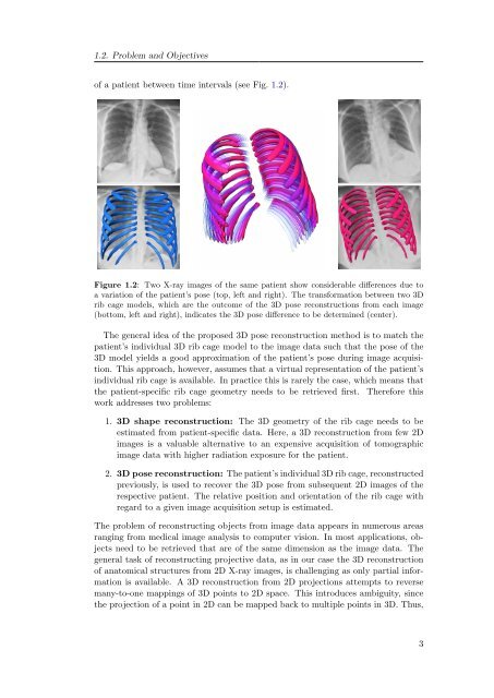
<strong>of</strong> a patient between time intervals (see Fig. 1.2).<br />
Figure 1.2: Two X-ray images <strong>of</strong> <strong>the</strong> same patient show considerable differences due to<br />
a variation <strong>of</strong> <strong>the</strong> patient’s pose (top, left and right). The transformation between two <strong>3D</strong><br />
rib cage models, which are <strong>the</strong> outcome <strong>of</strong> <strong>the</strong> <strong>3D</strong> pose reconstructions <strong>from</strong> each image<br />
(bottom, left and right), indicates <strong>the</strong> <strong>3D</strong> pose difference to be determined (center).<br />
The general idea <strong>of</strong> <strong>the</strong> proposed <strong>3D</strong> pose reconstruction method is to match <strong>the</strong><br />
patient’s individual <strong>3D</strong> rib cage model to <strong>the</strong> image data such that <strong>the</strong> pose <strong>of</strong> <strong>the</strong><br />
<strong>3D</strong> model yields a good approximation <strong>of</strong> <strong>the</strong> patient’s pose during image acquisition.<br />
This approach, however, assumes that a virtual representation <strong>of</strong> <strong>the</strong> patient’s<br />
individual rib cage is available. In practice this is rarely <strong>the</strong> case, which means that<br />
<strong>the</strong> patient-specific rib cage geometry needs to be retrieved first. Therefore this<br />
work addresses two problems:<br />
1. <strong>3D</strong> shape reconstruction: The <strong>3D</strong> geometry <strong>of</strong> <strong>the</strong> rib cage needs to be<br />
estimated <strong>from</strong> patient-specific data. Here, a <strong>3D</strong> reconstruction <strong>from</strong> few <strong>2D</strong><br />
images is a valuable alternative to an expensive acquisition <strong>of</strong> tomographic<br />
image data with higher radiation exposure for <strong>the</strong> patient.<br />
2. <strong>3D</strong> pose reconstruction: The patient’s individual <strong>3D</strong> rib cage, reconstructed<br />
previously, is used to recover <strong>the</strong> <strong>3D</strong> pose <strong>from</strong> subsequent <strong>2D</strong> images <strong>of</strong> <strong>the</strong><br />
respective patient. The relative position and orientation <strong>of</strong> <strong>the</strong> rib cage with<br />
regard to a given image acquisition setup is estimated.<br />
The problem <strong>of</strong> reconstructing objects <strong>from</strong> image data appears in numerous areas<br />
ranging <strong>from</strong> medical image analysis to computer vision. In most applications, objects<br />
need to be retrieved that are <strong>of</strong> <strong>the</strong> same dimension as <strong>the</strong> image data. The<br />
general task <strong>of</strong> reconstructing projective data, as in our case <strong>the</strong> <strong>3D</strong> reconstruction<br />
<strong>of</strong> anatomical structures <strong>from</strong> <strong>2D</strong> X-ray images, is challenging as only partial information<br />
is available. A <strong>3D</strong> reconstruction <strong>from</strong> <strong>2D</strong> projections attempts to reverse<br />
many-to-one mappings <strong>of</strong> <strong>3D</strong> points to <strong>2D</strong> space. This introduces ambiguity, since<br />
<strong>the</strong> projection <strong>of</strong> a point in <strong>2D</strong> can be mapped back to multiple points in <strong>3D</strong>. Thus,<br />
3