3D Reconstruction of the Human Rib Cage from 2D Projection ... - ZIB
3D Reconstruction of the Human Rib Cage from 2D Projection ... - ZIB
3D Reconstruction of the Human Rib Cage from 2D Projection ... - ZIB

You also want an ePaper? Increase the reach of your titles
YUMPU automatically turns print PDFs into web optimized ePapers that Google loves.
3.2. <strong>2D</strong>/<strong>3D</strong> <strong>Reconstruction</strong><br />
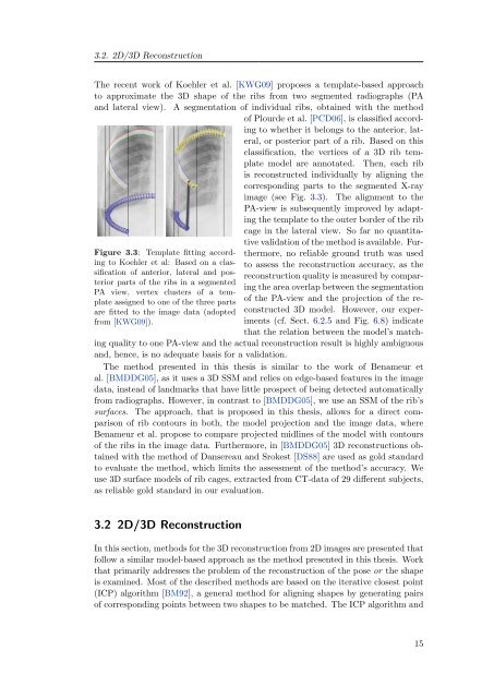
The recent work <strong>of</strong> Koehler et al. [KWG09] proposes a template-based approach<br />
to approximate <strong>the</strong> <strong>3D</strong> shape <strong>of</strong> <strong>the</strong> ribs <strong>from</strong> two segmented radiographs (PA<br />
and lateral view). A segmentation <strong>of</strong> individual ribs, obtained with <strong>the</strong> method<br />
Figure 3.3: Template fitting according<br />
to Koehler et al: Based on a classification<br />
<strong>of</strong> anterior, lateral and posterior<br />
parts <strong>of</strong> <strong>the</strong> ribs in a segmented<br />
PA view, vertex clusters <strong>of</strong> a template<br />
assigned to one <strong>of</strong> <strong>the</strong> three parts<br />
are fitted to <strong>the</strong> image data (adopted<br />
<strong>from</strong> [KWG09]).<br />
<strong>of</strong> Plourde et al. [PCD06], is classified according<br />
to whe<strong>the</strong>r it belongs to <strong>the</strong> anterior, lateral,<br />
or posterior part <strong>of</strong> a rib. Based on this<br />
classification, <strong>the</strong> vertices <strong>of</strong> a <strong>3D</strong> rib template<br />
model are annotated. Then, each rib<br />
is reconstructed individually by aligning <strong>the</strong><br />
corresponding parts to <strong>the</strong> segmented X-ray<br />
image (see Fig. 3.3). The alignment to <strong>the</strong><br />
PA-view is subsequently improved by adapting<br />
<strong>the</strong> template to <strong>the</strong> outer border <strong>of</strong> <strong>the</strong> rib<br />
cage in <strong>the</strong> lateral view. So far no quantitative<br />
validation <strong>of</strong> <strong>the</strong> method is available. Fur<strong>the</strong>rmore,<br />
no reliable ground truth was used<br />
to assess <strong>the</strong> reconstruction accuracy, as <strong>the</strong><br />
reconstruction quality is measured by comparing<br />
<strong>the</strong> area overlap between <strong>the</strong> segmentation<br />
<strong>of</strong> <strong>the</strong> PA-view and <strong>the</strong> projection <strong>of</strong> <strong>the</strong> reconstructed<br />
<strong>3D</strong> model. However, our experiments<br />
(cf. Sect. 6.2.5 and Fig. 6.8) indicate<br />
that <strong>the</strong> relation between <strong>the</strong> model’s match-<br />
ing quality to one PA-view and <strong>the</strong> actual reconstruction result is highly ambiguous<br />
and, hence, is no adequate basis for a validation.<br />
The method presented in this <strong>the</strong>sis is similar to <strong>the</strong> work <strong>of</strong> Benameur et<br />
al. [BMDDG05], as it uses a <strong>3D</strong> SSM and relies on edge-based features in <strong>the</strong> image<br />
data, instead <strong>of</strong> landmarks that have little prospect <strong>of</strong> being detected automatically<br />
<strong>from</strong> radiographs. However, in contrast to [BMDDG05], we use an SSM <strong>of</strong> <strong>the</strong> rib’s<br />
surfaces. The approach, that is proposed in this <strong>the</strong>sis, allows for a direct comparison<br />
<strong>of</strong> rib contours in both, <strong>the</strong> model projection and <strong>the</strong> image data, where<br />
Benameur et al. propose to compare projected midlines <strong>of</strong> <strong>the</strong> model with contours<br />
<strong>of</strong> <strong>the</strong> ribs in <strong>the</strong> image data. Fur<strong>the</strong>rmore, in [BMDDG05] <strong>3D</strong> reconstructions obtained<br />
with <strong>the</strong> method <strong>of</strong> Dansereau and Srokest [DS88] are used as gold standard<br />
to evaluate <strong>the</strong> method, which limits <strong>the</strong> assessment <strong>of</strong> <strong>the</strong> method’s accuracy. We<br />
use <strong>3D</strong> surface models <strong>of</strong> rib cages, extracted <strong>from</strong> CT-data <strong>of</strong> 29 different subjects,<br />
as reliable gold standard in our evaluation.<br />
3.2 <strong>2D</strong>/<strong>3D</strong> <strong>Reconstruction</strong><br />
In this section, methods for <strong>the</strong> <strong>3D</strong> reconstruction <strong>from</strong> <strong>2D</strong> images are presented that<br />
follow a similar model-based approach as <strong>the</strong> method presented in this <strong>the</strong>sis. Work<br />
that primarily addresses <strong>the</strong> problem <strong>of</strong> <strong>the</strong> reconstruction <strong>of</strong> <strong>the</strong> pose or <strong>the</strong> shape<br />
is examined. Most <strong>of</strong> <strong>the</strong> described methods are based on <strong>the</strong> iterative closest point<br />
(ICP) algorithm [BM92], a general method for aligning shapes by generating pairs<br />
<strong>of</strong> corresponding points between two shapes to be matched. The ICP algorithm and<br />
15