Deinotherium thraceiensis sp. nov. from the Miocene near Ezerovo ...
Deinotherium thraceiensis sp. nov. from the Miocene near Ezerovo ...
Deinotherium thraceiensis sp. nov. from the Miocene near Ezerovo ...

You also want an ePaper? Increase the reach of your titles
YUMPU automatically turns print PDFs into web optimized ePapers that Google loves.
wide and regularly curved. In its upper part <strong>the</strong>re is<br />
a wide deep groove, gradually becoming narrower<br />
and shallower with <strong>the</strong> curving of <strong>the</strong> sympnysis.<br />
Ramus ascendens wide and thick. All <strong>the</strong> area of<br />
proc. angularis is thick at <strong>the</strong> base and strongly protruding<br />
backwards. This thickness reaches as far as<br />
proc. articularis. The anterior part of ramus ascendens<br />
is significantly thinner, however, incl. proc. coronoideus<br />
itself. All <strong>the</strong> ramus ascendens in this part is<br />
laterally slightly concave. Foramen mandibulae wide<br />
and deep. Proc. coronoideus almost vertical in <strong>the</strong><br />
anteriror part. Tips of <strong>the</strong> processes curved backwards;<br />
at <strong>the</strong> posterior ends <strong>the</strong>re is a moderate concavity<br />
(incisure). Processus articularis thick and high.<br />
Perpendicularly positioned, with well shaped articular<br />
surfaces. Two rami of <strong>the</strong> mandible not parallel.<br />
At <strong>the</strong> level of M 1 a widening begins, rami coming<br />
closer again at <strong>the</strong> level of M 3 , <strong>the</strong>n going apart again.<br />
Most distant <strong>from</strong> each o<strong>the</strong>r at <strong>the</strong> posterior ends.<br />
The incisors (tusks) are a sequence of <strong>the</strong> symphysis,<br />
shaping <strong>the</strong> curve toge<strong>the</strong>r with it. Their basis<br />
starts deep inside <strong>the</strong> symphysis. There <strong>the</strong>y are hollow,<br />
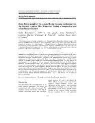
<strong>the</strong>ir alveoli are of semi-circular shape (Fig. 8).<br />
At <strong>the</strong> same time <strong>the</strong>y are becoming thicker inside.<br />
At <strong>the</strong> end of <strong>the</strong> symphysis <strong>the</strong> tusks are already<br />
wholly solid. They gradually become thinner<br />
and, curving in two directions – outwards and backwards,<br />
go apart <strong>from</strong> each o<strong>the</strong>r. Their tips are pointed,<br />
slightly smoo<strong>the</strong>d only at <strong>the</strong> foremost part of <strong>the</strong><br />
inner side, but <strong>the</strong>re is no clear flat surface. Their<br />
tips end just below <strong>the</strong> end of processus angulare.<br />
Lower cheek-teeth. (Pl. VI). The toothrows also have<br />
five teeth each – two premolars and three molars.<br />
Unlike <strong>the</strong> upper premolars, <strong>the</strong> lower are much<br />
narrower, and <strong>the</strong>ir structure is very different,<br />
e<strong>sp</strong>ecially P 3 .<br />
P 3 has a long and narrow crown, pointed at <strong>the</strong><br />
anterior end. One large longitudinal ridge is situated<br />
along its axis, and ano<strong>the</strong>r, transversal – in <strong>the</strong><br />
posterior part. They form <strong>the</strong> occlusial surface of<br />
<strong>the</strong> tooth. The longitudinal ridge is wider at its basis,<br />
gradually narrowing towards <strong>the</strong> apex of <strong>the</strong> crown.<br />
Its widest part is in <strong>the</strong> middle of <strong>the</strong> crown. There it<br />
is more worn out and part of <strong>the</strong> dentine is seen. On<br />
<strong>the</strong> inner part of <strong>the</strong> tooth, at <strong>the</strong> place where <strong>the</strong><br />
longitudinal ridge contacts <strong>the</strong> inner one, <strong>the</strong> first<br />
narrows abruptly thus shaping a large triangular<br />
valley. Outside <strong>the</strong> crown is smooth.<br />
P 4 dext has a larger crown, elongated and wide.<br />
The occlusal surface is moderately worn. It is formed<br />
by two transversal ridges, contacting at <strong>the</strong> outer part<br />
of <strong>the</strong> crown. At this place <strong>the</strong>y divide <strong>the</strong> valley between<br />
<strong>the</strong>m in two not quite equal parts. The anterior<br />
wall of <strong>the</strong> first ridge is strongly concave. The posterior<br />
part of <strong>the</strong> crown has a weak cingulum.<br />
M 1 dext has three transversal ridges. First two are<br />
more worn. Their structure is generally <strong>the</strong> same as<br />
in M 1 . All three ridges are wider on <strong>the</strong> outer side.<br />
The valleys dividing <strong>the</strong>m become gradually narrower<br />
towards <strong>the</strong> outer part. In this part <strong>the</strong> anterior walls<br />
of <strong>the</strong> second and third ridge are slightly convex but<br />
26<br />
Fig. 8. Shape of <strong>the</strong> tusks of <strong>Deino<strong>the</strong>rium</strong> thraceisensis <strong>sp</strong>. n.<br />
A – shape of <strong>the</strong> tusks at <strong>the</strong> basis of <strong>the</strong> alveolus; B – shape of<br />
<strong>the</strong> tusks at 115.0 mm <strong>from</strong> <strong>the</strong>ir basis in <strong>the</strong> symphysis; C –<br />
cross-section of I dext at 600.0 mm <strong>from</strong> <strong>the</strong> symphysis –<br />
natural size<br />
<strong>the</strong>y don’t touch <strong>the</strong> ridge in front of <strong>the</strong>m. On <strong>the</strong><br />
inner side, at <strong>the</strong> bottom of each valley <strong>the</strong>re is a weak<br />
tubercle, and on <strong>the</strong> anterior side – a small cingulum.<br />
Left M 1 resembles <strong>the</strong> right but is more worn.<br />
M 2 dext is large and tetragonal. It has two thick<br />
transversal ridges, wider on <strong>the</strong> outer side. There both<br />
ridges form a small concavity. The valley between<br />
<strong>the</strong>m is deep and free but on <strong>the</strong> inner side <strong>the</strong>re is a<br />
small tubercle. On its anterior and posterior sides <strong>the</strong><br />
crown has a cingulum.<br />
M 3 dext is large too but with an irregular tetragonal<br />
shape. Built by two transversal ridges. The first is<br />
wider. Both are widening outwards. At <strong>the</strong> ends –<br />
inner and outer – <strong>the</strong>y are slightly curved to <strong>the</strong> front,<br />
forming with <strong>the</strong>ir anterior walls shallow valleys. The<br />
valley between <strong>the</strong>m is deep and unblocked. The<br />
posterior talon on <strong>the</strong> inner side of <strong>the</strong> crown is formed<br />
by numerous tubercles of different size.<br />
Comparison. The structure of <strong>the</strong> described mandible<br />
is close to <strong>the</strong> o<strong>the</strong>r <strong>sp</strong>ecies (Fig. 9).<br />
The symphysis is moderately large. There are differences<br />
in <strong>the</strong> shape of ramus horizontalis, proc.<br />
articularis, proc. coronoideus and <strong>the</strong> tusks.<br />
1. The symphysis of D. bavaricum H. v. Meyer is more<br />
protruding. In D. levius Jourdan it is more rounded<br />
and curved inwards and in D. giganteum Kaup, as<br />
well as in D. gigantissimum Stefanescu and D. <strong>thraceiensis</strong><br />
<strong>sp</strong>. n. <strong>the</strong> curving is even stronger and more<br />
gradual.