Deinotherium thraceiensis sp. nov. from the Miocene near Ezerovo ...
Deinotherium thraceiensis sp. nov. from the Miocene near Ezerovo ...
Deinotherium thraceiensis sp. nov. from the Miocene near Ezerovo ...

Create successful ePaper yourself
Turn your PDF publications into a flip-book with our unique Google optimized e-Paper software.
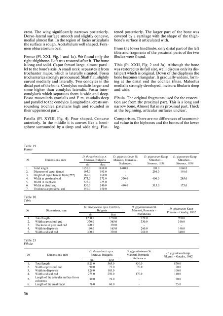
crest. The wing significantly narrows posteriorly.<br />
Dorso-lateral surface smooth and slightly concave,<br />
medial almost flat. In <strong>the</strong> region of facies articularis<br />
<strong>the</strong> surface is rough. Acetabulum well shaped. Foramen<br />
obturatorium oval.<br />
Femur (Pl. XXI, Fig. 1 and 1a). We found only <strong>the</strong><br />
right thighbone. Left was restored after it. The bone<br />
is long and solid. Caput femuri large, almost parallel<br />
to <strong>the</strong> bone’s axis. A small neck separates it <strong>from</strong><br />
trochanter major, which is laterally situated. Fossa<br />
trochanterica strongly pronounced. Shaft flat, slightly<br />
curved medially and laterally. Two condyles in <strong>the</strong><br />
distal part of <strong>the</strong> bone. Condylus medialis larger and<br />
some higher than condylus lateralis. Fossa intercondylaris<br />
which separates <strong>the</strong>m is wide and deep.<br />
Fossa muscularis cranialis and F. m. caudalis deep<br />
and parallel to <strong>the</strong> condyles. Longitudinal crests surrounding<br />
trochlea patellaris high and rounded in<br />
<strong>the</strong>ir uppermost part.<br />
Patella (Pl. XVIII, Fig. 4). Pear shaped. Concave<br />
anteriorly. In <strong>the</strong> middle it is convex like a hemi<strong>sp</strong>here<br />
surrounded by a deep and wide ring. Flat-<br />
Table 19<br />
Femur<br />
36<br />
№ Dimensions, mm<br />
D. <strong>thraceiensis</strong> <strong>sp</strong>.n.<br />
<strong>Ezerovo</strong>, Bulgaria<br />
sin dext<br />
D. gigantissimum St.<br />
Manzati, Romania –<br />
Stefanescu<br />
D. giganteum Kaup<br />
München –<br />
Stromer, 1938<br />
D. giganteum Kaup<br />
München –<br />
Stromer, 1938<br />
1. Total length 1650.0 1650.0 1440.0 188.0 1060.0<br />
2. Diameter of caput femuri 195.0 195.0 - 210.0 140.0<br />
3. Height of caput femuri <strong>from</strong> [???] 160.0 160.0 -<br />
4. Width at proximal end 375.0 375.0 350.0 400.0 295.0<br />
5. Width in diaphysis 225.0 225.0 -<br />
6. Width at distal end 330.0 340.0 440.0 315.0 175.0<br />
7. Thickness at proximal end 150.0 150.0<br />
Table 20<br />
Tibia<br />
№ Dimensions, mm<br />
D. <strong>thraceiensis</strong> <strong>sp</strong>.n. <strong>Ezerovo</strong>,<br />
Bulgaria<br />
sin dext<br />
D. gigantissimum St.<br />
Manzati, Romania –<br />
Stefanescu<br />
D. giganteum Kaup<br />
Pikermi – Gaudry, 1862<br />
1. Total length 1200.0 1250.0 920.0 950.0<br />
2. Width at proximal end 370.0 365.0 330.0 310.0<br />
3. Thickness at proximal end 320.0 320.0 – –<br />
4. Width in diaphysis 160.0 165.0 260.0 140.0<br />
5. Width at distal end 300.0 330.0 260.0 340.0<br />
Table 21<br />
Fibula<br />
№ Dimensions, mm<br />
D. <strong>thraceiensis</strong> <strong>sp</strong>.n.<br />
<strong>Ezerovo</strong>, Bulgaria<br />
sin dext<br />
tened posteriorly. The larger part of <strong>the</strong> bone was<br />
covered by a cartilage with <strong>the</strong> shape of <strong>the</strong> thighbone’s<br />
surface it articulated with.<br />
From <strong>the</strong> lower hindlimbs, only distal part of <strong>the</strong> left<br />
tibia and fragments of <strong>the</strong> proximal parts of <strong>the</strong> two<br />
fibulae were found.<br />
Tibia (Pl. XXII, Fig. 2 and 2a). Although <strong>the</strong> bone<br />
was restored to its full size, we’ll discuss only its distal<br />
part which is original. Down of <strong>the</strong> diaphysis <strong>the</strong><br />
bone becomes triangular. It gradually widens, forming<br />
at <strong>the</strong> distal end <strong>the</strong> cochlea tibiae. Maleolus<br />
medialis strongly developed, incisura fibularis deep<br />
and wide.<br />
Fibula. The original fragments used for <strong>the</strong> restoration<br />
are <strong>from</strong> <strong>the</strong> proximal part. This is a long and<br />
narrow bone. Almost flat in its proximal part. Thick<br />
at <strong>the</strong> beginning, articular surfaces unclear.<br />
Comparison. There are no differences of taxonomical<br />
value in <strong>the</strong> hipbones and <strong>the</strong> bones of <strong>the</strong> lower<br />
leg.<br />
D. gigantissimum St.<br />
Manzati, Romania –<br />
Stefanescu<br />
D. giganteum Kaup<br />
Pikermi – Gaudry, 1862<br />
1. Total length 1125.0 565.0 830.0 870.0<br />
2. Width at proximal end 90.0 72.0 70.0 70.0<br />
3. Width in diaphysis 126.0 103.0 - 100.0<br />
4. Width at distal end 275.0 250.0 170.0 140.0<br />
5.<br />
Length of <strong>the</strong> articular surface for os<br />
calcaneus<br />
80.0 72.0 - 70.0<br />
6. Length of <strong>the</strong> small facet 76.0 60.0 - 55.0