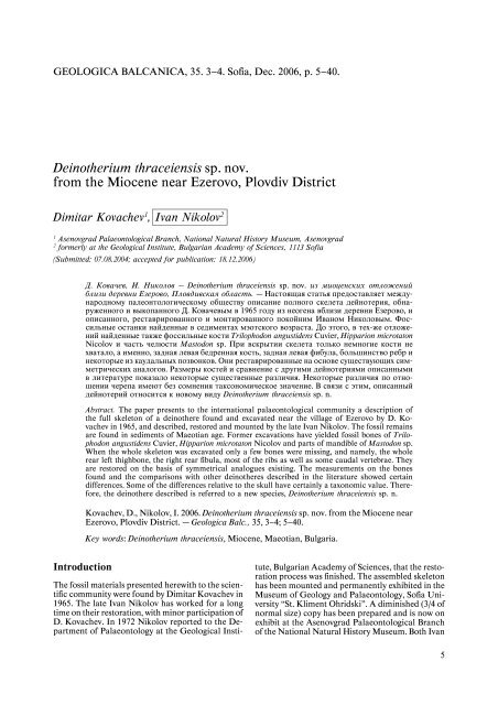
Deinotherium thraceiensis sp. nov. from the Miocene near Ezerovo ...
Deinotherium thraceiensis sp. nov. from the Miocene near Ezerovo ...
Deinotherium thraceiensis sp. nov. from the Miocene near Ezerovo ...

You also want an ePaper? Increase the reach of your titles
YUMPU automatically turns print PDFs into web optimized ePapers that Google loves.
GEOLOGICA BALCANICA, 35. 3—4. Sofia, Dec. 2006, p. 5—40.<br />
<strong>Deino<strong>the</strong>rium</strong> <strong>thraceiensis</strong> <strong>sp</strong>. <strong>nov</strong>.<br />
<strong>from</strong> <strong>the</strong> <strong>Miocene</strong> <strong>near</strong> <strong>Ezerovo</strong>, Plovdiv District<br />
Dimitar Kovachev 1 , Ivan Nikolov 2<br />
1 Ase<strong>nov</strong>grad Palaeontological Branch, National Natural History Museum, Ase<strong>nov</strong>grad<br />
2 formerly at <strong>the</strong> Geological Institute, Bulgarian Academy of Sciences, 1113 Sofia<br />
(Submitted: 07.08.2004; accepted for publication: 18.12.2006)<br />
Introduction<br />
Ä. Êîâà÷åâ, È. Íèêîëîâ – <strong>Deino<strong>the</strong>rium</strong> <strong>thraceiensis</strong> <strong>sp</strong>. <strong>nov</strong>. èç ìèîöåíñêèõ îòëîæåíèé<br />
áëèçè äåðåâíè Åçåðîâî, Ïëîâäèâñêàÿ îáëàñòü. – Íàñòîÿùàÿ ñòàòüÿ ïðåäîñòàâëÿåò ìåæäóíàðîäíîìó<br />
ïàëåîíòîëîãè÷åñêîìó îáùåñòâó îïèñàíèå ïîëíîãî ñêåëåòà äåéíîòåðèÿ, îáíàðóæåííîãî<br />
è âûêîïàííîãî Ä. Êîâà÷åâûì â 1965 ãîäó èç íåîãåíà âáëèçè äåðåâíè Åçåðîâî, è<br />
îïèñàííîãî, ðåñòàâðèðîâàííîãî è ìîíòèðîâàííîãî ïîêîéíèì Èâàíîì Íèêîëîâûì. Ôîññèëüíûå<br />
îñòàíêè íàéäåííûå â ñåäèìåíòàõ ìýîòñêîãî âîçðàñòà. Äî ýòîãî, â òåõ-æå îòëîæåíèé<br />
íàéäåííûå òàêæå ôîññèëüíûå êîñòè Trilophodon angustidens Cuvier, Hipparion microtaton<br />
Nicolov è ÷àñòü ÷åëþñòè Mastodon <strong>sp</strong>. Ïðè âñêðûòèè ñêåëåòà òîëüêî íåìíîãèå êîñòè íå<br />
õâàòàëî, à èìåííî, çàäíàÿ ëåâàÿ áåäðåííàÿ êîñòü, çàäíàÿ ëåâàÿ ôèáóëà, áîëüøèíñòâî ðåáð è<br />
íåêîòîðûå èç êàóäàëüíûõ ïîçâîíêîâ. Îíè ðåñòàâðèðîâàííûå íà îñíîâå ñóùåñòâóþùèõ ñèììåòðè÷åñêèõ<br />
àíàëîãîâ. Ðàçìåðû êîñòåé è ñðàâíåíèå ñ äðóãèìè äåéíîòåðèÿìè îïèñàííûìè<br />
â ëèòåðàòóðå ïîêàçàëî íåêîòîðûå ñóùåñòâåííûå ðàçëè÷èÿ. Íåêîòîðûå ðàçëè÷èÿ ïî îòíîøåíèè<br />
÷åðåïà èìåþò áåç ñîìíåíèÿ òàêñîíîìè÷åñêîå çíà÷åíèå.  ñâÿçè ñ ýòèì, îïèñàííûé<br />
äåéíîòåðèé îòíîñèòñÿ ê íîâîìó âèäó <strong>Deino<strong>the</strong>rium</strong> <strong>thraceiensis</strong> <strong>sp</strong>. n.<br />
Abstract. The paper presents to <strong>the</strong> international palaeontological community a description of<br />
<strong>the</strong> full skeleton of a deino<strong>the</strong>re found and excavated <strong>near</strong> <strong>the</strong> village of <strong>Ezerovo</strong> by D. Kovachev<br />
in 1965, and described, restored and mounted by <strong>the</strong> late Ivan Nikolov. The fossil remains<br />
are found in sediments of Maeotian age. Former excavations have yielded fossil bones of Trilophodon<br />
angustidens Cuvier, Hipparion microtaton Nicolov and parts of mandible of Mastodon <strong>sp</strong>.<br />
When <strong>the</strong> whole skeleton was excavated only a few bones were missing, and namely, <strong>the</strong> whole<br />
rear left thighbone, <strong>the</strong> right rear fibula, most of <strong>the</strong> ribs as well as some caudal vertebrae. They<br />
are restored on <strong>the</strong> basis of symmetrical analogues existing. The measurements on <strong>the</strong> bones<br />
found and <strong>the</strong> comparisons with o<strong>the</strong>r deino<strong>the</strong>res described in <strong>the</strong> literature showed certain<br />
differences. Some of <strong>the</strong> differences relative to <strong>the</strong> skull have certainly a taxonomic value. Therefore,<br />
<strong>the</strong> deino<strong>the</strong>re described is referred to a new <strong>sp</strong>ecies, <strong>Deino<strong>the</strong>rium</strong> <strong>thraceiensis</strong> <strong>sp</strong>. n.<br />
Kovachev, D., Nikolov, I. 2006. <strong>Deino<strong>the</strong>rium</strong> <strong>thraceiensis</strong> <strong>sp</strong>. <strong>nov</strong>. <strong>from</strong> <strong>the</strong> <strong>Miocene</strong> <strong>near</strong><br />
<strong>Ezerovo</strong>, Plovdiv District. – Geologica Balc., 35, 3—4; 5—40.<br />
Key words: <strong>Deino<strong>the</strong>rium</strong> <strong>thraceiensis</strong>, <strong>Miocene</strong>, Maeotian, Bulgaria.<br />
The fossil materials presented herewith to <strong>the</strong> scientific<br />
community were found by Dimitar Kovachev in<br />
1965. The late Ivan Nikolov has worked for a long<br />
time on <strong>the</strong>ir restoration, with minor participation of<br />
D. Kovachev. In 1972 Nikolov reported to <strong>the</strong> Department<br />
of Palaeontology at <strong>the</strong> Geological Insti-<br />
tute, Bulgarian Academy of Sciences, that <strong>the</strong> restoration<br />
process was finished. The assembled skeleton<br />
has been mounted and permanently exhibited in <strong>the</strong><br />
Museum of Geology and Palaeontology, Sofia University<br />
“St. Kliment Ohridski”. A diminished (3/4 of<br />
normal size) copy has been prepared and is now on<br />
exhibit at <strong>the</strong> Ase<strong>nov</strong>grad Palaeontological Branch<br />
of <strong>the</strong> National Natural History Museum. Both Ivan<br />
5
Nikolov and Dimitar Kovachev were fully aware of<br />
<strong>the</strong> importance of this very rare case of finding a<br />
whole skeleton of such a huge extinct animal, and<br />
<strong>the</strong>y estimated it to be a representative of a new <strong>sp</strong>ecies<br />
of genus <strong>Deino<strong>the</strong>rium</strong>. Ivan Nikolov, well-known<br />
for his thoroughness, wanted to make an exemplary<br />
publication but his early death stopped for a long<br />
time <strong>the</strong> work on it. The results of his enormous labour<br />
put into <strong>the</strong> description of <strong>the</strong> bones, <strong>the</strong> restoration<br />
and mounting of this unique skeleton, remained<br />
in <strong>the</strong> archives of <strong>the</strong> Geological Institute.<br />
After <strong>the</strong> sudden untimely death of Nikolov in 1984,<br />
<strong>the</strong> work on <strong>the</strong> publication remained unfinished.<br />
Now, D. Kovachev decided to prepare <strong>the</strong> materials<br />
for publication with <strong>the</strong> clear idea about <strong>the</strong> re<strong>sp</strong>onsibility<br />
undertaken, and that <strong>the</strong> whole blame for eventual<br />
flaws and errors should be addressed to himself.<br />
Such publication would be of importance for <strong>the</strong><br />
future studies on genus <strong>Deino<strong>the</strong>rium</strong>.<br />
Many scientists have helped <strong>the</strong> authors in different<br />
time. Prof. Dr. R. Dehm (Director of Universität-Institut<br />
für Paläontologie und historische Geologie<br />
– München) and Prof. Dr. H. Tobien (Director<br />
of Paläontologisches Institut der Johanes-Gutenberg<br />
Universität – Meinz) helped I. Nikolov during <strong>the</strong><br />
first studies and restoration. Academician T. Nikolov<br />
(Geological Institute, Bulgarian Academy of Sciences)<br />
gave valuable advice with <strong>the</strong> material. D. Kovachev<br />
wishes to express his gratitude to Dr. Marin<br />
Iva<strong>nov</strong> <strong>from</strong> <strong>the</strong> Sofia University for his re<strong>sp</strong>onsiveness<br />
to <strong>the</strong> problems of this research. Dr. N. Spassov<br />
(National Natural History Museum, Sofia) kindly<br />
helped with <strong>the</strong> literature I. Nikolov had used at <strong>the</strong><br />
library of <strong>the</strong> Museum.<br />
Studies on <strong>the</strong> genus <strong>Deino<strong>the</strong>rium</strong> –<br />
a historical review<br />
As early as <strong>the</strong> 17 century a place <strong>near</strong> Lyon, France,<br />
was known as “<strong>the</strong> field of <strong>the</strong> giants” because of <strong>the</strong><br />
large animal bones often found <strong>the</strong>re. Some of those<br />
bones came to Matsorier – a surgeon, who used to<br />
show <strong>the</strong>m for years in France and Germany as <strong>the</strong><br />
bones of king Töteboch. Much later, <strong>the</strong> real tomb of<br />
<strong>the</strong> king was found, and <strong>the</strong> deceit was exposed. The<br />
huge bones were transferred to <strong>the</strong> Natural History<br />
Museum in Paris. Probably <strong>the</strong>se are <strong>the</strong> first <strong>Deino<strong>the</strong>rium</strong><br />
remains found.<br />
One century later, in 1715, Réaumure admitted<br />
he could not refer <strong>the</strong> bones to any known animal.<br />
Kenedy supposed in 1775 that <strong>the</strong> materials were related<br />
to mammoths. G. Cuvier thought in 1779—1836<br />
that <strong>the</strong> animal had been a large tapir coexisting with<br />
mastodons. He called it Tapir gigantesque and<br />
thought that its tusks were curved upwards.<br />
Kaup created in 1829 <strong>the</strong> genus <strong>Deino<strong>the</strong>rium</strong> with<br />
<strong>the</strong> <strong>sp</strong>ecies D. giganteum upon <strong>the</strong> skull and mandible<br />
found <strong>near</strong> Eppelsheim, Germany. He thought<br />
this was an intermediary form between <strong>the</strong> sloth and<br />
<strong>the</strong> mastodons and referred <strong>the</strong>m to <strong>the</strong> larger taxonomic<br />
unit Curtognati. In 1841 and 1857 he de-<br />
6<br />
scribed fur<strong>the</strong>r remains of that genus found <strong>near</strong><br />
Eppelsheim and Westhofen. According to him, <strong>the</strong><br />
adult animals had five teeth in <strong>the</strong>ir upper and lower<br />
jaws. In 1833 he described <strong>the</strong> new <strong>sp</strong>ecies D. cuvieri,<br />
but because it was often found in Bavaria, H. v.<br />
Meyer called it D. bavaricum. This name is still used<br />
today. However, arguments about <strong>the</strong> exact place and<br />
<strong>the</strong> life environment of deino<strong>the</strong>res still continued.<br />
P. Gervais accepted in 1848 <strong>the</strong> opinion of Kenedy<br />
and Koch that <strong>the</strong> genera <strong>Deino<strong>the</strong>rium</strong> and Mastodon<br />
should be referred to Proboscidea. He recognized<br />
<strong>the</strong> existence of three <strong>sp</strong>ecies: D. giganteum Kaup =<br />
Tapir gigantesque Cuvier; D. intermedium and D. cuvieri<br />
Kaup. S. Pictet reported in 1853 about <strong>the</strong> finding<br />
<strong>near</strong> <strong>the</strong> village of Absdorf of a whole skeleton of D.<br />
giganteum Kaup. Only <strong>the</strong> head, <strong>the</strong> first two vertebrae,<br />
<strong>the</strong> corpus of a thoracic and a caudal vertebra and<br />
fragments of <strong>the</strong> limbs were preserved. These remains<br />
showed that it is close to <strong>the</strong> mastodon and belongs to<br />
Pachydermae. Four deino<strong>the</strong>re <strong>sp</strong>ecies were recognized<br />
in 1858—1859 by M. Lartet, and namely, <strong>Deino<strong>the</strong>rium</strong><br />
bavaricum H. v. Meyer, D. giganteum Kaup, D. <strong>sp</strong>. (an<br />
intermediary form between <strong>the</strong> first two), and D. cuvieri<br />
Kaup. The first <strong>sp</strong>ecies is known <strong>from</strong> <strong>the</strong> <strong>Miocene</strong><br />
of France and Bavaria, <strong>the</strong> second <strong>from</strong> <strong>the</strong> Late<br />
<strong>Miocene</strong> of France, Germany and Greece, <strong>the</strong> third<br />
and fourth <strong>from</strong> <strong>the</strong> <strong>Miocene</strong> of France. After Lartet,<br />
<strong>the</strong> dental formula of genus <strong>Deino<strong>the</strong>rium</strong> is: I 0-0/1-1;<br />
Pm 2-2/2-2 and M 3-3/3-3 for <strong>the</strong> permanent teeth,<br />
and for <strong>the</strong> deciduous – I 0-0/1-1; deciduous molars<br />
3-3/3-3. New fossil finds in Greece (Pikermi), India (Siwalik),<br />
Europe (France, Switzerland, Austria), etc. have<br />
been reported during <strong>the</strong> last quarter of <strong>the</strong> 19 th century<br />
by A. Gaudry, H. Hensel, B. A. Lydekker, E. Chantre,<br />
M. Vacek, V. Biber, C. Deperet, and o<strong>the</strong>r authors. Gr.<br />
Stefanescu first reported in 1891, and <strong>the</strong>n described<br />
(1895–1910) a deino<strong>the</strong>re skeleton found <strong>near</strong> Manzati,<br />
Romania. Because of <strong>the</strong> large dimensions of all<br />
its bones and teeth, he named it D. gigantissimum. Ehik<br />
(1930) published upper and lower molars and some<br />
finger bones, found in <strong>the</strong> Pliocene sediments of Hungary,<br />
as a new <strong>sp</strong>ecies – Prodino<strong>the</strong>rium hungaricum.<br />
Osborn (1936) changed <strong>the</strong> name of <strong>the</strong> genus (<strong>the</strong><br />
Latinized form Dino<strong>the</strong>rium) to <strong>the</strong> ancient Greek<br />
form <strong>Deino<strong>the</strong>rium</strong>. He revised all <strong>the</strong>n known<br />
deino<strong>the</strong>re materials, discussing in details each <strong>sp</strong>ecies,<br />
and illustrating skulls/mandibles and teeth <strong>from</strong><br />
D. giganteum Kaup, D. bavaricum H. v. Meyer, D.<br />
cuvieri Kaup, D. pentapotamiae Lydekker, D. indicum<br />
Falconer, etc. (Fig. 1).<br />
Numerous new finds of deino<strong>the</strong>res have been reported<br />
during <strong>the</strong> whole 20 th century <strong>from</strong> Austria,<br />
Bohemia, Bulgaria, France, Germany, Greece, Hungary,<br />
Macedonia, Moldova, Pakistan, Romania, Russia,<br />
Serbia, Ukraina, etc.<br />
Studies on <strong>the</strong> genus <strong>Deino<strong>the</strong>rium</strong><br />
in Bulgaria<br />
The first <strong>Deino<strong>the</strong>rium</strong> remains in Bulgaria have been<br />
found by G. Bonchev (Áîí÷åâ, 1897) who made ex-
Fig. 1. Distribution of genus <strong>Deino<strong>the</strong>rium</strong> in Sou<strong>the</strong>astern Europe and Asia Minor<br />
cavations in <strong>the</strong> Middle Sarmatian limestones <strong>near</strong><br />
Nessebar, studying <strong>the</strong> hipparion fauna. Three years<br />
later he reported new finds <strong>from</strong> <strong>the</strong> same locality.<br />
Áàêàëîâ (1911/1913) published <strong>the</strong> two deino<strong>the</strong>re<br />
mandibles excavated by G. Bonchev, toge<strong>the</strong>r with<br />
o<strong>the</strong>r single teeth <strong>from</strong> <strong>the</strong> village Sovolyano, <strong>near</strong><br />
Pleven, <strong>the</strong> village Archar and <strong>the</strong> village Mazgosh.<br />
They were all brought to <strong>the</strong> Institute of Geology by<br />
different people. From <strong>the</strong>se materials Bakalov determined<br />
two <strong>sp</strong>ecies – D. giganteum Kaup race mi-<br />
7
nor and D. giganteum Kaup race major. To <strong>the</strong> first<br />
he referred <strong>the</strong> smaller teeth, and to <strong>the</strong> second –<br />
<strong>the</strong> larger ones. Later on (Áàêàëîâ, 1949/1950)<br />
Bakalov described <strong>Deino<strong>the</strong>rium</strong> teeth found at <strong>the</strong><br />
“Meander of <strong>the</strong> Konska river” <strong>near</strong> <strong>the</strong> village Noevtsi;<br />
<strong>the</strong> village (now – a town; for some time called<br />
Bata<strong>nov</strong>tsi) Temelkovo (Pernik region) and Aksakovo,<br />
<strong>near</strong> Varna, as D. giganteum Kaup race major<br />
(<strong>from</strong> <strong>the</strong> first two localities, Pliocene) and D. giganteum<br />
Kaup race minor (<strong>from</strong> <strong>the</strong> Sarmatian limestones<br />
<strong>near</strong> Aksakovo). Íèêîëîâ (1960) reported a<br />
molar fragment of D. giganteum Kaup race major<br />
<strong>from</strong> <strong>the</strong> lignite coals <strong>near</strong> <strong>the</strong> village Hrabarsko (Sofia<br />
region), assuming a Pontian age. The whole fossil<br />
material belonging to genus <strong>Deino<strong>the</strong>rium</strong> <strong>the</strong>n known<br />
<strong>from</strong> Bulgaria has been published by Áàêàëîâ,<br />
Íèêîëîâ (1962). They recognize two races – minor<br />
and major, and refer <strong>the</strong> finds as follows: two mandibles,<br />
an upper jaw and three separate teeth to race<br />
minor, and a mandible, seven complete upper and<br />
lower teeth and three tooth fragments to race major.<br />
The age of both races varies <strong>from</strong> Sarmatian to Levantian.<br />
Íèêîëîâ (1962) referred <strong>the</strong> molar and fragment<br />
of a scapula found in <strong>the</strong> Levantian sediments<br />
<strong>near</strong> <strong>the</strong> village Popitsa, Vratsa region to D. giganteum<br />
Kaup. Íèêîëîâ, Êîâà÷åâ (1966) described a<br />
lower molar found by <strong>the</strong> latter author in <strong>the</strong> sands<br />
in <strong>the</strong> valley of Cherkezitsa river, <strong>near</strong> <strong>the</strong> village<br />
Ahmatovo. They assume <strong>the</strong> age to be Levantian because<br />
of <strong>the</strong> Anancus arvernensis Croizet et Jobert<br />
teeth found <strong>the</strong>re, referring <strong>the</strong> find to D. giganteum<br />
Kaup. Êîâà÷åâ (1966) reported <strong>the</strong> finding of an<br />
almost complete skeleton of <strong>Deino<strong>the</strong>rium</strong> <strong>near</strong> <strong>the</strong><br />
village <strong>Ezerovo</strong>, Plovdiv region. This same skeleton is<br />
<strong>the</strong> subject of <strong>the</strong> present paper. Öàíêîâ, Íèêîëîâ<br />
(1966) published a brief review of <strong>the</strong> proboscideans<br />
in Bulgaria, and gave also some preliminary information<br />
about <strong>the</strong> finding of Kovachev reporting<br />
metrical data on some bones.<br />
PLATE I<br />
8<br />
Distribution of genus <strong>Deino<strong>the</strong>rium</strong><br />
in Sou<strong>the</strong>ast Europe and parts of Asia<br />
Minor<br />
Until now, more than 80 localities (partly shown on<br />
Fig. 1) within <strong>the</strong> region have yielded deino<strong>the</strong>re remains,<br />
and <strong>the</strong>ir number is constantly growing.<br />
There are 16 localities <strong>from</strong> former Yugoslavia:<br />
1. Veles, Mokranje, Smederevska palanka, Resnik,<br />
Meduhana, Vracevic, Duboko, Tsiganski potok, and<br />
between <strong>the</strong> villages Viteževo and Porodin – D. giganteum<br />
Kaup. Near Veles and Ravanica, toge<strong>the</strong>r<br />
with D. giganteum Kaup, D. bavaricum H. v. Meyer<br />
is also present. The sediments <strong>near</strong> <strong>the</strong> villages Meduhana,<br />
Ravanica and those between Viteževo and<br />
Porodin are of Lower Sarmatian, and <strong>the</strong> rest – of<br />
Pontian age.<br />
2. Dre<strong>nov</strong>ica, Chukovica, Bresnica, Prebrezka, Zdarski<br />
and Kriva reka – D. bavaricum H. v. Meyer. The<br />
age cited is Helvetian-Tortonian, Lower Sarmatian,<br />
Sarmatian and Middle <strong>Miocene</strong>.<br />
In Greece D. giganteum Kaup is known <strong>from</strong> four<br />
localities: Pikermi, Thessaloniki and Tanagra (Pontian),<br />
and Samos (Middle Pontian). From <strong>the</strong> Sarmatian<br />
of Chios D. bavaricum H. v. Meyer has been<br />
described.<br />
From Turkey (Asia Minor), D. giganteum Kaup<br />
has been described <strong>from</strong> Küçükçekmece (Maeotian).<br />
From <strong>the</strong> territory of Romania and part of Bessarabia,<br />
two taxa are known – D. giganteum Kaup <strong>from</strong><br />
<strong>the</strong> Pontian and Dacian sediments <strong>near</strong> Vernesti,<br />
Culm, Lichtental, Pelineu, Telenesti, Visterniceni,<br />
Baimaclia, Cimislia, Comanesti, Curtea and Solcustria;<br />
and D. gigantissimum Stefanescu, – <strong>from</strong> <strong>the</strong> Pliocene<br />
sediments <strong>near</strong> Manzati, Breaza and Cimislia.<br />
From <strong>the</strong> territory of Moldova and Bessarabia, two<br />
taxa are known too: 1. D. giganteum Kaup <strong>near</strong> Taraklia,<br />
Ciobruciu, Sofijevka, Ta<strong>nov</strong>ka, Chernevo, Ti-<br />
<strong>Deino<strong>the</strong>rium</strong> thracåiensis <strong>sp</strong>. n.<br />
1, 2. Skull in lateral and ventral view. x 4.5% coll. SU.M. No. SU Pl 312/1; Locality: <strong>Ezerovo</strong>, <strong>near</strong> Plovdiv; Level: Maeotian<br />
PLATE II<br />
<strong>Deino<strong>the</strong>rium</strong> <strong>thraceiensis</strong> <strong>sp</strong>. n.<br />
Skull in dorsal view. Scale ca. 4.2% coll. SU. M. No. SU Pl 312/1; Locality: <strong>Ezerovo</strong>, <strong>near</strong> Plovdiv; Level: Maeotian<br />
PLATE III<br />
<strong>Deino<strong>the</strong>rium</strong> <strong>thraceiensis</strong> <strong>sp</strong>. n.<br />
1. Skull and mandible. Scale ca. 2.9% coll. SU.M. No. SU Pl 312/1, SU Pl 312/2<br />
2. Skull <strong>from</strong> behind – os occipitale. Scale ca 5.6% coll. SU.M. No. SU Pl 312/1; Locality: <strong>Ezerovo</strong>, <strong>near</strong> Plovdiv; Level: Maeotian<br />
PLATE IV<br />
→<br />
<strong>Deino<strong>the</strong>rium</strong> <strong>thraceiensis</strong> <strong>sp</strong>. n.<br />
1. Upper cheek teeth – P 3 to M 3 sin et dext. Scale ca. 25% coll. SU.M. No. SU Pl 312/1; Locality: <strong>Ezerovo</strong>, <strong>near</strong> Plovdiv; Level:<br />
Maeotian
PLATE I PLATE II<br />
PLATE III PLATE IV
PLATE VI<br />
PLATE V
PLATE VII<br />
PLATE VIII PLATE IX
PLATE X PLATE XI<br />
PLATE XII PLATE XIII
PLATE XIV PLATE XV<br />
PLATE XVI PLATE XVII
PLATE XVIII<br />
PLATE XXI<br />
PLATE XX<br />
PLATE XIX<br />
PLATE XXII
PLATE XXIII<br />
PLATE XXIV
Fig. 2. Distribution of genus <strong>Deino<strong>the</strong>rium</strong> in Bulgaria: 1. <strong>Ezerovo</strong>; 2. Nesebar; 3. Izgrev; 4. Parvomai; 5. Ahmatovo; 6. Lyubovishte;<br />
7. Sovolyano; 8. Bata<strong>nov</strong>tsi (Temelkovo); 9. Noevtsi; 10. Sofia; 11. Kremikovtsi; 12. Hrabarsko; 13. Aldomirovtsi; 14. Katina; 15.<br />
Kula; 16. Archar; 17. Mihaylovo; 18. Glozhene; 19. Popitsa; 20. Pleven; 21. Novo selo; 22. Aksakovo; 23. Yarebichna; 24. Breznik;<br />
25. Hadjidimovo; 26. Varna – Galata; 27. Kalimantsi; 28. Konyovo; 29. Rogozen; 30. Gabra – former open pit “Bolshevik”; 31.<br />
Mazgosh (now on Serbian territory)<br />
ras<strong>sp</strong>ol, Donau, Velikovo and Krivoy Rog. The sediments<br />
are of Maeotian, even Upper Sarmatian age.<br />
2. D. gigantissimum Stefanescu <strong>near</strong> Volchinec, Rahny-Lessovye<br />
and Pripecheni. The host sediments are<br />
also of Maeotian and Upper Sarmatian age.<br />
A considerable number of deino<strong>the</strong>re localities<br />
are known <strong>from</strong> <strong>the</strong> territory of Bulgaria (Fig. 2).<br />
Some of <strong>the</strong> first <strong>Deino<strong>the</strong>rium</strong> finds in Bulgaria<br />
came <strong>from</strong> <strong>the</strong> locality Mazgosh (now in Serbia).<br />
The larger number of localities in <strong>the</strong> western part<br />
of <strong>the</strong> country is a result <strong>from</strong> <strong>the</strong> fact that this<br />
part is better studied. Some of <strong>the</strong> more important<br />
finds come <strong>from</strong> Nessebar (by G. Bonchev), Noevtsi,<br />
locality “Meander of <strong>the</strong> Konska river” (by P.<br />
Bakalov), and <strong>the</strong> village <strong>Ezerovo</strong>, “Kolnu dere”<br />
(by D. Kovachev).<br />
Some of <strong>the</strong> materials have been found and<br />
brought to <strong>the</strong> <strong>sp</strong>ecialists by common people (farmers,<br />
workers, etc.). Therefore, <strong>the</strong> exact location has<br />
been mentioned only approximately, and <strong>the</strong>re is no<br />
stratigraphic profile. In such cases, a probable age<br />
←<br />
PLATE V<br />
<strong>Deino<strong>the</strong>rium</strong> <strong>thraceiensis</strong> <strong>sp</strong>. n.<br />
1. Mandible. Scale ca. 8.3% coll. SU.M. No. SU Pl 312/2; Locality: <strong>Ezerovo</strong>, <strong>near</strong> Plovdiv; Level: Maeotian<br />
PLATE VI<br />
<strong>Deino<strong>the</strong>rium</strong> <strong>thraceiensis</strong> <strong>sp</strong>. n.<br />
1. Lower cheek teeth – P 3 to M 3 . Scale ca. 25% coll. SU.M. No. SU Pl 312/2; Locality: <strong>Ezerovo</strong>, <strong>near</strong> Plovdiv; Level: Maeotian<br />
2 Geologica Balcanica, 3-4/2006<br />
has been assumed after <strong>the</strong> age of <strong>the</strong> sediments in<br />
<strong>the</strong> noted areas.<br />
The vertical distribution of deino<strong>the</strong>res in Bulgaria<br />
starts in <strong>the</strong> Sarmatian, includes <strong>the</strong> Maeotian<br />
and Pontian and ends in <strong>the</strong> Pliocene. The remains<br />
of D. bavaricum H. v. Meyer are found only in Sarmatian<br />
sediments, while D. giganteum Kaup is present<br />
<strong>from</strong> <strong>the</strong> Middle Sarmatian up to <strong>the</strong> Romanian.<br />
Stratigraphic notes on <strong>the</strong> sediments<br />
<strong>near</strong> <strong>Ezerovo</strong>, Plovdiv region<br />
The village of <strong>Ezerovo</strong> is situated ca. 30 km east of<br />
Plovdiv (Fig. 3). Sediments of Eocene, <strong>Miocene</strong>,<br />
Pliocene and Pleistocene Age crop out in <strong>the</strong> area<br />
(Fig. 4).<br />
The Upper Eocene (Priabonian) is represented by<br />
light organogenic limestones, rich of nummulites and<br />
corals. South of <strong>the</strong> village <strong>the</strong> dam wall is built onto<br />
<strong>the</strong>m. The thickness exposed is about 50 m. In a bore-<br />
9
10<br />
Fig. 3. Position of <strong>the</strong> locality of <strong>Ezerovo</strong> in <strong>the</strong> Upper Thracian plain<br />
Fig. 4. Stratigraphic profile of <strong>the</strong> locality <strong>Ezerovo</strong>
hole (No 16; Áðúíêèí, Ñòàí÷åâà, 1965) situated<br />
<strong>near</strong> <strong>the</strong> village of <strong>Ezerovo</strong>, <strong>the</strong>se rocks are covered<br />
by massive limestones, marls, and well-cemented<br />
quartz sandstones. They are referred to <strong>the</strong> Oligocene<br />
on <strong>the</strong> basis of rich foraminifer and ostracod fauna.<br />
These rocks pass into <strong>the</strong> Lower <strong>Miocene</strong> dark gray<br />
aleurolites and marls with foraminifer and ostracod<br />
fauna similar to <strong>the</strong> Aquitanian-Burdigalian faunas<br />
reported in France (Áðúíêèí, Ñòàí÷åâà, 1965). The<br />
maximum thickness of <strong>the</strong> Lower <strong>Miocene</strong> is about<br />
180 m.<br />
The Palaeogene and Lower <strong>Miocene</strong> marine sediments<br />
are unconformably covered by fluvial sediments<br />
referred to <strong>the</strong> Ahmatovo Formation (Êîþìäæèåâà,<br />
Äðàãîìàíîâ, 1979). They crop out at numerous<br />
places in <strong>the</strong> valley of <strong>the</strong> river Cherkezitsa.<br />
At <strong>the</strong> basis <strong>the</strong>re are aleurolites and shales covered<br />
by sands of light rusty colour, alternating with conglomerate<br />
lenses. At places <strong>the</strong> sand layers are crossbedded.<br />
Petrified trees occur in <strong>the</strong> sands. The thickness<br />
of <strong>the</strong> sand layers varies <strong>from</strong> 1.5 m to 7.5 m;<br />
and that of <strong>the</strong> conglomerates is up to 1.5 m. The<br />
whole thickness of <strong>the</strong> sands has been revealed only<br />
by drillings and is up to 515 m. Ahmatovo Formation<br />
is referred to <strong>the</strong> Maeotian, Pontian and Dacian<br />
regional Paratethys stages. Three macrocycles<br />
have been distinguished (Êîþìäæèåâà, Äðàãîìàíîâ,<br />
1979). The first macrocycle is referred to <strong>the</strong><br />
Maeotian (possibly also upper parts of <strong>the</strong> Sarmatian)<br />
on <strong>the</strong> basis of Lower to Middle Turolian (“Pikermian”)<br />
mammal fauna. The Pontian age of <strong>the</strong><br />
lower parts of <strong>the</strong> second cycle is determined by Upper<br />
Pikermian (= Upper Turolian) fauna, and <strong>the</strong> upper<br />
parts of <strong>the</strong> second cycle are referred to <strong>the</strong> Dacian<br />
Regional Stage by Ruscinian fossil remains. The<br />
fossil locality of <strong>Deino<strong>the</strong>rium</strong> <strong>thraceiensis</strong> <strong>sp</strong>. n. belongs<br />
to <strong>the</strong> first cycle of <strong>the</strong> Ahmatovo Formation<br />
(Nikolov, 1985, p. 58).<br />
About 800 m west of <strong>the</strong> village of <strong>Ezerovo</strong>, right<br />
<strong>from</strong> <strong>the</strong> road to <strong>the</strong> dam, at <strong>the</strong> fork with <strong>the</strong> road<br />
for <strong>the</strong> pumping station, in light medium-grained<br />
sands, D. Kovachev found in 1965 <strong>the</strong> skeleton of<br />
<strong>Deino<strong>the</strong>rium</strong> <strong>thraceiensis</strong> <strong>sp</strong>. n. described in <strong>the</strong><br />
present paper. It was lying at a depth of ca. 40 m<br />
relatively to <strong>the</strong> level of borehole No 16. At <strong>the</strong> same<br />
level, but 50 m to <strong>the</strong> east, V. Tsankov and I. Vaptzarov<br />
have found in 1956 a part of a skull, determined by<br />
P. Bakalov as Trilophodon angustidens Cuvier. In 1962<br />
St. Stoykov and I. Nikolov found 4—5 m below <strong>the</strong><br />
find described here, teeth of Hipparion microtaton<br />
Nikolov and a part of a mandible of Mastodon <strong>sp</strong>.<br />
They all indicate a Maeotian age. Above <strong>the</strong><br />
deino<strong>the</strong>re have been found teeth of Hipparion mediterraneum<br />
Gervais and Tetralophodon longirostris<br />
Kaup. They unequivocally prove a Pontian age.<br />
After 1965, D. Kovachev collected <strong>the</strong> following<br />
fossils during numerous field trips:<br />
1. A skull, a mandible and bones <strong>from</strong> <strong>the</strong> limbs of<br />
Mastodon <strong>sp</strong>. In <strong>the</strong> structure of its molars it resembles<br />
T. angustidens Cuvier, but <strong>the</strong> tusks and <strong>the</strong> skull<br />
are different <strong>from</strong> this <strong>sp</strong>ecies.<br />
2. A whole mandible with a preserved symphysis and<br />
M 3 dext et sin of Choerolophodon <strong>sp</strong>.<br />
3. Two maxillary fragments with M 3 also belonging<br />
to Choerolophodon <strong>sp</strong>.<br />
4. M 3 sin et M 2 dext – Choerolophodon <strong>sp</strong>.<br />
5. Right semimandible with M 3 – Tetralophodon longirostris<br />
Kaup.<br />
6. M 3 sin – Anancus <strong>sp</strong>.<br />
7. M 2 and M 3 dext and M 3 sin – Zygolophodon borsoni<br />
Hays.<br />
8. M 3 dext – Zygolophodon borsoni Hays.<br />
9. M 3 sin <strong>Deino<strong>the</strong>rium</strong> giganteum Kaup.<br />
10. Right semimandible – Microstonyx major.<br />
They will be subject of o<strong>the</strong>r publications.<br />
The Neogene sands are covered by a layer of soil<br />
1.0 to 1.5 m thick including isolated rock fragments<br />
of different size. These soils are referred to <strong>the</strong> Pleistocene.<br />
Palaeontological part<br />
Order Proboscidea Illiger, 1821<br />
Suborder Deino<strong>the</strong>rioidea Osborn, 1921<br />
Family Deino<strong>the</strong>riidae Bonaparte, 1845<br />
Genus <strong>Deino<strong>the</strong>rium</strong> Kaup, 1829<br />
Type <strong>sp</strong>ecies of <strong>the</strong> genus <strong>Deino<strong>the</strong>rium</strong> giganteum<br />
Kaup, 1829<br />
<strong>Deino<strong>the</strong>rium</strong> <strong>thraceiensis</strong> <strong>sp</strong>. n.<br />
(Pl. I – XXII)<br />
Derivatio nominis: <strong>thraceiensis</strong> <strong>from</strong> Greek “Thraceia”<br />
– Thrace, <strong>the</strong> region of <strong>the</strong> find.<br />
Holotype. As such we design all <strong>the</strong> bones of <strong>the</strong> skeleton<br />
– No. 312/1 to No. 312/23-30, collection of <strong>the</strong><br />
Museum of Geology and Palaeontology of <strong>the</strong> Sofia<br />
University “St. Kliment Ohridski”.<br />
Locality. Light, medium-grained sands (Ahmatovo<br />
Formation, first cycle) <strong>near</strong> <strong>Ezerovo</strong>, Plovdiv region,<br />
just at <strong>the</strong> turn for <strong>the</strong> pumping station <strong>from</strong> <strong>the</strong><br />
road to <strong>the</strong> dam.<br />
Age. Maeotian.<br />
Diagnosis. Large animals, skull short and high. Nasal<br />
bones short, fusing at <strong>the</strong> anterior ends, curved<br />
and slightly flattened laterally. Large external nares.<br />
Ear area also large and with trapezoid shape. Eye<br />
sockets almost separated <strong>from</strong> <strong>the</strong> ear areas. Forehead<br />
high and, compared to <strong>the</strong> external nares –<br />
short. The occipital bone is high and wide. Symphysis<br />
of <strong>the</strong> mandible curved downwards and backwards.<br />
Incisors of moderate length, slightly curving<br />
outwards. Their tips end exactly beneath processus<br />
angularis. Neck short, with seven thin cervical vertebrae.<br />
First ones lack processus <strong>sp</strong>inosus, in <strong>the</strong> last<br />
ones it is weakly developed. Skull and mandible, relatively<br />
to <strong>the</strong> size of <strong>the</strong> whole skeleton, are much<br />
smaller than in <strong>the</strong> o<strong>the</strong>r proboscideans. Fingers high,<br />
with large phalanxes – hoofs, strongly developed on<br />
<strong>the</strong> third finger.<br />
11
12<br />
Fig. 5. <strong>Deino<strong>the</strong>rium</strong> <strong>thraceiensis</strong> <strong>sp</strong>. n.<br />
Description and comparison<br />
Skull. (Pl. I, fig. 1 and 2; Pl. II, fig. 1 and 2; pl. III, fig.<br />
2; Fig. 5 – A, B, C and D).<br />
The skull described here is entirely preserved. It is<br />
very short and its posterior part – high (Fig. 5).<br />
The premaxillary bones (os premaxilarae), which are<br />
strongly developed in Proboscidea, do not merge at<br />
<strong>the</strong>ir anterior ends and are slightly curved downwards.<br />
There are no upper tusks and alveoli for <strong>the</strong>m in<br />
<strong>the</strong>se bones. They are widening in front of <strong>the</strong> eye<br />
sockets. All <strong>the</strong>ir surface is smooth, without any rugosity.<br />
Nasal bones (os nasale) are short, high in <strong>the</strong>ir anterior<br />
part, rounded and laterally flattened. On <strong>the</strong><br />
inner side, in <strong>the</strong> region of <strong>the</strong> maxillary bones, <strong>the</strong><br />
nasals are slightly concave, so that toge<strong>the</strong>r with <strong>the</strong><br />
premaxillary bones <strong>the</strong>y have a rounded, wedge-like<br />
shape (Fig. 5, A). They are connected by a strong suture.<br />
The nasal canal has an inner width of 70.0 mm<br />
and length to <strong>the</strong> end of <strong>the</strong> external nares 710.0 mm.<br />
External nares (Fig. 5, ex. n., and Pl. II, figs 1 and 2)<br />
pear-shaped and ra<strong>the</strong>r large. They start almost at<br />
<strong>the</strong> beginning of os nasale, gradually becoming wider<br />
and deeper. By <strong>the</strong> beginning of <strong>the</strong> eye socket<br />
<strong>the</strong>y narrow, <strong>the</strong>n becoming straight and ending with<br />
a regular, arch-shaped curve just at <strong>the</strong> beginning<br />
of <strong>the</strong> ear area. They penetrate for 70–80 mm more<br />
and end in <strong>the</strong> oscular area with <strong>the</strong> dorsal nasal<br />
opening, which is small and oval, parallel to <strong>the</strong> forehead.<br />
On <strong>the</strong> bottom, parallel to <strong>the</strong> nasal cavity, cris-<br />
ta conchalis dorsalis is outlined. (Measurements of<br />
<strong>the</strong> external nares in Table 1).<br />
The calvaria – os frontale and os parietale (frontoparietal<br />
part) starts against <strong>the</strong> beginning of <strong>the</strong> ear<br />
area ends with os occipitale. In <strong>the</strong> middle it is slightly<br />
concave, and in <strong>the</strong> posterior part – strongly convex<br />
(measurements in Table 1).<br />
Occipital bone (os occipitale). (Fig. 5 A, C, D ocs;<br />
Pl. III, Fig. 2). High and wide, laterally slightly rounded,<br />
under <strong>the</strong> condyles and by <strong>the</strong> parietal bone almost<br />
straight and parallel. On both sides its wings<br />
are curved backwards, and in <strong>the</strong> region of <strong>the</strong><br />
condyles and above <strong>the</strong>m it is slightly convex. In relation<br />
to <strong>the</strong> forehead this bone is almost perpendicularly<br />
situated, at an angle of 80° (measurements in<br />
Table 1).<br />
Occipital condyles. (Fig. 5; Pl. I, Fig. 1 and 2).<br />
They are situated in <strong>the</strong> middle and in <strong>the</strong> lower part<br />
of <strong>the</strong> occipital bone. Their shape is irregular and<br />
oval. Wider and convex in <strong>the</strong>ir upper part. They<br />
surround foramen magnum – fm, which has an oval<br />
shape and is situated perpendicularly to <strong>the</strong> occipital<br />
bone (measurements in Table 1).<br />
Eye socket (orbita) (Fig. 5 A orb.) is preserved only<br />
on <strong>the</strong> right side of <strong>the</strong> skull. In front it is limited by<br />
<strong>the</strong> massive and laterally strongly protruding os lacrimale,<br />
and in <strong>the</strong> back – by <strong>the</strong> well pronounced<br />
processus postorbitale, which is long and thin. From<br />
below it is limited by os zygomaticum. Thus <strong>the</strong> eye<br />
socket is almost entirely separated <strong>from</strong> <strong>the</strong> ear area<br />
(measurements in Table 1).
Table 1<br />
Skulls – Crania<br />
D.bavaricum<br />
H. v. Meyer<br />
Bavaria<br />
D.gigantissimum<br />
Stefanescu Manzati<br />
D.giganteum Kaup<br />
Munich<br />
D.giganteum Kaup<br />
Eppelsheim<br />
№ Dimensions, mm D.<strong>thraceiensis</strong><br />
<strong>Ezerovo</strong><br />
1. Total length 1320.0 121.0 1035.0 1260.0 860.0<br />
2. Length <strong>from</strong> os nasale to cond. occipitalis 1290.0 900.0 950.0 1118.0 780.0<br />
3. Length <strong>from</strong> os nasale to os occipitale 1012.0 840.0 - - -<br />
4. Width at <strong>the</strong> basis of os occipitale 165.0 240.0 252.0 270.0 -<br />
5. Width at <strong>the</strong> basis of <strong>the</strong> external nares (nasale) 570.0 - 108.0 - -<br />
6. Width in <strong>the</strong> middle of <strong>the</strong> external nares (nasale) 680.0 - 170.0 -<br />
7. Width in eye sockets 730.0 - 500.0 - -<br />
8. Width in ear area 540.0 570.0 - - -<br />
9. Height of <strong>the</strong> skull at M 1 715.0 - 360.0 - -<br />
10. Height of <strong>the</strong> skull at M 3 805.0 550.0 380.0 630.0 385.0<br />
11. Length of ear area 380.0 530.0 - 540.0 260.0<br />
12. Height of eye socket 185.0<br />
13. Width of eye socket 120.0<br />
14. Depth of eye socket 160.0 -<br />
15. Length of os nasale to <strong>the</strong> anterior end of P 3 350.0 324.0 330.0 430.0 300.0<br />
16. Width of os nasale in premaxillaria 260.0<br />
17. Width of os nasale to <strong>the</strong> anterior end of P 3 130.0<br />
18. Width of os nasale in <strong>the</strong> middle between <strong>the</strong>se two points 185.0<br />
19. Minimum height of os nasale 250.0 160.0 108.0 210.0 -<br />
20. Beginning of external nares <strong>from</strong> <strong>the</strong> beginning of os nasale 150.0<br />
21. Width of external nares at <strong>the</strong> first curve 160.0<br />
22. Width of external nares at <strong>the</strong> second curve 300.0<br />
23. Width of external nares at <strong>the</strong> eye socket 360.0<br />
24. Depth of <strong>the</strong> external nares at <strong>the</strong> first curve 154.0<br />
25. Depth of <strong>the</strong> external nares at <strong>the</strong> second curve 165.0<br />
26. Depth of <strong>the</strong> external nares in <strong>the</strong> middle 500.0<br />
27. Length of os frontale 308.0 - - - -<br />
28. Minimum width of os frontale 500.0 - - - -<br />
29. Maximum width of os frontale 540.0 - - - -<br />
30. Length of os occipitale 850.0 - - - -<br />
31. Height of os occipitale 700.0 450.0 - 420.0 450.0<br />
32. Height of condylus occipitalis 196-sin<br />
176-dext<br />
33. Maximum width of condylus occipitalis 126-sin<br />
134-dext<br />
34. Maximum height of foramen magnum 155.0<br />
35. Maximum width of foramen magnum 75.0<br />
36. Length of arcus zygomaticus 440.0<br />
37. Width of arcus zygomaticus 95-100<br />
38. Maximum depth of palatum durum 176.0<br />
39. Width of palatum durum at P 3 550.0<br />
40. Length of tooth row P 3 -M 3 510.0 470.0 390.0 540.0 252.0<br />
13
Ear area. (Fig. 5 A, C or. r and Pl. I, Fig. 2). It is high,<br />
wide and deep. Begins immediately behind <strong>the</strong> eye<br />
socket. In <strong>the</strong> posteriror part it borders os occipitale.<br />
Maxillary bone (Maxilla) (Fig. 5 B m and Pl. I, Fig. 2).<br />
Wide, slightly concave at <strong>the</strong> palatum. Length<br />
400.0 mm, height at M 1 – 250 mm. Facial crest and<br />
infraorbital canal not pronounced.<br />
Palatum durum. (Fig. 5 B; Pl. I, Fig. 2). Long and<br />
very narrow. Strongly concave in <strong>the</strong> middle (measurements<br />
in Table 1).<br />
Upper cheek teeth (Pl. I, Fig. 2 and Pl. IV, Fig. 1).<br />
Both toothrows are entirely preserved. Only <strong>the</strong> first<br />
molars are slightly worn out, so that part of <strong>the</strong> dentin<br />
is seen. There is no difference in <strong>the</strong> structure of<br />
<strong>the</strong> left and <strong>the</strong> right ones. Their sizes differ with 1 to<br />
3 mm (see Table 2) but this is assumed to be normal.<br />
We shall discuss only <strong>the</strong> morphological differences,<br />
where <strong>the</strong>re are such.<br />
The third premolar (P 3 dext) is large, of almost<br />
regular trapezoid shape. It has one large outer ridge<br />
and two inner, situated diagonally to it. Their inner<br />
ends touch each o<strong>the</strong>r, and <strong>the</strong> outer ends – <strong>the</strong><br />
anterior and <strong>the</strong> posterior part of <strong>the</strong> outer ridge corre<strong>sp</strong>ondingly.<br />
Thus <strong>the</strong> three ridges surround a deep<br />
triangular valley. The anterior inner ridge is wider,<br />
with a triangular shape, slightly worn out at <strong>the</strong> tip.<br />
Its inner side is concave and, toge<strong>the</strong>r with <strong>the</strong> cingulum<br />
on this side of <strong>the</strong> crown, surrounds a small<br />
and shallow triangular valley. On <strong>the</strong> inner side it is<br />
slightly more worn out. The cingulum is well developed<br />
on <strong>the</strong> whole anterior and <strong>the</strong> posterior outer<br />
part of <strong>the</strong> crown.<br />
The fourth premolar (P 4 dext) has an almost tetragonal<br />
shape. It consists of one outer and two inner<br />
ridges, situated perpendicularly to <strong>the</strong> outer. The<br />
anterior inner ridge is wider than <strong>the</strong> posterior and<br />
less worn out. It has <strong>the</strong> shape of a isosceles triangle,<br />
<strong>the</strong> base of which touches <strong>the</strong> inner anterior part of<br />
<strong>the</strong> outer ridge. At this place <strong>the</strong> two ridges surround<br />
a shallow valley. The posterior ridge is narrower and<br />
less worn out. By touching <strong>the</strong> outer ridge it is divided<br />
in two parts, and V-shaped. Thus <strong>the</strong> ridges surround<br />
a deep triangular valley with steep walls. On<br />
<strong>the</strong> inner and outer posterior side and on <strong>the</strong> anterior<br />
side of <strong>the</strong> crown <strong>the</strong>re is a slightly developed cingulum.<br />
The left P 4 has a weaker cingulum on its anterior<br />
side.<br />
The first molar (M 1 dext) is long and narrow. It<br />
consists of three ridges. First two are more worn out.<br />
The three ridges on <strong>the</strong> inner side are wider. They<br />
gradually become thinner toward <strong>the</strong> outer wall of<br />
<strong>the</strong> crown. At <strong>the</strong> very end <strong>the</strong>y are slightly curved<br />
downwards but <strong>the</strong>y don’t touch each o<strong>the</strong>r. They<br />
are separated by small valleys narrowing in <strong>the</strong>ir middles.<br />
The second and third ridges are slightly concave<br />
on <strong>the</strong> posterior side, while <strong>the</strong> anterior side of<br />
<strong>the</strong> third ridge is more convex. A tubercle at <strong>the</strong> base<br />
of each valley, but only <strong>from</strong> <strong>the</strong> inner side, closes<br />
<strong>the</strong>m. The valleys are open outwards. In front, <strong>the</strong><br />
crown has a cingulum which, toge<strong>the</strong>r with <strong>the</strong> anterior<br />
wall of <strong>the</strong> first ridge, surrounds a long and<br />
shallow valley.<br />
14<br />
The second molar (M 2 dext) is large and almost<br />
square. It is built by two thick and high ridges which<br />
are slightly curved backwards on <strong>the</strong> outer side. Thus<br />
on <strong>the</strong> posterior wall of <strong>the</strong> ridge <strong>the</strong>re is a small<br />
concavity, while in front <strong>the</strong>y are straight. A deep<br />
valley with steep walls separates <strong>the</strong>m. Both teeth (left<br />
and right) have a clear cingulum on <strong>the</strong> anterior<br />
and <strong>the</strong> posterior sides. In <strong>the</strong> anterior part of <strong>the</strong><br />
crown <strong>the</strong> cingulum is something like an anterior<br />
talon and has a worn-out tip.<br />
The last molar (M 3 dext) is <strong>the</strong> largest. It has two<br />
thick ridges, slightly convex forwardly. A deep and<br />
wide valley separates <strong>the</strong>m. The ridges are wider on<br />
<strong>the</strong> inner side and slightly curved backwards on <strong>the</strong><br />
outer side. They form a small concavity on <strong>the</strong> posterior<br />
side. The valley between <strong>the</strong> two ridges is free.<br />
The cingulum is well developed both at <strong>the</strong> anterior<br />
and <strong>the</strong> posterior part, shaping an anterior and posterior<br />
talon.<br />
Comparison<br />
According to <strong>the</strong> literature, <strong>the</strong>re are only two completely<br />
preserved skulls found up till now. The first<br />
has been found <strong>near</strong> Eppelsheim in 1835, described<br />
by Andrews (1921) as D. giganteum Kaup and is<br />
stored at <strong>the</strong> British Museum. The second has been<br />
found <strong>near</strong> <strong>the</strong> village Gussiatin, Tiras<strong>sp</strong>ol region,<br />
by a palaeontological expedition <strong>from</strong> <strong>the</strong> Institute<br />
of Zoology, Academy of Sciences of <strong>the</strong> Ukrainian<br />
SSR in 1963, described by Svistun in 1974 and referred<br />
by him to D. levius Jourdan. In Vienna <strong>the</strong>re<br />
is a mounted skeleton of D. bavaricum H. v. Meyer<br />
but its skull is entirely reconstructed (see Pl. XXII).<br />
Similar is <strong>the</strong> case with D. gigantissimum Stefanescu<br />
in Bucharest. From <strong>the</strong> skull of <strong>the</strong> deino<strong>the</strong>re skeleton<br />
recently mounted in Kishinev only part of <strong>the</strong><br />
maxilla with <strong>the</strong> upper teeth is preserved.<br />
In conclusion, it seems that <strong>the</strong> only possible comparisons<br />
are with <strong>the</strong> skulls <strong>from</strong> Eppelsheim and<br />
Gussiatin. The main differences between <strong>the</strong>m and<br />
<strong>the</strong> skull described by us (cf. Figs. 5, 6 and 7) are as<br />
follows:<br />
1. Relatively to <strong>the</strong> body size, <strong>the</strong> skull described here<br />
is short, but high. Compared to <strong>the</strong> o<strong>the</strong>r two, <strong>the</strong><br />
<strong>Ezerovo</strong> skull is highest, and <strong>the</strong> one <strong>from</strong> Gussiatin<br />
– <strong>the</strong> lowest.<br />
2. The premaxillary bones have interesting differences<br />
(Figs. 5, 6 and 7 pmx). In <strong>the</strong> skull described by us<br />
<strong>the</strong>y are curved downwards as in <strong>the</strong> Eppelsheim skull,<br />
but are not so strongly flattened, <strong>the</strong> part not knit<br />
toge<strong>the</strong>r is much smaller and <strong>the</strong> incisure between<br />
<strong>the</strong>m is narrower and shallower. In <strong>the</strong> Gussetin skull<br />
<strong>the</strong> curving is weaker. In this <strong>sp</strong>ecies, in <strong>the</strong>ir preorbital<br />
part <strong>the</strong> lateral processes are much stronger than<br />
in both our and <strong>the</strong> Eppelsheim skull. Besides, in <strong>the</strong><br />
Gussiatin skull <strong>the</strong> premaxillary bones are coarsely rugged<br />
with a tuberosity protruding forward. On it, according<br />
to Svistun 1974, <strong>the</strong>re could be horn-like structures<br />
substituting for <strong>the</strong> upper tusks. There is no such<br />
thing in our material, nei<strong>the</strong>r in <strong>the</strong> Eppelsheim skull.
Table 2<br />
Upper cheek-teeth<br />
D. giganteum Kaup<br />
D. thraceensis<br />
D. gigantissimum<br />
Measurements in mm<br />
<strong>sp</strong>.n. <strong>Ezerovo</strong>, Bulgaria Stefanescu, 1909, Romania racemjor Bakalov, Nikolov,<br />
raceminor, Bakalov,<br />
1962, Bulgaria<br />
Nikolov, 1962, Bulgaria<br />
sin dext sin dext sin dext sin dext sin dext<br />
Length of <strong>the</strong> tooth 100.0 100.0 - - 102.0 - - - - 36.0<br />
Width of <strong>the</strong> tooth 105.0 106.5 - - 100.0 - - - - 30.0<br />
P 3 Width of first ridge 90.5 91.8 - - 97.0 - - - - 17.0<br />
Width of second ridge 106.5 106.0 - - 106.0 - - - - 36.0<br />
Enamel thickness 4.0 4.2 - - - - - - - -<br />
Length of <strong>the</strong> tooth 75.0 81.5 87.0 - 80.0 - - - - 51.0<br />
Width of <strong>the</strong> tooth 100.8 98.5 - - - - - - - 50.0<br />
P 4 Width of first ridge 96.3 87.0 96.0 - 68.0 - - - - 42.5<br />
Width of second ridge 100.5 102.5 - - 62.0 - - - - 50.0<br />
Enamel thickness 4.2 4.3 - - - - - - - -<br />
Length of <strong>the</strong> tooth 110.0 110.0 119.0 115.0 99.0 103.0 92.6 - - 74.0<br />
Width of <strong>the</strong> first ridge at <strong>the</strong> base 94.6 100.0 100.0 93.0 82.0 73.0 75.0 - - 52.0<br />
Width of <strong>the</strong> first ridge at <strong>the</strong> top 70.0 73.0 - - - - - - - 29.5<br />
Width of <strong>the</strong> second ridge at <strong>the</strong> base 94.0 97.0 98.0 92.0 65.0 70.0 61.0 - - 57.0<br />
M 1 Width of <strong>the</strong> second ridge at <strong>the</strong> top 69.0 71.0 - - - - - - - 33.0<br />
Width of <strong>the</strong> third ridge at <strong>the</strong> base 83.5 84.5 86.0 83.0 - - 53.0 - - 48.5<br />
Width of <strong>the</strong> third ridge at <strong>the</strong> top 63.5 62.0 - - - - - - - 31.0<br />
Width of posterior talon - - - - - - - - - -<br />
Enamel thickness 4.3 4.3 - - - - - - - -<br />
Length of <strong>the</strong> tooth 107.7 106.5 116.0 114.0 103.0 92.0 86.0 91.0 70.0 71.0<br />
Width of <strong>the</strong> first ridge at <strong>the</strong> base 113.6 115.0 129.0 111.0 103.0 98.0 96.0 101.0 62.0 70.5<br />
Width of <strong>the</strong> first ridge at <strong>the</strong> top 88.8 87.5 - - - - - - 44.0 52.0<br />
M 2 Width of <strong>the</strong> second ridge at <strong>the</strong> base 110.0 110.0 117.0 114.0 102.0 90.0 84.5 83.0 59.0 73.0<br />
Width of <strong>the</strong> second ridge at <strong>the</strong> top 85.3 84.5 - - - - - - 38.0 51.5<br />
Width of posterior talon - - - - - - - - - -<br />
Enamel thickness 4.7 4.6 - - - - - - - -<br />
Length of <strong>the</strong> tooth 110.0 108.0 115.0 107.0 96.6 105.0 89.0 96.0 - -<br />
Width of <strong>the</strong> first ridge at <strong>the</strong> base 118.0 115.0 114.0 114.0 103.0 107.0 95.0 105.6 - -<br />
Width of <strong>the</strong> first ridge at <strong>the</strong> top 100.0 98.0 - - - - - - - -<br />
M 3 Width of <strong>the</strong> second ridge at <strong>the</strong> base 107.2 107.0 107.0 104.0 98.0 101.0 89.0 89.0 - -<br />
Width of <strong>the</strong> second ridge at <strong>the</strong> top 87.0 89.0 - - - - - - - -<br />
Width of posterior talon 78.0 78.0 - - - - - - - -<br />
Enamel thickness 5.1 5.2 - - - - - - - -<br />
15
Fig. 6. <strong>Deino<strong>the</strong>rium</strong> levius Jourdan<br />
Fig. 7. <strong>Deino<strong>the</strong>rium</strong> giganteum Kaup<br />
3. The external nares (Figs. 5, 6, and 7 – exn) in D.<br />
<strong>thraceiensis</strong> <strong>sp</strong>. n. is large, deep and pear-shaped. It<br />
starts <strong>from</strong> <strong>the</strong> very beginning of <strong>the</strong> nasal bones,<br />
gradually widening and taking two thirds of <strong>the</strong><br />
length of <strong>the</strong> whole skull. In <strong>the</strong> o<strong>the</strong>r <strong>sp</strong>ecies it is<br />
shorter and almost of <strong>the</strong> same width in its anterior<br />
and posterior parts.<br />
4. The nasal bones in D. <strong>thraceiensis</strong> <strong>sp</strong>. n. are short,<br />
narrow, frontally rounded and fused. In <strong>the</strong> o<strong>the</strong>r<br />
<strong>sp</strong>ecies <strong>the</strong>y are longer, wider, almost flat for most of<br />
<strong>the</strong>ir length, and separated. In <strong>the</strong> Gussiatin <strong>sp</strong>ecies<br />
<strong>the</strong>y protrude above <strong>the</strong> external nares. This is not<br />
<strong>the</strong> case with <strong>the</strong> two o<strong>the</strong>r skulls (Figs. 5, 6, and 7).<br />
5. The calvaria of <strong>the</strong> skull (fronto-parietal part, Figs.<br />
5, 6, and 7 – Ic in D. <strong>thraceiensis</strong> <strong>sp</strong>. n.) is high and<br />
wide). In <strong>the</strong> o<strong>the</strong>r two <strong>sp</strong>ecies it is lower and shorter.<br />
18<br />
In <strong>the</strong> Gussiatin <strong>sp</strong>ecies it is even strongly concave<br />
in its anterior part.<br />
6. The eye socket (Figs. 5, 6, and 7 – orb) in <strong>the</strong><br />
<strong>Ezerovo</strong> skull is large and separated <strong>from</strong> <strong>the</strong> ear<br />
area. In D. giganteum Kaup <strong>the</strong>y are almost fused,<br />
and in D. levius Jourdan <strong>from</strong> Gussiatin <strong>the</strong> situation<br />
is closer to our case.<br />
7. The zygomatic arch (Figs. 5, 6, and 7 – zyg) of D.<br />
<strong>thraceiensis</strong> <strong>sp</strong>. n. with its developed proc. postorbitalis<br />
resembles <strong>the</strong> one of D. levius Jourdan, but in its<br />
posterior part, behind its connection with <strong>the</strong> temporal<br />
bone, it lacks <strong>the</strong> processus that Svistun notes.<br />
He regards this processus as related to <strong>the</strong> development<br />
of <strong>the</strong> lower tusks. No comparison with <strong>the</strong> zygomatic<br />
arch of <strong>the</strong> Eppelsheim skull is possible,<br />
because it is not preserved.
8. There are significant differences between <strong>the</strong> three<br />
skulls in <strong>the</strong> occipital region. Os occipitale in D. <strong>thraceiensis</strong><br />
<strong>sp</strong>. n. is high, wide and at an angle of 80°<br />
toward <strong>the</strong> forehead, and of ca. 70° to <strong>the</strong> plane of<br />
<strong>the</strong> teeth. In D. giganteum Kaup <strong>the</strong>se angles are 70°<br />
and 50° corre<strong>sp</strong>ondingly; in D. levius Jourdan <strong>the</strong><br />
declination of <strong>the</strong> occipital bone is even larger, so<br />
<strong>the</strong> angle is only 60°. As a whole, <strong>the</strong> skull of D. levius<br />
Jourdan is very flat and low. Os occipitale is visibly<br />
concave and very wide, forming two lateral wings<br />
(Fig. 6 C and D – OS). Those wings are almost lacking<br />
in D. <strong>thraceiensis</strong> <strong>sp</strong>. n. and in D. giganteum<br />
Kaup <strong>the</strong>y are much less developed.<br />
9. The position of <strong>the</strong> occipital condyles is ra<strong>the</strong>r<br />
different (Figs. 5, 6, and 7 – oc). In D. <strong>thraceiensis</strong><br />
<strong>sp</strong>. n. <strong>the</strong>y are situated in <strong>the</strong> middle and in <strong>the</strong> lower<br />
part of <strong>the</strong> occipital bone, almost <strong>the</strong> same is <strong>the</strong>ir<br />
position in D. giganteum Kaup, and in D. levius Jourdan<br />
<strong>the</strong>y are on <strong>the</strong> upper half of <strong>the</strong> noted bone.<br />
According to Svistun, this permitted <strong>the</strong> animal to<br />
raise its head almost at right angle to its neck, thus<br />
fully using its back curved lower tusks.<br />
10. Worth mentioning is a characteristic peculiarity<br />
of <strong>the</strong> Gussiatin skull lacking in all <strong>the</strong> o<strong>the</strong>rs,<br />
including <strong>the</strong> skull <strong>from</strong> <strong>Ezerovo</strong>. This skull has a<br />
double articulation with <strong>the</strong> mandible. Once by an<br />
articular surface on <strong>the</strong> zygomatic process of <strong>the</strong><br />
temporal bone and <strong>the</strong>n by a second surface on <strong>the</strong><br />
petromastoideum. Between those two surfaces is <strong>the</strong><br />
external meatus of <strong>the</strong> auditory canal. How and<br />
when this second articular surface was used, is<br />
unclear.<br />
After comparing <strong>the</strong>ir anatomy we can see that<br />
deino<strong>the</strong>res could be divided in two groups. One includes<br />
D. giganteum Kaup, D. gigantissimum Stefanescu,<br />
D. <strong>thraceiensis</strong> <strong>sp</strong>. n., D. levius Jourdan and<br />
D. indicus Falconer, which have larger teeth, and <strong>the</strong><br />
second – D. bavaricum H. v. Meyer, D. pentapotamiae<br />
Lydekker and o<strong>the</strong>rs, all with smaller teeth.<br />
As a whole, <strong>the</strong> teeth are ra<strong>the</strong>r similar and differences<br />
are seen mainly in <strong>the</strong> two premolars P 3 and P 4 .<br />
In D. giganteum Kaup P 3 is more rounded on <strong>the</strong><br />
inner side. The two inner ridges are more perpendicular<br />
to <strong>the</strong> outer one. The anterior inner ridge is not<br />
connected with <strong>the</strong> outer and is at an acuter angle to it<br />
than in o<strong>the</strong>r taxa. They almost don’t touch each o<strong>the</strong>r<br />
on <strong>the</strong> inner side. If anything like that is observed in<br />
<strong>the</strong>se <strong>sp</strong>ecies, than it is down at <strong>the</strong> very basis. The<br />
ridges merge when <strong>the</strong>y are worn. The cingulum is<br />
more clearly seen here. It consists of numerous large<br />
and small tubercles on all sides of <strong>the</strong> crown.<br />
No comparison with <strong>the</strong> third premolars of D. gigantissimum<br />
Stefanescu is possible, because <strong>the</strong>y have<br />
been restored after D. giganteum Kaup.<br />
In D. levius Jourdan <strong>the</strong> anterior inner ridge of P 3<br />
is situated diagonally to <strong>the</strong> outer one and touches<br />
it. The posterior one is wider and V-shaped in its<br />
upper part, with a shorter anterior part. It is also diagonal<br />
to <strong>the</strong> outer ridge, but does not touch it.<br />
In D. indicum Falconer <strong>the</strong> inner ridges are some<br />
more diagonally situated, compared to D. giganteum<br />
Kaup, but <strong>the</strong>ir tips do not contact.<br />
D. bavaricum H. v. Meyer has small P 3 s. Their<br />
inner ridges are some more perpendicular to <strong>the</strong> outer<br />
one. This is ra<strong>the</strong>r clear with <strong>the</strong> anterior inner ridge.<br />
The valley between <strong>the</strong>m is wider and deeper.<br />
In D. pentapotamiae Lydekker, D. levius Jourdan<br />
and D. indicum Falconer <strong>the</strong> differences are almost<br />
<strong>the</strong> same, concerning <strong>the</strong> position of <strong>the</strong> two inner<br />
ridges and <strong>the</strong> size of <strong>the</strong> tooth and its cingulum.<br />
The fourth premolar of D. <strong>thraceiensis</strong> <strong>sp</strong>. n. has<br />
inner ridges perpendicular to <strong>the</strong> outer one. The<br />
posterior is narrower and longer. The triangular valley<br />
surrounded by <strong>the</strong> posterior inner wall of <strong>the</strong> outer<br />
ridge and <strong>the</strong> inner wall of <strong>the</strong> posterior ridge is<br />
clearer than in all <strong>the</strong> o<strong>the</strong>r <strong>sp</strong>ecies.<br />
D. gigantissimum Stefanescu has a longer P 4 . Its<br />
transverse ridges are at a larger distance <strong>from</strong> each<br />
o<strong>the</strong>r. Besides, it has a strong cingulum on <strong>the</strong> anterior<br />
and <strong>the</strong> posterior side, lacking in our <strong>sp</strong>ecies.<br />
In D. bavaricum H. v. Meyer P 4 s are smaller and<br />
of a more regular square shape. The transverse ridges<br />
are more distant at <strong>the</strong> inner side of <strong>the</strong> crown,<br />
and <strong>the</strong> valley surrounded by <strong>the</strong>m and <strong>the</strong> outer<br />
ridge is wider and larger.<br />
In D. levius Jourdan <strong>the</strong> posterior inner ridge is<br />
also attached to <strong>the</strong> outer one with its longer posterior<br />
branch.<br />
In D. indicum Falconer and D. pentapotamiae<br />
Lydekker <strong>the</strong> last premolars have a square shape and<br />
are smaller. Their transverse ridges are parallel and<br />
of equal size. The valley between <strong>the</strong>m is larger and<br />
everywhere equally wide.<br />
In <strong>the</strong> M 1 s of D. giganteum Kaup, unlike D. <strong>thraceiensis</strong><br />
<strong>sp</strong>. n., <strong>the</strong> three ridges are almost of equal<br />
size, with an equally developed cingulum on both<br />
sides of <strong>the</strong> valleys, blocking <strong>the</strong>m entirely on <strong>the</strong><br />
outer side. The latter is of significant taxonomical<br />
value.<br />
In D. gigantissimum Stefanescu has a developed<br />
cingulum on <strong>the</strong> outer anterior and on <strong>the</strong> posterior<br />
side of <strong>the</strong> crown, resembling a chain of large tubercles,<br />
closely arranged along <strong>the</strong> end of <strong>the</strong> tooth.<br />
There are no significant differences in <strong>the</strong> structure<br />
of <strong>the</strong> first molars of D. bavaricum H. v. Meyer,<br />
except in <strong>the</strong>ir size.<br />
The o<strong>the</strong>r molars (M 2 and M 3 ) of D. <strong>thraceiensis</strong><br />
are not very different <strong>from</strong> those of <strong>the</strong> o<strong>the</strong>r <strong>sp</strong>ecies.<br />
Mandible. (Pl. III, Fig. 1 and Pl. V).<br />
Like <strong>the</strong> skull, it is entirely preserved. Compared to<br />
<strong>the</strong> size of <strong>the</strong> body, it is not large (measurements in<br />
Table 3). Ramus horizontalis long, high, wide and<br />
laterally slightly flattened. Below P 3 <strong>the</strong>re is a well<br />
shaped double opening – foramen mentale. The<br />
highest point of <strong>the</strong> mandible is at <strong>the</strong> symphysis, it<br />
becomes lower towards <strong>the</strong> posterior end (see Plate<br />
V). Because of this, <strong>the</strong> last molar is not horizontal,<br />
but inclined backwards. Processus angularis is strongly<br />
developed, and fossa masseterica is wide but not<br />
very deep. The most typical feature of <strong>the</strong> animals<br />
belonging to this group is <strong>the</strong> shape of <strong>the</strong> symphysis.<br />
Toge<strong>the</strong>r with <strong>the</strong> tusks, it is curved down and<br />
back. In D. <strong>thraceiensis</strong> <strong>sp</strong>. n. <strong>the</strong> symphysis is solid,<br />
19
16<br />
Table 2 A<br />
Upper cheek-teeth<br />
D. giganteum<br />
Deperet, 1887<br />
D. giganteum Kaup<br />
D. bavaricum H. v. Meyer,<br />
Germany<br />
D. <strong>thraceiensis</strong><br />
<strong>sp</strong>.n. <strong>Ezerovo</strong>,<br />
after<br />
after<br />
after<br />
<strong>from</strong> <strong>from</strong><br />
Bulgaria<br />
Петронjевиh,<br />
А. Белокрые, Stromer,<br />
Athanasia, 1907,<br />
Seint Eppel<br />
1954,<br />
1960, Krivoi Münhen<br />
Romania<br />
Yean sheim<br />
Yugoslavia<br />
Rog, Moldova<br />
sin dext sin dext sin dext sin dext sin dext sin dext<br />
Measurements in mm<br />
Length of <strong>the</strong> tooth 100.0 100.0 - 73.5 87.0 - 98.0 96.0 72.0 71.0 85.0 83.0 66.0 66.0 52.0 53.0<br />
Width of <strong>the</strong> tooth 105.0 106.5 - 69.0 100.0 - - - - - - - 61.0 61.0 33.0 -<br />
Width of second ridge 90.5 91.8 - 68.2 98.0 - 97.0 95.0 69.0 71.0 - - - - 47.5 -<br />
Width of second ridge 106.5 106.0 - 70.4 70.0 - - - - - - - - - - -<br />
Enamel thickness 4.0 4.2 - - 5.0 - - - - - - - - - - -<br />
Length of <strong>the</strong> tooth 75.0 81.5 - 67.5 62.0 - 88.0 86.0 68.0 63.0 77.0 78.0 61.0 61.0 50.0 -<br />
Width of <strong>the</strong> tooth 100.8 98.5 - 79.0 50.0 - - - - - - - 66.0 66.0 51.0 -<br />
Width of first ridge 96.3 87.0 - - 55.0 - 99.0 98.0 74.0 73.0 - - - - - -<br />
Width of second ridge 100.5 102.5 - 75.0 37.0 - - - - - - - - - - -<br />
Enamel thickness 4.2 4.3 - - - - - - - - - - - - - -<br />
Length of <strong>the</strong> tooth 110.0 110.0 61.2 - 119.0 - 104.0 - 81.0 82.0 - 95.0 75.0 75.0 72.0 -<br />
Width of <strong>the</strong> first ridge at <strong>the</strong> base 94.6 100.0 50.5 - 100.0 - 93.0 - 68.0 68.0 - - 62.0 62.0 54.0 -<br />
Width of <strong>the</strong> first ridge at <strong>the</strong> top 70.0 73.0 - - 48.0 - - - - - - - - - - -<br />
Width of <strong>the</strong> second ridge at <strong>the</strong> base 94.0 97.0 70.8 - 98.0 - - - - - - - - - 54.0 -<br />
Width of <strong>the</strong> second ridge at <strong>the</strong> top 69.0 71.0 - - 45.0 - - - - - - - - - - -<br />
Width of <strong>the</strong> third ridge at <strong>the</strong> base 83.5 84.5 - - 86.0 - - - - - - - - - 44.0 -<br />
Width of <strong>the</strong> third ridge at <strong>the</strong> top 63.5 62.0 - - - - - - - - - - - - - -<br />
Width of posterior talon - - - - - - - - - - - - - - - -<br />
Enamel thickness 4.3 4.3 - - - - - - - - - - - - - -<br />
Length of <strong>the</strong> tooth 107.7 106.5 79.5 - - 115.0 99.0 - 80.0 78.0 - 91.0 68.0 68.0 60.0 64.0<br />
Width of <strong>the</strong> first ridge at <strong>the</strong> base 113.6 115.0 80.5 - - 88.0 - - 82.0 81.0 - - 66.0 66.0 - 61.5<br />
Width of <strong>the</strong> first ridge at <strong>the</strong> top 88.8 87.5 - - - - - - - - - - - - - -<br />
Width of <strong>the</strong> second ridge at <strong>the</strong> base 110.0 110.0 79.0 - - 89.0 108.0 - - - - - - - - 61.0<br />
Width of <strong>the</strong> second ridge at <strong>the</strong> top 85.3 84.5 - - - - - - - - - - - - - -<br />
Width of posterior talon - - - - - 75.0 - - - - - - - - - -<br />
Enamel thickness 4.7 4.6 - - - - - - - - - - - - - -<br />
Length of <strong>the</strong> tooth 110.0 108.0 - 70.0 - - - - 81.0 82.0 - 91.0 67.0 63.0 62.0 63.0<br />
Width of <strong>the</strong> first ridge at <strong>the</strong> base 118.0 115.0 - 73.0 - - - - 89.0 89.0 - - 70.0 70.0 - 63.0<br />
Width of <strong>the</strong> first ridge at <strong>the</strong> top 100.0 98.0 - - - - - - - - - - - - - -<br />
Width of <strong>the</strong> second ridge at <strong>the</strong> base 107.2 107.0 - 69.0 - - - - - - - - - - - 56.0<br />
Width of <strong>the</strong> second ridge at <strong>the</strong> top 87.0 89.0 - - - - - - - - - - - - - -<br />
Width of posterior talon 78.0 78.0 - - - - - - - - - - - - - -<br />
Enamel thickness 5.1 5.2 - - - - - - - - - - - - - -<br />
P 3<br />
P 4<br />
M 1<br />
M 2<br />
M 3
Table 2 B<br />
Upper cheek-teeth<br />
Rhone – Deperet, 1887 after Lydekker, 1880<br />
D. pentapotamiae<br />
D. <strong>thraceiensis</strong> <strong>sp</strong>.n.<br />
after Dehm 1963,<br />
Measurements in mm<br />
<strong>Ezerovo</strong>, Bulgaria D. levius D. giganteum D. cuvieri D.<br />
D. indicum Fal.<br />
Pakistan<br />
Jordan Kaup Kaup pentapotamiae Narbada, India<br />
sin dext<br />
sin dext sin<br />
Length of <strong>the</strong> tooth 100.0 100.0 66.0 70.0 53.0 - - 50.0 51.0 58.4<br />
Width of <strong>the</strong> tooth 105.0 106.5 - - - - - 45.0 51.0 58.2<br />
P 3 Width of second ridge 90.5 91.8 - - - - - 51.0 52.0 60.0<br />
Width of second ridge 106.5 106.0 - - - - - - - -<br />
Enamel thickness 4.0 4.2 - - - - - - - -<br />
Length of <strong>the</strong> tooth 75.0 81.5 78.0 - - - - - 52.4<br />
Width of <strong>the</strong> tooth 100.8 98.5 - - - - - - 64.4<br />
P 4 Width of first ridge 96.3 87.0 - - - - - - 62.2<br />
Width of second ridge 100.5 102.5 - - - - - - -<br />
Enamel thickness 4.2 4.3 - - - - - - -<br />
Length of <strong>the</strong> tooth 110.0 110.0 95.0 72.0 2.3 3.9 72.0 70.0 68.4<br />
Width of <strong>the</strong> first ridge at <strong>the</strong> base 94.6 100.0 - - 2.1 2.5 62.0 52.2 62.2<br />
Width of <strong>the</strong> first ridge at <strong>the</strong> top 70.0 73.0 - - - - - - -<br />
Width of <strong>the</strong> second ridge at <strong>the</strong> base 94.0 97.0 - - - - 60.3 - 62.2<br />
M 1 Width of <strong>the</strong> second ridge at <strong>the</strong> top 69.0 71.0 - - - - - - -<br />
Width of <strong>the</strong> third ridge at <strong>the</strong> base 83.5 84.5 - - - - - - -<br />
Width of <strong>the</strong> third ridge at <strong>the</strong> top 63.5 62.0 - - - - - - -<br />
Width of posterior talon - - - - - - - - -<br />
Enamel thickness 4.3 4.3 - - - - - - -<br />
Length of <strong>the</strong> tooth 107.7 106.5 91.0 60.0 2.1 2.5 - - 71.3<br />
Width of <strong>the</strong> first ridge at <strong>the</strong> base 113.6 115.0 - - 2.5 2.4 - - 73.2<br />
Width of <strong>the</strong> first ridge at <strong>the</strong> top 88.8 87.5 - - - - - - -<br />
M 2 Width of <strong>the</strong> second ridge at <strong>the</strong> base 110.0 110.0 - - - - - - 71.8<br />
Width of <strong>the</strong> second ridge at <strong>the</strong> top 85.3 84.5 - - - - - - -<br />
Width of posterior talon - - - - - - - - -<br />
Enamel thickness 4.7 4.6 - - - - - - -<br />
Length of <strong>the</strong> tooth 110.0 108.0 91.0 62.0 2.75 - 67.0 66.0 68.4<br />
Width of <strong>the</strong> first ridge at <strong>the</strong> base 118.0 115.0 - - 2.4 - - 70.0 74.0<br />
Width of <strong>the</strong> first ridge at <strong>the</strong> top 100.0 98.0 - - - - - - -<br />
M 3 Width of <strong>the</strong> second ridge at <strong>the</strong> base 107.2 107.0 - - - - 69.0 59.6 64.3<br />
Width of <strong>the</strong> second ridge at <strong>the</strong> top 87.0 89.0 - - - - - - -<br />
Width of posterior talon 78.0 78.0 - - - - - - -<br />
Enamel thickness 5.1 5.2 - - - - - - -<br />
3 Geologica Balcanica, 3-4/2006<br />
Note: In <strong>the</strong> columns after Lydekker, 1880, <strong>the</strong> size is in inches.<br />
17
Fig. 6. <strong>Deino<strong>the</strong>rium</strong> levius Jourdan<br />
Fig. 7. <strong>Deino<strong>the</strong>rium</strong> giganteum Kaup<br />
3. The external nares (Figs. 5, 6, and 7 – exn) in D.<br />
<strong>thraceiensis</strong> <strong>sp</strong>. n. is large, deep and pear-shaped. It<br />
starts <strong>from</strong> <strong>the</strong> very beginning of <strong>the</strong> nasal bones,<br />
gradually widening and taking two thirds of <strong>the</strong><br />
length of <strong>the</strong> whole skull. In <strong>the</strong> o<strong>the</strong>r <strong>sp</strong>ecies it is<br />
shorter and almost of <strong>the</strong> same width in its anterior<br />
and posterior parts.<br />
4. The nasal bones in D. <strong>thraceiensis</strong> <strong>sp</strong>. n. are short,<br />
narrow, frontally rounded and fused. In <strong>the</strong> o<strong>the</strong>r<br />
<strong>sp</strong>ecies <strong>the</strong>y are longer, wider, almost flat for most of<br />
<strong>the</strong>ir length, and separated. In <strong>the</strong> Gussiatin <strong>sp</strong>ecies<br />
<strong>the</strong>y protrude above <strong>the</strong> external nares. This is not<br />
<strong>the</strong> case with <strong>the</strong> two o<strong>the</strong>r skulls (Figs. 5, 6, and 7).<br />
5. The calvaria of <strong>the</strong> skull (fronto-parietal part, Figs.<br />
5, 6, and 7 – Ic in D. <strong>thraceiensis</strong> <strong>sp</strong>. n.) is high and<br />
wide). In <strong>the</strong> o<strong>the</strong>r two <strong>sp</strong>ecies it is lower and shorter.<br />
18<br />
In <strong>the</strong> Gussiatin <strong>sp</strong>ecies it is even strongly concave<br />
in its anterior part.<br />
6. The eye socket (Figs. 5, 6, and 7 – orb) in <strong>the</strong><br />
<strong>Ezerovo</strong> skull is large and separated <strong>from</strong> <strong>the</strong> ear<br />
area. In D. giganteum Kaup <strong>the</strong>y are almost fused,<br />
and in D. levius Jourdan <strong>from</strong> Gussiatin <strong>the</strong> situation<br />
is closer to our case.<br />
7. The zygomatic arch (Figs. 5, 6, and 7 – zyg) of D.<br />
<strong>thraceiensis</strong> <strong>sp</strong>. n. with its developed proc. postorbitalis<br />
resembles <strong>the</strong> one of D. levius Jourdan, but in its<br />
posterior part, behind its connection with <strong>the</strong> temporal<br />
bone, it lacks <strong>the</strong> processus that Svistun notes.<br />
He regards this processus as related to <strong>the</strong> development<br />
of <strong>the</strong> lower tusks. No comparison with <strong>the</strong> zygomatic<br />
arch of <strong>the</strong> Eppelsheim skull is possible,<br />
because it is not preserved.
8. There are significant differences between <strong>the</strong> three<br />
skulls in <strong>the</strong> occipital region. Os occipitale in D. <strong>thraceiensis</strong><br />
<strong>sp</strong>. n. is high, wide and at an angle of 80°<br />
toward <strong>the</strong> forehead, and of ca. 70° to <strong>the</strong> plane of<br />
<strong>the</strong> teeth. In D. giganteum Kaup <strong>the</strong>se angles are 70°<br />
and 50° corre<strong>sp</strong>ondingly; in D. levius Jourdan <strong>the</strong><br />
declination of <strong>the</strong> occipital bone is even larger, so<br />
<strong>the</strong> angle is only 60°. As a whole, <strong>the</strong> skull of D. levius<br />
Jourdan is very flat and low. Os occipitale is visibly<br />
concave and very wide, forming two lateral wings<br />
(Fig. 6 C and D – OS). Those wings are almost lacking<br />
in D. <strong>thraceiensis</strong> <strong>sp</strong>. n. and in D. giganteum<br />
Kaup <strong>the</strong>y are much less developed.<br />
9. The position of <strong>the</strong> occipital condyles is ra<strong>the</strong>r<br />
different (Figs. 5, 6, and 7 – oc). In D. <strong>thraceiensis</strong><br />
<strong>sp</strong>. n. <strong>the</strong>y are situated in <strong>the</strong> middle and in <strong>the</strong> lower<br />
part of <strong>the</strong> occipital bone, almost <strong>the</strong> same is <strong>the</strong>ir<br />
position in D. giganteum Kaup, and in D. levius Jourdan<br />
<strong>the</strong>y are on <strong>the</strong> upper half of <strong>the</strong> noted bone.<br />
According to Svistun, this permitted <strong>the</strong> animal to<br />
raise its head almost at right angle to its neck, thus<br />
fully using its back curved lower tusks.<br />
10. Worth mentioning is a characteristic peculiarity<br />
of <strong>the</strong> Gussiatin skull lacking in all <strong>the</strong> o<strong>the</strong>rs,<br />
including <strong>the</strong> skull <strong>from</strong> <strong>Ezerovo</strong>. This skull has a<br />
double articulation with <strong>the</strong> mandible. Once by an<br />
articular surface on <strong>the</strong> zygomatic process of <strong>the</strong><br />
temporal bone and <strong>the</strong>n by a second surface on <strong>the</strong><br />
petromastoideum. Between those two surfaces is <strong>the</strong><br />
external meatus of <strong>the</strong> auditory canal. How and<br />
when this second articular surface was used, is<br />
unclear.<br />
After comparing <strong>the</strong>ir anatomy we can see that<br />
deino<strong>the</strong>res could be divided in two groups. One includes<br />
D. giganteum Kaup, D. gigantissimum Stefanescu,<br />
D. <strong>thraceiensis</strong> <strong>sp</strong>. n., D. levius Jourdan and<br />
D. indicus Falconer, which have larger teeth, and <strong>the</strong><br />
second – D. bavaricum H. v. Meyer, D. pentapotamiae<br />
Lydekker and o<strong>the</strong>rs, all with smaller teeth.<br />
As a whole, <strong>the</strong> teeth are ra<strong>the</strong>r similar and differences<br />
are seen mainly in <strong>the</strong> two premolars P 3 and P 4 .<br />
In D. giganteum Kaup P 3 is more rounded on <strong>the</strong><br />
inner side. The two inner ridges are more perpendicular<br />
to <strong>the</strong> outer one. The anterior inner ridge is not<br />
connected with <strong>the</strong> outer and is at an acuter angle to it<br />
than in o<strong>the</strong>r taxa. They almost don’t touch each o<strong>the</strong>r<br />
on <strong>the</strong> inner side. If anything like that is observed in<br />
<strong>the</strong>se <strong>sp</strong>ecies, than it is down at <strong>the</strong> very basis. The<br />
ridges merge when <strong>the</strong>y are worn. The cingulum is<br />
more clearly seen here. It consists of numerous large<br />
and small tubercles on all sides of <strong>the</strong> crown.<br />
No comparison with <strong>the</strong> third premolars of D. gigantissimum<br />
Stefanescu is possible, because <strong>the</strong>y have<br />
been restored after D. giganteum Kaup.<br />
In D. levius Jourdan <strong>the</strong> anterior inner ridge of P 3<br />
is situated diagonally to <strong>the</strong> outer one and touches<br />
it. The posterior one is wider and V-shaped in its<br />
upper part, with a shorter anterior part. It is also diagonal<br />
to <strong>the</strong> outer ridge, but does not touch it.<br />
In D. indicum Falconer <strong>the</strong> inner ridges are some<br />
more diagonally situated, compared to D. giganteum<br />
Kaup, but <strong>the</strong>ir tips do not contact.<br />
D. bavaricum H. v. Meyer has small P 3 s. Their<br />
inner ridges are some more perpendicular to <strong>the</strong> outer<br />
one. This is ra<strong>the</strong>r clear with <strong>the</strong> anterior inner ridge.<br />
The valley between <strong>the</strong>m is wider and deeper.<br />
In D. pentapotamiae Lydekker, D. levius Jourdan<br />
and D. indicum Falconer <strong>the</strong> differences are almost<br />
<strong>the</strong> same, concerning <strong>the</strong> position of <strong>the</strong> two inner<br />
ridges and <strong>the</strong> size of <strong>the</strong> tooth and its cingulum.<br />
The fourth premolar of D. <strong>thraceiensis</strong> <strong>sp</strong>. n. has<br />
inner ridges perpendicular to <strong>the</strong> outer one. The<br />
posterior is narrower and longer. The triangular valley<br />
surrounded by <strong>the</strong> posterior inner wall of <strong>the</strong> outer<br />
ridge and <strong>the</strong> inner wall of <strong>the</strong> posterior ridge is<br />
clearer than in all <strong>the</strong> o<strong>the</strong>r <strong>sp</strong>ecies.<br />
D. gigantissimum Stefanescu has a longer P 4 . Its<br />
transverse ridges are at a larger distance <strong>from</strong> each<br />
o<strong>the</strong>r. Besides, it has a strong cingulum on <strong>the</strong> anterior<br />
and <strong>the</strong> posterior side, lacking in our <strong>sp</strong>ecies.<br />
In D. bavaricum H. v. Meyer P 4 s are smaller and<br />
of a more regular square shape. The transverse ridges<br />
are more distant at <strong>the</strong> inner side of <strong>the</strong> crown,<br />
and <strong>the</strong> valley surrounded by <strong>the</strong>m and <strong>the</strong> outer<br />
ridge is wider and larger.<br />
In D. levius Jourdan <strong>the</strong> posterior inner ridge is<br />
also attached to <strong>the</strong> outer one with its longer posterior<br />
branch.<br />
In D. indicum Falconer and D. pentapotamiae<br />
Lydekker <strong>the</strong> last premolars have a square shape and<br />
are smaller. Their transverse ridges are parallel and<br />
of equal size. The valley between <strong>the</strong>m is larger and<br />
everywhere equally wide.<br />
In <strong>the</strong> M 1 s of D. giganteum Kaup, unlike D. <strong>thraceiensis</strong><br />
<strong>sp</strong>. n., <strong>the</strong> three ridges are almost of equal<br />
size, with an equally developed cingulum on both<br />
sides of <strong>the</strong> valleys, blocking <strong>the</strong>m entirely on <strong>the</strong><br />
outer side. The latter is of significant taxonomical<br />
value.<br />
In D. gigantissimum Stefanescu has a developed<br />
cingulum on <strong>the</strong> outer anterior and on <strong>the</strong> posterior<br />
side of <strong>the</strong> crown, resembling a chain of large tubercles,<br />
closely arranged along <strong>the</strong> end of <strong>the</strong> tooth.<br />
There are no significant differences in <strong>the</strong> structure<br />
of <strong>the</strong> first molars of D. bavaricum H. v. Meyer,<br />
except in <strong>the</strong>ir size.<br />
The o<strong>the</strong>r molars (M 2 and M 3 ) of D. <strong>thraceiensis</strong><br />
are not very different <strong>from</strong> those of <strong>the</strong> o<strong>the</strong>r <strong>sp</strong>ecies.<br />
Mandible. (Pl. III, Fig. 1 and Pl. V).<br />
Like <strong>the</strong> skull, it is entirely preserved. Compared to<br />
<strong>the</strong> size of <strong>the</strong> body, it is not large (measurements in<br />
Table 3). Ramus horizontalis long, high, wide and<br />
laterally slightly flattened. Below P 3 <strong>the</strong>re is a well<br />
shaped double opening – foramen mentale. The<br />
highest point of <strong>the</strong> mandible is at <strong>the</strong> symphysis, it<br />
becomes lower towards <strong>the</strong> posterior end (see Plate<br />
V). Because of this, <strong>the</strong> last molar is not horizontal,<br />
but inclined backwards. Processus angularis is strongly<br />
developed, and fossa masseterica is wide but not<br />
very deep. The most typical feature of <strong>the</strong> animals<br />
belonging to this group is <strong>the</strong> shape of <strong>the</strong> symphysis.<br />
Toge<strong>the</strong>r with <strong>the</strong> tusks, it is curved down and<br />
back. In D. <strong>thraceiensis</strong> <strong>sp</strong>. n. <strong>the</strong> symphysis is solid,<br />
19
20<br />
Table 3<br />
Mandibles – mandibulae<br />
D. <strong>thraceiensis</strong> <strong>sp</strong>.n. D. gigantissimum D. giganteum Kaup D. bavaricum H. v. Mayer<br />
№ Dimensions, mm<br />
<strong>Ezerovo</strong><br />
Romania<br />
Moldova München H. v. Meyer München<br />
1. Total length – <strong>from</strong> <strong>the</strong> beginning to <strong>the</strong> end of ramus ascendens 1120.0 1200.0 1000.0 1120.0 820.0 840.0<br />
2. Length of symphysis – horizontal 360.0 420.0 - 320.0 230.0 235.0<br />
3. Length of symphysis – vertical 435.0 - - - - -<br />
4. Diameter of <strong>the</strong> symphysis at <strong>the</strong> angle with <strong>the</strong> mandible 310.0 - -<br />
5. Diameter of <strong>the</strong> symphysis at <strong>the</strong> beginning of I 280.0 - -<br />
6. Width of <strong>the</strong> through of <strong>the</strong> symphysis 145.0 - -<br />
7. Length of symphysis and ramus ascendens 1100.0 1150.0 -<br />
8. Height of <strong>the</strong> mandible at P3 235.0 - -<br />
9. Height of <strong>the</strong> mandible at M1 230.0 190.0 230.0 210.0 160.0 154.0<br />
10. Height of <strong>the</strong> mandible at M3 185.0 - 192.0<br />
11. Height of ramus ascendens at proc. articularis 685.0 680.0 455.0<br />
12. Height of ramus ascendens at proc. coronoideus 505.0 - -<br />
13. Width – middle of ramus ascendens 470.0 482.0 -<br />
14. Distance between <strong>the</strong> two semimandibles at P3 105.0 - -<br />
15. Distance between <strong>the</strong> two semimandibles at M1 150.0 - -<br />
16. Distance between <strong>the</strong> two semimandibles at M3 120.0 - -<br />
17. Length of <strong>the</strong> incisors – I 855.0 608.0 900.0 795.0 530.0 525.0<br />
18. Diameter of I – at <strong>the</strong> basis 132.0 172.0 142.0<br />
19. Diameter of I – in <strong>the</strong> middle 125.0 120.0 125.0<br />
20. Distance between <strong>the</strong> points of I 470.0 - -<br />
21. Distance between <strong>the</strong> point of I and ramus ascendens 895.0 - -<br />
22. Length of <strong>the</strong> entire tooth row P3-M1 510.0 540.0 470.0 485.0 370.0 365.0
Table 4<br />
Lower cheek teeth<br />
D. giganteum Kaup<br />
D. <strong>thraceiensis</strong><br />
D. gigantissimum<br />
<strong>sp</strong>.n. <strong>Ezerovo</strong>, Bulgaria Stefanescu, 1909, Romania<br />
Bakalov, Nikolov, 1962,<br />
raceminor, Bakalov,<br />
Measurements in mm<br />
Bulgaria<br />
Nikolov, 1962, Bulgaria<br />
sin dext sin dext sin dext sin dext sin dext<br />
Length of <strong>the</strong> tooth 80.5 75.5 - - - 80.0 - - - -<br />
Width of <strong>the</strong> tooth 68.0 67.0 - - - - - - - -<br />
P 3 Width of first ridge 58.0 55.0 - - - 68.0 - - - -<br />
Width of second ridge 70.0 73.0 - - - 62.0 - - - -<br />
Enamel thickness 4.0 3.8 - - - - - - - -<br />
Length of <strong>the</strong> tooth 89.0 89.0 90.0 - - 92.0 - - - -<br />
Width of <strong>the</strong> tooth 80.0 81.0 - - - - - - - -<br />
P 4 Width of first ridge 76.0 78.0 72.0 - - 88.0 - - - -<br />
Width of second ridge 81.1 83.0 73.0 - - 86.0 - - - -<br />
Enamel thickness 4.0 4.0 - - - - - - - -<br />
Length of <strong>the</strong> tooth 107.5 107.0 111.0 109.0 - 111.0 - - 109.0 73.0<br />
Width of <strong>the</strong> first ridge at <strong>the</strong> base 82.0 81.5 72.0 77.0 - 85.0 - - 81.0 73.0<br />
Width of <strong>the</strong> first ridge at <strong>the</strong> top - - - - - - - - - -<br />
Width of <strong>the</strong> second ridge at <strong>the</strong> base 81.0 81.5 72.0 - - 82.0 - - 76.0 37.0<br />
M 1 Width of <strong>the</strong> second ridge at <strong>the</strong> top - - - - - - - - - -<br />
Width of <strong>the</strong> third ridge at <strong>the</strong> base 75.0 75.5 71.0 70.0 - 75.0 - - - -<br />
Width of <strong>the</strong> third ridge at <strong>the</strong> top - - - - - - - - - -<br />
Width of posterior talon - - - - - - - - - -<br />
Enamel thickness 4.3 4.2 - - - - - - - -<br />
Length of <strong>the</strong> tooth 115.5 112.5 111.0 - 106.0 104.0 - - 71.0 72.0<br />
Width of <strong>the</strong> first ridge at <strong>the</strong> base 103.0 110.5 99.0 - 84.0 103.0 - - 70.5 67.0<br />
Width of <strong>the</strong> first ridge at <strong>the</strong> top 80.0 81.0 - - - - - - - -<br />
M 2 Width of <strong>the</strong> second ridge at <strong>the</strong> base 97.5 107.0 97.0 - 82.0 99.5 - - 73.0 76.0<br />
Width of <strong>the</strong> second ridge at <strong>the</strong> top 83.0 75.5 - - - - - - - -<br />
Width of posterior talon - - - - - - - - - -<br />
Enamel thickness 4.6 4.5 - - - - - - - -<br />
Length of <strong>the</strong> tooth 120.0 118.0 115.0 - 100.5 110.0 - 115.5 - -<br />
Width of <strong>the</strong> first ridge at <strong>the</strong> base 106.0 105.0 114.0 - 84.0 110.0 - 92.0 - -<br />
Width of <strong>the</strong> first ridge at <strong>the</strong> top 85.0 82.0 - - 68.0 - - - - -<br />
M 3 Width of <strong>the</strong> second ridge at <strong>the</strong> base 95.0 100.0 107.0 - 83.0 93.0 - 96.0 - -<br />
Width of <strong>the</strong> second ridge at <strong>the</strong> top 78.5 80.0 - - 66.5 - - - - -<br />
Width of posterior talon 64.0 67.0 - - 65.0 - - 56.0 - -<br />
Enamel thickness 5.2 5.3 - - - - - - - -<br />
21
22<br />
Table 4 A<br />
Lower cheek teeth<br />
D. giganteum Kaup<br />
D. <strong>thraceiensis</strong><br />
Simionescu.<br />
Петронjевиh, 1954, А. Белокрые, 1960, Stromer, 1938, R. Deim, 1949, South Bavaria,<br />
Measurements in mm<br />
<strong>sp</strong>.n. <strong>Ezerovo</strong>,<br />
1939,<br />
Yugoslavia Krivoi Rog, Moldova München<br />
Museum of München<br />
Bulgaria<br />
Romania<br />
sin dext sin dext sin dext sin dext sin dext sin dext sin<br />
Length of <strong>the</strong> tooth 80.5 75.5 58.2 40.0 81.0 - 60.0 - 62.0 - 58.0 - -<br />
Width of <strong>the</strong> tooth 68.0 67.0 - - 64.0 - - - 55.0 - 47.0 - -<br />
P 3 Width of first ridge 58.0 55.0 38.5 28.0 - - - - - - - - -<br />
Width of second ridge 70.0 73.0 49.1 - - - 55.0 - - - - - -<br />
Enamel thickness 4.0 3.8 - - - - - - - - - - -<br />
Length of <strong>the</strong> tooth 89.0 89.0 64.4 50.5 - 88.0 68.0 - 68.0 57.0 69.0 67.0 -<br />
Width of <strong>the</strong> tooth 80.0 81.0 - - - 99.0 - - 60.0 47.0 62.0 61.0 -<br />
P 4 Width of first ridge 76.0 78.0 57.5 39.0 - - 55.0 - - - - - 21.5<br />
Width of second ridge 81.1 83.0 - 40.0 - - 56.0 - - - - - 27.5<br />
Enamel thickness 4.0 4.0 - - - - - - - - - - -<br />
Length of <strong>the</strong> tooth 107.5 107.0 - - - - 75.0 - 80.0 - 83.0 82.0 -<br />
Width of <strong>the</strong> first ridge at <strong>the</strong> base 82.0 81.5 - - - - 55.0 - - - - - 22.0<br />
Width of <strong>the</strong> first ridge at <strong>the</strong> top - - - - - - - - - - - - -<br />
Width of <strong>the</strong> second ridge at <strong>the</strong> base 81.0 81.5 59.7 - - - - - - - 62.0 - 28.0<br />
M 1 Width of <strong>the</strong> second ridge at <strong>the</strong> top - - - - - - - - - - - - -<br />
Width of <strong>the</strong> third ridge at <strong>the</strong> base 75.0 75.5 - - - 52.0 - 60.0 - - - -<br />
Width of <strong>the</strong> third ridge at <strong>the</strong> top - - - - - - - - - - - -<br />
Width of posterior talon - - - - - - - - - - - -<br />
Enamel thickness 4.3 4.2 - - - - - - - - - -<br />
Length of <strong>the</strong> tooth 115.5 112.5 76.7 102.0 - 79.0 - 82.0 76.0 80.0 78.0 -<br />
Width of <strong>the</strong> first ridge at <strong>the</strong> base 103.0 110.5 68.7 - 86.0 71.0 - 76.0 68.0 76.0 74.0 -<br />
Width of <strong>the</strong> first ridge at <strong>the</strong> top 80.0 81.0 - - - - - - - - - -<br />
M 2 Width of <strong>the</strong> second ridge at <strong>the</strong> base 97.5 107.0 66.0 - - 70.0 - - - 76.0 76.0 -<br />
Width of <strong>the</strong> second ridge at <strong>the</strong> top 83.0 75.5 - - - - - - - - - -<br />
Width of posterior talon - - - - - - - - - - - -<br />
Enamel thickness 4.6 4.5 - - - - - - - - - -<br />
Length of <strong>the</strong> tooth 120.0 118.0 - 105.0 107.0 100.0 - 93.0 81.0 86.0 83.0 -<br />
Width of <strong>the</strong> first ridge at <strong>the</strong> base 106.0 105.0 - 93.0 100.0 ? - 78.0 68.0 76.0 76.0 -<br />
Width of <strong>the</strong> first ridge at <strong>the</strong> top 85.0 82.0 - - - - - - - - - -<br />
M 3 Width of <strong>the</strong> second ridge at <strong>the</strong> base 95.0 100.0 62.0 - - - - - - 76.0 76.0 -<br />
Width of <strong>the</strong> second ridge at <strong>the</strong> top 78.5 80.0 - - - - - - - - - -<br />
Width of posterior talon 64.0 67.0 - - - - - - - - - -<br />
Enamel thickness 5.2 5.3 - - - - - - - - - -
Table 4 B<br />
Lower cheek teeth<br />
D. <strong>thraceiensis</strong><br />
Rhone<br />
<strong>from</strong> Siwaliku Narbada,<br />
D. bavaricum H.v.<br />
D. pentapotamiae<br />
<strong>sp</strong>.n. <strong>Ezerovo</strong>,<br />
Deperet, 1887<br />
after Lydekker, 1889, India<br />
Meyer, R.Dehm, 1949,<br />
Lydekker, after<br />
Measurements in mm<br />
Bulgaria<br />
D. D.<br />
D.<br />
South Bavaria, Museum of<br />
D. cuvieri D.<br />
R. Dehm, 1963,<br />
levius giganteum<br />
Lydekker indicum<br />
sin dext<br />
Münhen<br />
Kaup pentapotamiae<br />
Pakistan<br />
Jordan Kaup<br />
Falconer<br />
Length of <strong>the</strong> tooth 80.5 75.5 44.0 43.0 - 55.0 63.0 43.0 1.8 1.47 - - 54.0<br />
Width of <strong>the</strong> tooth 68.0 67.0 - 36.0 - - - - 2.1 2.05 - - 33.0<br />
P 3 Width of first ridge 58.0 55.0 - - - - - - - - - - 42.5<br />
Width of second ridge 70.0 73.0 - - - - - - - - - - -<br />
Enamel thickness 4.0 3.8 - - - - - - - - - - -<br />
Length of <strong>the</strong> tooth 89.0 89.0 43.0 50.0 57.0 70.0 70.0 48.0 1.8 - 2.9 56.0 -<br />
Width of <strong>the</strong> tooth 80.0 81.0 - 42.0 - - - - 1.7 - 2.6 42.0 -<br />
P 4 Width of first ridge 76.0 78.0 - - 47.0 - - - - - - - -<br />
Width of second ridge 81.1 83.0 - - - - - - - - - 45.8 -<br />
Enamel thickness 4.0 4.0 - - - - - - - - - - -<br />
Length of <strong>the</strong> tooth 107.5 107.0 72.0 67.0 68.0 60.0 2.34 1.8 4.0 67.0 -<br />
Width of <strong>the</strong> first ridge at <strong>the</strong> bas 82.0 81.5 47.0 - - - 2.4 2.1 2.8 47.0 -<br />
Width of <strong>the</strong> first ridge at <strong>the</strong> top - - - - - - - - - - -<br />
Width of <strong>the</strong> second ridge at <strong>the</strong> base 81.0 81.5 50.0 41.0 45.0 - - - - - -<br />
M 1 Width of <strong>the</strong> second ridge at <strong>the</strong> top - - - - - - - - - - -<br />
Width of <strong>the</strong> third ridge at <strong>the</strong> base 75.0 75.5 - - - - - - - - -<br />
Width of <strong>the</strong> third ridge at <strong>the</strong> top - - - - - - - - - - -<br />
Width of posterior talon - - - - - - - - - - -<br />
Enamel thickness 4.3 4.2 - - - - - - - - -<br />
Length of <strong>the</strong> tooth 115.5 112.5 67.0 64.0 71.0 59.0 2.4 2.90 3.9 73.0 -<br />
Width of <strong>the</strong> first ridge at <strong>the</strong> base 103.0 110.5 57.0 54.0 - - 2.15 2.15 3.5 65.0 -<br />
Width of <strong>the</strong> first ridge at <strong>the</strong> top 80.0 81.0 - - 56.0 - - - - - -<br />
M 2 Width of <strong>the</strong> second ridge at <strong>the</strong> base 97.5 107.0 58.5 53.0 - - - - - 58.0 -<br />
Width of <strong>the</strong> second ridge at <strong>the</strong> top 83.0 75.5 - - - - - - - - -<br />
Width of posterior talon - - - - - - - - - - -<br />
Enamel thickness 4.6 4.5 - - - - - - - - -<br />
Length of <strong>the</strong> tooth 120.0 118.0 68.0 70.0 77.0 72.0 2.9 3.18 - 65.0 73.0<br />
Width of <strong>the</strong> first ridge at <strong>the</strong> base 106.0 105.0 59.5 55.0 - - 2.25 2.45 - - -<br />
Width of <strong>the</strong> first ridge at <strong>the</strong> top 85.0 82.0 - - - - - - - - -<br />
M 3 Width of <strong>the</strong> second ridge at <strong>the</strong> base 95.0 100.0 - 51.0 - - - - - - -<br />
Width of <strong>the</strong> second ridge at <strong>the</strong> top 78.5 80.0 - - - - - - - - -<br />
Width of posterior talon 64.0 67.0 - - - - - - - - -<br />
Enamel thickness 5.2 5.3 - - - - - - - - -<br />
Note: In <strong>the</strong> columns after Lydekker, 1880, <strong>the</strong> size is in inches.<br />
23
PLATE VII<br />
<strong>Deino<strong>the</strong>rium</strong> <strong>thraceiensis</strong> <strong>sp</strong>. n.<br />
1. Part of vertebral column (vertebrae, ribs). Scale ca. 5%<br />
Locality: <strong>Ezerovo</strong>, <strong>near</strong> Plovdiv; Level: Maeotian<br />
PLATE VIII<br />
<strong>Deino<strong>the</strong>rium</strong> <strong>thraceiensis</strong> <strong>sp</strong>. n.<br />
1—3. Atlas. Scale ca. 13.6%. Coll. SU.M. No. SU Pl 312/3-1<br />
4, 5. Axis. Scale ca. 13.6%. Coll. SU.M. No. SU Pl 312/3-2<br />
Locality: <strong>Ezerovo</strong>, <strong>near</strong> Plovdiv; Level: Maeotian<br />
PLATE IX<br />
<strong>Deino<strong>the</strong>rium</strong> <strong>thraceiensis</strong> <strong>sp</strong>. n.<br />
1. Third cervical vertebra. Coll. SU.M. No. SU Pl 312/3-3<br />
2. Fourth cervical vertebra. Coll. SU.M. No. SU Pl 312/ 3-4<br />
3. Fifth cervical vertebra. Coll. SU.M. No. SU Pl 312/3-5<br />
4. Second thoracic vertebra. Coll. SU.M. No. SU Pl 312/3-9<br />
5. Sixth thoracic vertebra. Coll. SU.M. No. SU Pl 312/3-13<br />
Scale of all figures ca. 11.9%; Locality: <strong>Ezerovo</strong>, <strong>near</strong> Plovdiv; Level: Maeotian<br />
PLATE X<br />
<strong>Deino<strong>the</strong>rium</strong> <strong>thraceiensis</strong> <strong>sp</strong>. n.<br />
1. Ninth thoracic vertebra. Coll. SU.M. No. SU Pl 312/ 3-16<br />
2. Tenth thoracic vertebra. Coll. SU.M. No. SU Pl 312/ 3-17<br />
3. Eleventh thoracic vertebra. Coll. SU.M. No. SU Pl 312/3-18<br />
4. Twelfth thoracic vertebra. Coll. SU.M. No. SU Pl 312/3-19<br />
5. Thirteenth thoracic vertebra. Coll. SU.M. No. SU Pl 312/3-20<br />
6. Fourteenth thoracic vertebra. Coll. SU.M. No. SU Pl 312/3-21<br />
Scale of all figures ca. 11.8%; Locality: <strong>Ezerovo</strong>, <strong>near</strong> Plovdiv; Level: Maeotian<br />
PLATE XI<br />
<strong>Deino<strong>the</strong>rium</strong> <strong>thraceiensis</strong> <strong>sp</strong>. n.<br />
1—3. Sacrum. Coll. SU.M. No. SU Pl 312/3-32<br />
4. First lumbar vertebra. Coll. SU.M. No. SU Pl 312/3-22<br />
Scale for all figures ca. 12.9%; Locality: <strong>Ezerovo</strong>, <strong>near</strong> Plovdiv; Level: Maeotian<br />
PLATE XII<br />
<strong>Deino<strong>the</strong>rium</strong> <strong>thraceiensis</strong> <strong>sp</strong>. n.<br />
1, 1a. Right scapula in medial and lateral view. Coll. SU. M. No. SU Pl 312/6<br />
2, 2a. Left scapula in medial and lateral view. Coll. SU.M. No. SU Pl 312/5<br />
Scale ca. 4.6%; Locality: <strong>Ezerovo</strong>, <strong>near</strong> Plovdiv; Level: Maeotian<br />
PLATE XIII<br />
<strong>Deino<strong>the</strong>rium</strong> <strong>thraceiensis</strong> <strong>sp</strong>. n.<br />
1, 1a. Right humerus in lateral and cranial view. Coll. SU.M. No. SU Pl 312/8<br />
2. Left humerus in cranial view. Coll. SU.M. No. SU Pl 312/7<br />
Scale ca. 5.4%; Locality: <strong>Ezerovo</strong>, <strong>near</strong> Plovdiv; Level: Maeotian<br />
PLATE XIV<br />
<strong>Deino<strong>the</strong>rium</strong> <strong>thraceiensis</strong> <strong>sp</strong>. n.<br />
1, 1a. Right ulna in lateral and medial view. Coll. SU.M. No. SU Pl 312/10<br />
2, 2a. Right radius in lateral and medial view. Coll. SU.M. No. SU Pl 312/12<br />
Scale ca. 6.2%; Locality: <strong>Ezerovo</strong>, <strong>near</strong> Plovdiv; Level: Maeotian<br />
PLATE XV<br />
<strong>Deino<strong>the</strong>rium</strong> <strong>thraceiensis</strong> <strong>sp</strong>. n.<br />
Carpals<br />
1. Cuneiforme (ulnare) sin. Scale ca. 15.6%. Coll. SU.M. No. SU Pl 312/13-1<br />
2. Lunare sin. Scale ca. 15.6%. Coll. SU.M. No. SU Pl 312/13-2<br />
3. Magnum sin. Scale ca. 15.6%. Coll. SU.M. No. SU Pl 312/13-7<br />
4. Unciforme sin. Scale ca. 15.6%. Coll. SU.M. No. SU Pl 312/13-8<br />
5. Pisiforme sin. Scale ca. 15.6%. Coll. SU.M. No. SU Pl 312/13-4<br />
Locality: <strong>Ezerovo</strong>, <strong>near</strong> Plovdiv; Level: Maeotian<br />
24
PLATE VII<br />
PLATE VIII PLATE IX
PLATE X PLATE XI<br />
PLATE XII PLATE XIII
PLATE XIV PLATE XV<br />
PLATE XVI PLATE XVII
PLATE XVIII<br />
PLATE XXI<br />
PLATE XX<br />
PLATE XIX<br />
PLATE XXII
PLATE XXIII<br />
PLATE XXIV
PLATE XVI<br />
<strong>Deino<strong>the</strong>rium</strong> <strong>thraceiensis</strong> <strong>sp</strong>. n.<br />
1, 1a. Metacarpus I of <strong>the</strong> first digit in frontal and lateral view. Scale ca. 18.6%. Coll. SU.M. No. SU Pl 312/14-1<br />
2, 2a. Metacarpus II of <strong>the</strong> second digit in frontal and lateral view. Scale ca. 18.6%. Coll. SU.M. No. SU Pl 312/14-2<br />
3, 3a. Second phalanx of <strong>the</strong> second digit in frontal and lateral view. Scale ca. 14.1%. Coll. SU.M. No. SU Pl 312/15-2<br />
Locality: <strong>Ezerovo</strong>, <strong>near</strong> Plovdiv; Level: Maeotian<br />
PLATE XVII<br />
<strong>Deino<strong>the</strong>rium</strong> <strong>thraceiensis</strong> <strong>sp</strong>. n.<br />
1, 1a. Metacarpus V sin in frontal and lateral view. Scale ca. 22.2%. Coll. SU.M. No. SU Pl 312/14-5<br />
2, 2a. Hoof of <strong>the</strong> third digit <strong>from</strong> above and <strong>from</strong> below. Scale ca. 65%. Coll. SU.M. No. SU Pl 312/15-10.<br />
3, 3a. Second phalanx of <strong>the</strong> fourth digit in frontal and lateral view. Scale ca. 22.2%. Coll. SU.M. No. SU Pl 312/15-8<br />
4. Patella. Scale ca. 22.2%. Coll. SU.M. No. SU Pl 312/18-2<br />
Locality: <strong>Ezerovo</strong>, <strong>near</strong> Plovdiv; Level: Maeotian<br />
PLATE XVIII<br />
<strong>Deino<strong>the</strong>rium</strong> <strong>thraceiensis</strong> <strong>sp</strong>. n.<br />
1. Left hand. Scale ca. 7.7%. Coll. SU.M. No. SU Pl 312/13<br />
Locality: <strong>Ezerovo</strong>, <strong>near</strong> Plovdiv; Level: Maeotian<br />
PLATE XIX<br />
<strong>Deino<strong>the</strong>rium</strong> <strong>thraceiensis</strong> <strong>sp</strong>. n.<br />
1. Pelvic bones with <strong>the</strong> sacrum. Scale ca. 4.0%. Coll. SU.M. No. SU Pl 312/16-1 and 2; SU Pl 312/3-32<br />
Locality: <strong>Ezerovo</strong>, <strong>near</strong> Plovdiv; Level: Maeotian<br />
PLATE XX<br />
<strong>Deino<strong>the</strong>rium</strong> <strong>thraceiensis</strong> <strong>sp</strong>. n.<br />
1. Pelvis in lateral view. Scale ca. 4.0%. Coll. SU.M. No. SU Pl 312/16-1<br />
Locality: <strong>Ezerovo</strong>, <strong>near</strong> Plovdiv; Level: Maeotian<br />
PLATE XXI<br />
<strong>Deino<strong>the</strong>rium</strong> <strong>thraceiensis</strong> <strong>sp</strong>. n.<br />
1, 1a. Right femur in dorsal and caudal view. Coll. SU.M. No. SU Pl 312/17-2 Figs. 2 and 2a. Left femur (restored) in caudal view.<br />
coll. SU.M. No. SU Pl 312/17-1<br />
Scale ca. 6.8%; Locality: <strong>Ezerovo</strong>, <strong>near</strong> Plovdiv; Level: Maeotian<br />
PLATE XXII<br />
<strong>Deino<strong>the</strong>rium</strong> <strong>thraceiensis</strong> <strong>sp</strong>. n.<br />
2, 2a. Left tibia (restored) in dorsal and caudal view. Coll. SU.M. No. SU Pl 312/19-1<br />
3. Right tibia (restored) in dorsal view. Coll. SU.M. No SU Pl 312/19-2<br />
Scale ca. 8.0%; Locality: <strong>Ezerovo</strong>, <strong>near</strong> Plovdiv; Level: Maeotian<br />
PLATE XXIII<br />
<strong>Deino<strong>the</strong>rium</strong> <strong>thraceiensis</strong> <strong>sp</strong>. n.<br />
1, 1a. Pes (restored). Scale ca. 9.0%. Coll. SU.M. No. SU Pl 312/14<br />
2. Calcaneus sin. Scale ca 20.3%. Coll. SU.M. No SU Pl 312/21-1<br />
Locality: <strong>Ezerovo</strong>, <strong>near</strong> Plovdiv; Level: Maeotian.<br />
PLATE XXIV<br />
<strong>Deino<strong>the</strong>rium</strong> <strong>thraceiensis</strong> <strong>sp</strong>. n.<br />
Restored skeleton<br />
Coll. SU.M. No SU Pl 312<br />
Locality: <strong>Ezerovo</strong>, <strong>near</strong> Plovdiv; Level: Maeotian<br />
4 Geologica Balcanica, 3-4/2006<br />
25
wide and regularly curved. In its upper part <strong>the</strong>re is<br />
a wide deep groove, gradually becoming narrower<br />
and shallower with <strong>the</strong> curving of <strong>the</strong> sympnysis.<br />
Ramus ascendens wide and thick. All <strong>the</strong> area of<br />
proc. angularis is thick at <strong>the</strong> base and strongly protruding<br />
backwards. This thickness reaches as far as<br />
proc. articularis. The anterior part of ramus ascendens<br />
is significantly thinner, however, incl. proc. coronoideus<br />
itself. All <strong>the</strong> ramus ascendens in this part is<br />
laterally slightly concave. Foramen mandibulae wide<br />
and deep. Proc. coronoideus almost vertical in <strong>the</strong><br />
anteriror part. Tips of <strong>the</strong> processes curved backwards;<br />
at <strong>the</strong> posterior ends <strong>the</strong>re is a moderate concavity<br />
(incisure). Processus articularis thick and high.<br />
Perpendicularly positioned, with well shaped articular<br />
surfaces. Two rami of <strong>the</strong> mandible not parallel.<br />
At <strong>the</strong> level of M 1 a widening begins, rami coming<br />
closer again at <strong>the</strong> level of M 3 , <strong>the</strong>n going apart again.<br />
Most distant <strong>from</strong> each o<strong>the</strong>r at <strong>the</strong> posterior ends.<br />
The incisors (tusks) are a sequence of <strong>the</strong> symphysis,<br />
shaping <strong>the</strong> curve toge<strong>the</strong>r with it. Their basis<br />
starts deep inside <strong>the</strong> symphysis. There <strong>the</strong>y are hollow,<br />
<strong>the</strong>ir alveoli are of semi-circular shape (Fig. 8).<br />
At <strong>the</strong> same time <strong>the</strong>y are becoming thicker inside.<br />
At <strong>the</strong> end of <strong>the</strong> symphysis <strong>the</strong> tusks are already<br />
wholly solid. They gradually become thinner<br />
and, curving in two directions – outwards and backwards,<br />
go apart <strong>from</strong> each o<strong>the</strong>r. Their tips are pointed,<br />
slightly smoo<strong>the</strong>d only at <strong>the</strong> foremost part of <strong>the</strong><br />
inner side, but <strong>the</strong>re is no clear flat surface. Their<br />
tips end just below <strong>the</strong> end of processus angulare.<br />
Lower cheek-teeth. (Pl. VI). The toothrows also have<br />
five teeth each – two premolars and three molars.<br />
Unlike <strong>the</strong> upper premolars, <strong>the</strong> lower are much<br />
narrower, and <strong>the</strong>ir structure is very different,<br />
e<strong>sp</strong>ecially P 3 .<br />
P 3 has a long and narrow crown, pointed at <strong>the</strong><br />
anterior end. One large longitudinal ridge is situated<br />
along its axis, and ano<strong>the</strong>r, transversal – in <strong>the</strong><br />
posterior part. They form <strong>the</strong> occlusial surface of<br />
<strong>the</strong> tooth. The longitudinal ridge is wider at its basis,<br />
gradually narrowing towards <strong>the</strong> apex of <strong>the</strong> crown.<br />
Its widest part is in <strong>the</strong> middle of <strong>the</strong> crown. There it<br />
is more worn out and part of <strong>the</strong> dentine is seen. On<br />
<strong>the</strong> inner part of <strong>the</strong> tooth, at <strong>the</strong> place where <strong>the</strong><br />
longitudinal ridge contacts <strong>the</strong> inner one, <strong>the</strong> first<br />
narrows abruptly thus shaping a large triangular<br />
valley. Outside <strong>the</strong> crown is smooth.<br />
P 4 dext has a larger crown, elongated and wide.<br />
The occlusal surface is moderately worn. It is formed<br />
by two transversal ridges, contacting at <strong>the</strong> outer part<br />
of <strong>the</strong> crown. At this place <strong>the</strong>y divide <strong>the</strong> valley between<br />
<strong>the</strong>m in two not quite equal parts. The anterior<br />
wall of <strong>the</strong> first ridge is strongly concave. The posterior<br />
part of <strong>the</strong> crown has a weak cingulum.<br />
M 1 dext has three transversal ridges. First two are<br />
more worn. Their structure is generally <strong>the</strong> same as<br />
in M 1 . All three ridges are wider on <strong>the</strong> outer side.<br />
The valleys dividing <strong>the</strong>m become gradually narrower<br />
towards <strong>the</strong> outer part. In this part <strong>the</strong> anterior walls<br />
of <strong>the</strong> second and third ridge are slightly convex but<br />
26<br />
Fig. 8. Shape of <strong>the</strong> tusks of <strong>Deino<strong>the</strong>rium</strong> thraceisensis <strong>sp</strong>. n.<br />
A – shape of <strong>the</strong> tusks at <strong>the</strong> basis of <strong>the</strong> alveolus; B – shape of<br />
<strong>the</strong> tusks at 115.0 mm <strong>from</strong> <strong>the</strong>ir basis in <strong>the</strong> symphysis; C –<br />
cross-section of I dext at 600.0 mm <strong>from</strong> <strong>the</strong> symphysis –<br />
natural size<br />
<strong>the</strong>y don’t touch <strong>the</strong> ridge in front of <strong>the</strong>m. On <strong>the</strong><br />
inner side, at <strong>the</strong> bottom of each valley <strong>the</strong>re is a weak<br />
tubercle, and on <strong>the</strong> anterior side – a small cingulum.<br />
Left M 1 resembles <strong>the</strong> right but is more worn.<br />
M 2 dext is large and tetragonal. It has two thick<br />
transversal ridges, wider on <strong>the</strong> outer side. There both<br />
ridges form a small concavity. The valley between<br />
<strong>the</strong>m is deep and free but on <strong>the</strong> inner side <strong>the</strong>re is a<br />
small tubercle. On its anterior and posterior sides <strong>the</strong><br />
crown has a cingulum.<br />
M 3 dext is large too but with an irregular tetragonal<br />
shape. Built by two transversal ridges. The first is<br />
wider. Both are widening outwards. At <strong>the</strong> ends –<br />
inner and outer – <strong>the</strong>y are slightly curved to <strong>the</strong> front,<br />
forming with <strong>the</strong>ir anterior walls shallow valleys. The<br />
valley between <strong>the</strong>m is deep and unblocked. The<br />
posterior talon on <strong>the</strong> inner side of <strong>the</strong> crown is formed<br />
by numerous tubercles of different size.<br />
Comparison. The structure of <strong>the</strong> described mandible<br />
is close to <strong>the</strong> o<strong>the</strong>r <strong>sp</strong>ecies (Fig. 9).<br />
The symphysis is moderately large. There are differences<br />
in <strong>the</strong> shape of ramus horizontalis, proc.<br />
articularis, proc. coronoideus and <strong>the</strong> tusks.<br />
1. The symphysis of D. bavaricum H. v. Meyer is more<br />
protruding. In D. levius Jourdan it is more rounded<br />
and curved inwards and in D. giganteum Kaup, as<br />
well as in D. gigantissimum Stefanescu and D. <strong>thraceiensis</strong><br />
<strong>sp</strong>. n. <strong>the</strong> curving is even stronger and more<br />
gradual.
Fig. 9. Comparison of deino<strong>the</strong>re mandibles: a – <strong>Deino<strong>the</strong>rium</strong><br />
bavaricum H. v. Meyer; b – <strong>Deino<strong>the</strong>rium</strong> levius Jourdan;<br />
c – <strong>Deino<strong>the</strong>rium</strong> giganteum Kaup; d – <strong>Deino<strong>the</strong>rium</strong> thraciensis<br />
<strong>sp</strong>. n.<br />
2. In D. <strong>thraceiensis</strong> <strong>sp</strong>. n. ramus horizontalis (in <strong>the</strong><br />
region of proc. angualris) strongly protrudes backwards<br />
with a pronounced curve. It almost reaches<br />
<strong>the</strong> level of incisura mandibulae.<br />
3. Ramus ascendens is almost perpendicular to ramus<br />
horizontalis in its anterior part.<br />
4. Processus articularis is high and abruptly separated<br />
<strong>from</strong> ramus ascendens, forming with it an obtuse<br />
angle in its posterior part. Anteriorly it descends<br />
steeply toward incisura mandibulae.<br />
5. Processus coronoideus is high too and anteriorly<br />
almost vertical, <strong>near</strong>ly at <strong>the</strong> same level as proc. articularis.<br />
6. Incisura mandibulae is deep and almost horizontal<br />
in relation to <strong>the</strong> mandible, perpendicular to ramus<br />
ascendens.<br />
In D. gigantissimum Stefanescu <strong>the</strong> curve at proc.<br />
angularis is weaker. Poc. articularis is not so abruptly<br />
separated <strong>from</strong> it. Proc. coronoideus posterioly<br />
turns into incisura mandibulae. The latter is very<br />
shallow. The symphysis is some more elongated, but<br />
it has been restored. No P 3 is known for this <strong>sp</strong>ecies.<br />
7. The third premolar – P 3 in D. <strong>thraceiensis</strong> <strong>sp</strong>. n. is<br />
built mainly by one longitudinal ridge and one weak<br />
transversal ridge on <strong>the</strong> posterior inner side. The longitudinal<br />
crest touches <strong>the</strong> inner one. No cingulum.<br />
In D. giganteum Kaup <strong>the</strong>re is a bending tubercle<br />
on <strong>the</strong> anterior inner side of P 3 which surrounds a<br />
deep valley toge<strong>the</strong>r with <strong>the</strong> longitudinal ridge. A<br />
high cingulum encircles <strong>the</strong> crown.<br />
In D. bavaricum H. v. Meyer P 3 is smaller and<br />
built by two smaller transversal ridges and a large<br />
longitudinal one, positioned on <strong>the</strong> outer side of <strong>the</strong><br />
crown.<br />
8. P 4 in D. <strong>thraceiensis</strong> <strong>sp</strong>. n. has two transversal ridges<br />
connected on <strong>the</strong> outer side. Cingulum is weak<br />
and only on <strong>the</strong> posterior side.<br />
In D. giganteum Kaup <strong>the</strong> structure of <strong>the</strong> tooth is<br />
<strong>the</strong> same, but with well developed cingulum on <strong>the</strong><br />
inner side of <strong>the</strong> crown.<br />
In D. gigantissimum Stefanescu P 4 has <strong>the</strong> same<br />
structure as D. giganteum Kaup but without <strong>the</strong> large<br />
cingulum.<br />
P 4 in D. bavaricum H. v. Meyer has two ridges.<br />
The posterior is larger, with two parts at a right angle<br />
to each o<strong>the</strong>r. It is elongated and touches <strong>the</strong> posterior<br />
wall of <strong>the</strong> anterior ridge. The latter is “Cshaped”.<br />
It surrounds a tetragonal valley anteriorly.<br />
Strong cingulum.<br />
As a whole, <strong>the</strong> fourth premolars in <strong>the</strong> studied<br />
taxa are ra<strong>the</strong>r close in <strong>the</strong>ir structure.<br />
9. M 1 , M 2 and M 3 in <strong>the</strong> <strong>sp</strong>ecies described show no<br />
significant differences when compared to <strong>the</strong> o<strong>the</strong>r<br />
studied <strong>sp</strong>ecies. A small exception is <strong>the</strong> fact that<br />
our <strong>sp</strong>ecies lacks a developed cingulum. The di<strong>sp</strong>lacement<br />
of <strong>the</strong> third ridge in M 3 is not regarded as<br />
a distinct taxonomic peculiarity by us.<br />
We had no opportunity to compare <strong>the</strong> material<br />
to <strong>the</strong> <strong>sp</strong>ecies D. cuvieri Kaup, D. levius Jourdan, D.<br />
indicum Falconer, D. pentapotamiae Lydekker.<br />
Vertebral column (Pl. VII).<br />
In D. <strong>thraceiensis</strong> <strong>sp</strong>. n. it consists of 7 cervical, probably<br />
14 thoracic, probably 14 lumbar, 3 sacral and<br />
probably 6 caudal vertebrae.<br />
Atlas (Pl. VIII, Figs. 1, 2 and 3).<br />
Almost entirely preserved. Processes are only partially<br />
broken, without losing <strong>the</strong> shape of <strong>the</strong> vertebra. It<br />
consists of two arches – dorsal and ventral, limiting<br />
a large foramen vertebrae. The foramen widens downwards<br />
and is slightly constricted in <strong>the</strong> middle. There’s<br />
no corpus vertebrae. In fact <strong>the</strong> atlas is a wide bone<br />
ring. The transverse processes have become large bone<br />
plates – wings of <strong>the</strong> atlas. There is no <strong>sp</strong>inous process.<br />
The atlas articulates with <strong>the</strong> skull by wide oval<br />
concavities, cranially positioned on <strong>the</strong> arches. Caudally,<br />
on <strong>the</strong> ventral arch, <strong>the</strong>re is an articular surface<br />
for <strong>the</strong> dens of <strong>the</strong> axis. Foramen ovale and for.<br />
ventrale lateralis on both sides of <strong>the</strong> vertebra and<br />
large (measurements in Table 5).<br />
Axis (Pl. VIII, Fig. 4 and 5). Body narrow and oval,<br />
<strong>sp</strong>inous process high, wide and long. Two strong<br />
27
Table 5<br />
Cervicals<br />
Table 6<br />
Thoracics<br />
№ Dimensions<br />
1 2 3 4 5 6<br />
THORACICS<br />
7 8 9 10 11 12 13 14<br />
1. Diameter of corpus<br />
vertebrae<br />
250.0 240.0 230.0 220.0 210.0 200.0 196.0 192.0 188.0 186.0 182.0 180.5 180.0 179.0<br />
2. Thickness of corpus<br />
vertebrae<br />
102.0 96.0 92.0 89.0 85.0 86.0 87.0 88.0 89.0 90.0 90.5 91.0 92.0<br />
3. Length at proc. <strong>sp</strong>inosus 400.0 396.0 390.0 384.0 380.0 362.0 340.0 320.0 310.0 295.0 290.0 287.0 280.0<br />
4. Length of proc. <strong>sp</strong>inosus 205.0 610.0 580.0 550.0 532.0 510.0 500.0 460.0 400.0 340.0 290.0 289.0 288.0 285.0<br />
5. Length of proc.<br />
transversus<br />
93.0 102.0 112.0 117.0 120.0 116.0 112.0 110.0 106.0 100.0 96.0 94.0 87.0<br />
6. Length of foramen<br />
vertebrae<br />
110.0 97.0 92.0 88.0 83.0 80.0 70.0 62.0 54.0 46.0 40.0 43.5 47.0 58.0<br />
7. Width of foramen<br />
vertebrae<br />
78.0 70.0 62.0 50.0 41.0 30.0 38.0 45.0 52.0 57.0 60.0 60.0 60.5 59.0<br />
crests on tip with a deep furrow between <strong>the</strong>m. Dens<br />
epistrophei connects <strong>the</strong> axis with <strong>the</strong> atlas. Foramen<br />
vertebrae much smaller than in atlas, tetragonal<br />
(measurements in Table 5).<br />
Third, fourth and fifth cervicals (Pl. IX, Figs. 1, 2<br />
and 3). All have similar structure. Narrow bodies.<br />
Cranially slightly convex, caudally slightly concave.<br />
Transverse processes small and rounded in <strong>the</strong> ends.<br />
Transverse foramina limited by <strong>the</strong> bodies and <strong>the</strong><br />
transverse processes. Well pronounced articular surfaces<br />
on <strong>the</strong> cranial and caudal processes. Foramen<br />
vertebrae wide and high. Spinous processes not pronounced.<br />
Sixth and seventh cervicals are built in <strong>the</strong> same<br />
way as <strong>the</strong> previous three, but <strong>the</strong>y have <strong>sp</strong>inous processes,<br />
largest on <strong>the</strong> seventh. Below <strong>the</strong> transverse<br />
processes <strong>the</strong> seventh cervical has a well shaped fovea<br />
costalis – a concavity for <strong>the</strong> head of <strong>the</strong> first rib<br />
which is between <strong>the</strong> seventh and <strong>the</strong> eighth vertebra<br />
(measurements of all cervicals in Table 5).<br />
Thoracic vertebrae (Pl. IX, Fig. 4 and 5, Pl. X, Fig. 1,<br />
2, 3, 4, 5, and 6).<br />
The first thoracic vertebra is very similar to <strong>the</strong> last<br />
cervical, only its <strong>sp</strong>inous process is larger. Thus it<br />
resembles <strong>the</strong> second thoracic vertebra, where this<br />
process is most developed. The second thoracic vertebra<br />
(Pl. IX, Fig. 4) has a distinct structure. Its body<br />
is small, cranially convex, caudally – slightly rounded.<br />
Its cranial articular processes are less develped<br />
than <strong>the</strong> caudal. Fovea costalis cranialis larger than<br />
f. c. caudalis. Spinous process strong, widening at<br />
<strong>the</strong> tip, tetragonal. Foramen vertebrae elongated to-<br />
28<br />
№ Dimensions, mm<br />
Atlas Axis 3<br />
CERVICSLES<br />
4 5 6 7<br />
1. Height of <strong>the</strong> vertebra 300.0 340.0 330.0 340.0 343.0 350.0 352.0<br />
2. Width of <strong>the</strong> vertebra 400.0 320.0 310.0 315.0 218.0 224.0 226.0<br />
3. Length of corpus vertebrae 212.0 230.0 238.0 240.0 246.0 250.0<br />
4. Width of corpus vertebrae 170.0 195.0 202.0 206.0 208.0 210.0<br />
5. Length of articular surfaces 100.0 103.0 105.0 106.0 108.0<br />
6. Height of processus <strong>sp</strong>inosus 60.0 150.0<br />
7. Length of foramen vertebrae 200.5 165.0 60.0 80.0<br />
8. Width of foramen vertebrae 145.0 93.0<br />
wards <strong>the</strong> <strong>sp</strong>inous process. All o<strong>the</strong>r thoracic vertebrae<br />
resemble <strong>the</strong> second. There are differences only<br />
in <strong>the</strong> size of proc. transversus and proc. <strong>sp</strong>inosus,<br />
which are smaller in each next vertebra. From <strong>the</strong><br />
first to <strong>the</strong> twelfth <strong>the</strong> size of <strong>the</strong> corpus and foramina<br />
decreases, <strong>the</strong>n increases to <strong>the</strong> last. In <strong>the</strong> fifteenth<br />
and all <strong>the</strong> following <strong>the</strong>re is no fovea costalis.<br />
So, <strong>the</strong>re were no ribs attached to <strong>the</strong>se vertebrae<br />
(measurements in Table 6).<br />
Lumbar vertebrae (Pl. XI, Fig. 4)<br />
Resemble <strong>the</strong> last thoracic vertebrae. Here, too, fovea<br />
costalis is missing. The diameter and thickness of<br />
corpus vertebrae gradually decreases <strong>from</strong> <strong>the</strong> first<br />
to <strong>the</strong> last. Spinous processes become shorter. As in<br />
<strong>the</strong> thoracic vertebrae, <strong>the</strong>y are inclined backwards.<br />
In <strong>the</strong> o<strong>the</strong>r vertebrates <strong>the</strong>se processes on <strong>the</strong> lumbars<br />
are pointing anteriorly. This peculiarity shouldn’t<br />
be of taxonomic importance for genus <strong>Deino<strong>the</strong>rium</strong><br />
because it is present in <strong>the</strong> o<strong>the</strong>r proboscideans as<br />
well. The transverse processes are not long as should<br />
be expected for lumbars so we cannot <strong>sp</strong>eak of proc.<br />
costarius. These peculiarities in <strong>the</strong> development of<br />
<strong>the</strong> processes of <strong>the</strong> lumbars and <strong>the</strong> lack of fovea<br />
costalis in <strong>the</strong> last thoracic vertebrae make it difficult<br />
for us to decide unequivocally where one group<br />
of vertebrae ends and <strong>the</strong> o<strong>the</strong>r begins. This is why<br />
we cannot say for sure how many <strong>the</strong> thoracic and<br />
corre<strong>sp</strong>ondingly <strong>the</strong> limbar vertebrae are. The diameters<br />
of foramen vertebrae increase as in <strong>the</strong> last thoracic<br />
vertebrae. They are largest in <strong>the</strong> last vertebra<br />
(Table 7). This character too creates difficulties in<br />
<strong>the</strong> determination of <strong>the</strong> two groups of vertebrae but
it probably has its functional meaning. It should be<br />
related to <strong>the</strong> use of <strong>the</strong> hindlimbs. The accumulation<br />
of nerve tissue close to <strong>the</strong> pelvis probably facilitated<br />
<strong>the</strong> movements of <strong>the</strong>se huge “columns” – <strong>the</strong><br />
hindlimbs.<br />
Sacral vertebrae (Pl. XI, figs. 1, 2 and 3). Three, fused,<br />
building a single bone – <strong>the</strong> sacrum. Body of <strong>the</strong><br />
sacrum large, with wide, slightly rounded wings. Two<br />
foramina sacralia dorsalia on each of <strong>the</strong> two sides<br />
of <strong>the</strong> wings. Spinous processes of <strong>the</strong> first two<br />
vertebrae smaller and narrower, on third – highest<br />
and widening in its upper part. Two foramina on<br />
each side ventrally, by <strong>the</strong> attachment of <strong>the</strong><br />
vertebrae. Of <strong>the</strong>se four ventrally situated foramina,<br />
<strong>the</strong> first two are wider, penetrating inside to <strong>the</strong><br />
sacral canal. Body of <strong>the</strong> sacrum oval, anteriorly<br />
slightly concave and protruding in front of <strong>the</strong><br />
wings.<br />
Caudal vertebrae. Preserved are only six original caudals,<br />
but on <strong>the</strong> mounted skeleton <strong>the</strong>re are 21. The<br />
first, those after <strong>the</strong> sacrum, resemble <strong>the</strong> last lumbars,<br />
but <strong>the</strong>ir processes are gradually decreasing in<br />
size. In <strong>the</strong> first, a chanal is still present, which grad-<br />
Table 7<br />
Lumbars<br />
Table 8<br />
Sacrum<br />
№ Dimensions mm<br />
1. Total length 315.0<br />
2. Width at <strong>the</strong> wings 405.0<br />
3. Anterior width of <strong>the</strong> corpus (at first fused vertebra) 180.0<br />
4. Posterior width of <strong>the</strong> corpus (at third fused vertebra) 140.0<br />
5. Anterior height of <strong>the</strong> corpus (at first fused vertebra) 118.0<br />
6. Posterior height of <strong>the</strong> corpus (at third fused vertebra) 50.0<br />
7. Height of processus <strong>sp</strong>inosus 155.0<br />
8. Anterior width of canalis sacralis (at first fused vertebra) 162.0<br />
9. Posterior height of canalis sacralis (at third fused vertebra) 64.0<br />
Table 9<br />
Caudal vertebrae – vert. coccigiae<br />
ually disappears. The body of <strong>the</strong> caudals is elongated,<br />
narrow and convex on both sides.<br />
Comparison. Many of <strong>the</strong> vertebrae in <strong>the</strong> skeletons<br />
we compare our materials with are reconstructed<br />
which makes <strong>the</strong> comparison difficult. Never<strong>the</strong>less,<br />
one can see that in our <strong>sp</strong>ecimen <strong>the</strong> <strong>sp</strong>inous processes<br />
of <strong>the</strong> cervical vertebrae are weaker. These processes<br />
are strong and high in all known skeletons. A<br />
difference in D. <strong>thraceiensis</strong> <strong>sp</strong>. n. is <strong>the</strong> large tetragonal<br />
widening of <strong>the</strong> tip of this process on <strong>the</strong> second<br />
thoracic. There are no o<strong>the</strong>r visible differences.<br />
The number of <strong>the</strong> vertebrae (mainly of <strong>the</strong> thoracics<br />
and <strong>the</strong> caudals) varies. In fact <strong>the</strong>ir number is unknown.<br />
As far as we know, <strong>the</strong>re are no finds of a<br />
whole tail.<br />
Ribs (costae). Fragments of seven pairs of ribs were<br />
found. As far as <strong>the</strong> fifteenth vertebra lacks fovea<br />
costalis, <strong>the</strong> ribs must have been 14. First rib is wide<br />
and thick. One half of caput costae articulating with<br />
<strong>the</strong> last cervical is oval, <strong>the</strong> o<strong>the</strong>r half – smaller and<br />
<strong>sp</strong>herical. Caput costae of <strong>the</strong> second vertebra has<br />
<strong>the</strong> same structure but its neck is narrow and short.<br />
Its body is wide and flat. The third rib is narrower,<br />
№ Dimensions 1 2 3 4 5 6 7 8 9 10<br />
1. Diameter of corpus vertebrae 178.0 177.0 176.0 174.0 172.0 170.0 168.0 165.0 162.0 160.0<br />
2. Thickness of corpus vertebrae 93.7 95.0 97.0 97.5 98.0 100.0 100.0 98.0 96.0 94.0<br />
3. Length at proc. transversus 274.0 269.0 264.0 258.0 250.0 240.0 240.0 242.0 243.0 245.0<br />
4. Length of proc. <strong>sp</strong>inosus 282.7 280.5 277.8 275.5 273.0 270.0 268.0 266.5 262.0 258.0<br />
5. Length of proc. transversus 82.0 77.0 73.0 69.8 66.5 62.0 63.0 63.0 64.5 66.0<br />
6. Length of foramen vertebrae 64.0 75.0 81.0 90.0 102.0 110.0 120.0 130.0 140.0 150.0<br />
7. Width of foramen vertebrae 58.5 60.0 61.0 59.5 60.0 60.0 61.0 61.5 62.0 63.0<br />
№ Dimensions, mm 1 2 3 5 7 8<br />
1. Height of <strong>the</strong> vertebra 180 175 155 150 130 130<br />
2. Width of <strong>the</strong> vertebra 290 240 180 190 190 190<br />
3. Diameter of corpus vertebrae 110 109 109 100 100 100<br />
4. Thickness of corpus vertebrae 106 104 94 85 90 90<br />
5. Height of processus <strong>sp</strong>inosus 100 90 - - - -<br />
6. Vertical diameter of foram. vertebrae 60 60 60 40 - -<br />
7. Horizontal diameter of foram. vertebrae 100 100 60 50 - -<br />
29
30<br />
Table 10<br />
Ribs – costae<br />
№ Dimensions, mm 1 2 3 4 5 6 7 8 9 10 11 12 13 14<br />
1. Total length 1245.0 1390.0 1450.0 472.0 148.0 1430.0 1376.0 1322.0 1274.0 1230.0 1195.0 1102.0 950.0 840.0<br />
2. Diameter of caput costae 80.0 82.0 96.0 68.0 69.9 70.5 68.5 67.0 64.0 62.3 60.0 57.2 54.0 50.0<br />
3. Diameter at <strong>the</strong><br />
98.0 105.0 81.0 85.0 67.0 54.5 52.0 56.3 48.1 46.5 45.0 46.0 45.5 45.0<br />
thickening<br />
4. Maximum width of <strong>the</strong> 80.0 153.0 114.0 91.0 75.0 60.0 60.5 60.9 61.3 61.7 62.0 64.5 67.3 70.0<br />
rib<br />
5. Distance between <strong>the</strong> 50.0 140.0 120.0 136.0 162.0 194.0 198.0 203.5 206.0 213.0 220.0 195.0 180.0 160.0<br />
beginning of caput<br />
costae to <strong>the</strong> thickening<br />
Note: The measurments in <strong>the</strong> table are taken for <strong>the</strong> left ribs<br />
Table 11<br />
Scapula<br />
D. giganteum Kaup<br />
Munchen - Stromer<br />
D. giganteum Kaup<br />
Munchen - Stromer<br />
D. giganteum<br />
Kaup<br />
D. giganteum Kaup<br />
Pikermi<br />
D. gigantissimum<br />
St. Manzati, Romania<br />
Stefanescu<br />
D. <strong>thraceiensis</strong> <strong>sp</strong>.n.<br />
<strong>Ezerovo</strong>, Bulgaria<br />
№ Dimensions<br />
sin dext<br />
1. Total length 1135.0 1135.0 970.0 730.0 - 756.0 -<br />
2. Width c-d 500.0 500.0 300.0 525.0 - 570.0 -<br />
3. Maximum width m-l 895.0 895.0 650.0 - -<br />
4. Maximum width of l 650.0 650.0 - -<br />
5. Maximum width of m 245.0 245.0 205.0 225.0<br />
6. Diameter at <strong>the</strong> neck 254.0 245.0 260.0 - -<br />
7. Diameter at <strong>the</strong> articular surface 260.0 260.0 280.0 170.0 185.0 170.0 250.0<br />
8. Height at <strong>the</strong> articular surface 96.0 110.0 110.0 160.0<br />
9. Length of tuberositas <strong>sp</strong>inae 635.0 635.0 150.0 - -<br />
10. Height of acromion 215.0 215.0 90.0 - -<br />
11. Angle at l 85 o
also with a short and narrow neck as <strong>the</strong> first two<br />
ribs. The body of <strong>the</strong> fourth is long and almost round.<br />
Caput costae <strong>sp</strong>herical, divided by a well pronounced<br />
furrow in <strong>the</strong> middle into two surfaces – facies articularis<br />
and facies articularis condilis, <strong>the</strong> first of<br />
which is bigger. Near its distal end <strong>the</strong> rib gradually<br />
becomes thinner. At <strong>the</strong> very end it becomes wider<br />
ending with a rough surface for <strong>the</strong> attachment of<br />
<strong>the</strong> cartilage. The o<strong>the</strong>r ribs are similar to <strong>the</strong> fourth.<br />
Their length gradually decreases (measurements in<br />
Table 10).<br />
Sternum. Only a fragment preserved, with a rough<br />
surface and places for <strong>the</strong> attachment of <strong>the</strong> rib cartilages.<br />
Comparison. The most di<strong>sp</strong>utable problem concerning<br />
<strong>the</strong> ribs is <strong>the</strong>ir number. There is still no explicit<br />
opinion on that matter and we don’t know <strong>the</strong>ir precise<br />
number. D. gigantissimum Stefanescu has 32 pairs<br />
of ribs restored, reaching up to <strong>the</strong> sacrum. What<br />
was <strong>the</strong> reason for this number, we do not know. By<br />
<strong>the</strong> restoration of D. bavaricum H. v. Meyer 11 pairs<br />
of ribs were mounted. As we can see <strong>from</strong> Plate XXIV,<br />
<strong>the</strong>y had 5th, 9th, 17th and 18th thoracic vertebrae.<br />
It seems <strong>the</strong>y restricted <strong>the</strong>mselves to <strong>the</strong> 12th.<br />
Scapula (Pl. XII, Figs. 1 and 1a, 2 and 2a). Only <strong>the</strong><br />
right is entirely preserved. There are separate frag-<br />
Table 12<br />
Humerus<br />
№ Dimensions, mm<br />
D. <strong>thraceiensis</strong><br />
<strong>sp</strong>.n. <strong>Ezerovo</strong>,<br />
Bulgaria<br />
sin dext<br />
D. gigantissimum<br />
St. Manzati,<br />
Romania<br />
ments <strong>from</strong> <strong>the</strong> left, and it was restored after <strong>the</strong> right.<br />
Dorsally strongly flattened, becoming thinner at <strong>the</strong><br />
anterior and posterior edge. Spina scapulae and <strong>the</strong><br />
edges surround two concavities of unequal size. The<br />
crest takes almost <strong>the</strong> entire length of <strong>the</strong> scapula. It<br />
is divided in two <strong>near</strong> <strong>the</strong> tubersitas <strong>sp</strong>inae, forming<br />
a wide long groove starting immediately behind <strong>the</strong><br />
acromion and ending in <strong>the</strong> dorsal thickening. The<br />
acromion itself is high and curved towards <strong>the</strong> neck<br />
of <strong>the</strong> scapula. Fossa infra<strong>sp</strong>ina shallower but wider<br />
than fossa supra<strong>sp</strong>ina. Margo thoracales long and<br />
pointing outwards. It has a regular triangular shape.<br />
The articular surface for <strong>the</strong> attachment to <strong>the</strong> humerus<br />
is shallow.<br />
Humerus (Pl. XIII, Figs. 1. 1a and 2). Both humeri<br />
entirely preserved. Caput humeri large and curved,<br />
with no distinct neck. Tuberculum majus protrudes<br />
upwards, above caput humeri. Tuberculum minus is<br />
much smaller. Positioned cranio-medially. Between<br />
<strong>the</strong> two tubercles <strong>the</strong>re is a deep and wide furrow –<br />
sulcus intertubercularis. Shaft of <strong>the</strong> bone round<br />
and smooth with weak sulcus <strong>sp</strong>iralis. It starts on <strong>the</strong><br />
dorsal surface above <strong>the</strong> caput and ends in front on<br />
<strong>the</strong> distal part over <strong>the</strong> joint. In <strong>the</strong> distal part of <strong>the</strong><br />
bone is trochlea humeri, with two large projections,<br />
<strong>the</strong> lateral being <strong>the</strong> higher. In front, above <strong>the</strong>m, is<br />
<strong>the</strong> wide and shallow fossa radialis, and on <strong>the</strong> posterior<br />
side – fossa olecrani. It is deeper and wider.<br />
D. giganteum<br />
Kaup Saint<br />
Yean – Lartet<br />
D. giganteum<br />
Kaup München –<br />
Stromer<br />
D. giganteum Kaup<br />
München – Stromer<br />
№ 495<br />
1. Total length 1142.0 1146.0 1100.0 740.0 1090.0 900.0<br />
2. Width of caput humeri 295.0 292.0 170.0 120.0 - -<br />
3. Width in proximal part 325.0 320.0 370.0 280.0 - -<br />
4. Width in diaphysis 190.0 195.0 - 124.0 115.0 90.0<br />
5. Width in distal part 355.0 350.0 310.0 240.0 - -<br />
6. Width at epicondyles 290.0 296.0 300.0 316.0 290.0 200.0<br />
7. Diameter of caput<br />
humeri<br />
200.0 200.0 - - 260.0 190.0<br />
8. Diameter of trochanter 202.0 207.0 260.0 108.0 260.0 210.0<br />
major<br />
9. Diameter of trochanter<br />
minor<br />
Table 13<br />
Ulna<br />
185.0 190.0 140.0<br />
№ Dimensions, mm<br />
D. thraceiensic<br />
<strong>sp</strong>.n. <strong>Ezerovo</strong>,<br />
Bulgaria<br />
sin dext<br />
D. gigantissimum<br />
St. Manzati,<br />
D. giganteum<br />
Kaup Saint Yean<br />
Romania –<br />
– Deperet, 1892<br />
Stefanescu<br />
D. giganteum<br />
Kaup München<br />
№ 495 – Stromer,<br />
1938<br />
D. giganteum<br />
Kaup München –<br />
Stromer, 1938<br />
1. Total length 1150.0 1130.0 1050.0 - 850.0 1020.0<br />
2. Width at proximal end proc. olecrani 320.0 300.0 300.0 320.0 - -<br />
3. Height <strong>from</strong> proc. olecrani to proc.<br />
styloideus<br />
150.0 140.0 - - - -<br />
4. Height of inc. semilunaris 127.0 120.0 - 150.0 - -<br />
5. Width at inc. semilunaris – prox. end 96.0 90.0 - 50.0 120.0 115.0<br />
6. Width at inc. semilunaris – dist. end 265.0 60.0 - - 260.0 225.0<br />
7. Width in diaphysis 88.0 86.0 - 130.0 115.0 90.0<br />
8. Width at distal end 255.0 50.0 -<br />
31
Table 14<br />
Radius<br />
The lateral concavity for <strong>the</strong> sinews is larger than<br />
<strong>the</strong> medial.<br />
Ulna (Pl. XIV, Fig. 1 and 1a). Both bones are entirely<br />
preserved. They are wholly identical. Tuber olecrani<br />
large and curved medially. Processus anconeus slightly<br />
twisted and situated posteriorly, incisura semilunaris<br />
shallow and wide. Lateral coronoid processes<br />
strongly protruding sideways. Their upper surfaces<br />
form <strong>the</strong> whole semilunar incisure. Between <strong>the</strong>m is<br />
incisura radialis, with coarse surface.<br />
According to V. M. Svistun, such a structure of<br />
<strong>the</strong> elbow joint permitted different movements when<br />
<strong>the</strong> forelimb was in flexion.<br />
The diaphysis is flat, <strong>the</strong> bone ending distally with<br />
well shaped processus styloideus ulnae and cicumferentia<br />
articularis.<br />
Radius (Pl. XIV, Fig. 2 and 2a). This bone is much<br />
smaller than <strong>the</strong> ulna. Slightly widening in its proximal<br />
part, with well pronounced articular surface.<br />
Slightly rounded in <strong>the</strong> diaphysis. Ends with a clear-<br />
32<br />
№ Dimensions, mm<br />
D.<strong>thraceiensis</strong> <strong>sp</strong>.n.<br />
<strong>Ezerovo</strong>, Bulgaria<br />
sin dext<br />
D.gigantissimum<br />
St. Manzati,<br />
Romania Stefanescu<br />
D.giganteum Kaup Pikermi –<br />
Gaudry, 1862<br />
1. Total length 928.0 925.0 680.0 920.0<br />
2. Width at proximal end 144.0 142.0 130.0 150.0<br />
3. Width in diaphysis 95.0 95.0 40.0 -<br />
4. Width at distal end 210.0 214.0 - 210.0<br />
5.<br />
Length of <strong>the</strong> articular<br />
surfaces for inc. semilunaris<br />
160.0 160.0 90.0 -<br />
6. Length of same surface 130.0 132.0 - -<br />
Fig. 10. Manus of <strong>Deino<strong>the</strong>rium</strong> thraciensis <strong>sp</strong>. n. A – left manus; B – right manus<br />
ly outlined articular surface. With this part it articulates<br />
with <strong>the</strong> ulna. Slightly convex laterally, in <strong>the</strong><br />
distal end.<br />
Comparison. There are no significant differences<br />
between <strong>the</strong> taxa of genus <strong>Deino<strong>the</strong>rium</strong> concerning<br />
<strong>the</strong> structure of <strong>the</strong> scapula, humerus, ulna and radius.<br />
According to Svistun, <strong>the</strong> ulna of <strong>the</strong> skeleton<br />
he describes has a triangular shape of <strong>the</strong> diaphysis<br />
and is fused with <strong>the</strong> radius at ca. 1/3 of its length.<br />
There is no such thing in D. <strong>thraceiensis</strong> <strong>sp</strong>. n.<br />
Carpus (Pl. XV, Figs. 1–6; Fig. 10 A, B).<br />
Made up of nine bones, arranged in two rows – proximal<br />
and distal. Five bones in <strong>the</strong> proximal: scaphoideum,<br />
lunare, cuneiforme, pisiforme and trapezium;<br />
four in <strong>the</strong> distal: trapezoideum, magnum, unciforme<br />
and centrale. Trapezium and trapezoideum are missing<br />
and have been reconstructed. More bones are<br />
missing <strong>from</strong> <strong>the</strong> right wrist, and have been restored<br />
after <strong>the</strong> left. Here we’ll describe only <strong>the</strong> left bones<br />
(measurements in Table 15).
Table 15<br />
Carpals – carpus sin et dext<br />
5 Geologica Balcanica, 3-4/2006<br />
<strong>Deino<strong>the</strong>rium</strong> <strong>thraceiensis</strong> <strong>sp</strong>.n.<br />
№ Dimensions, mm<br />
scaphoid lunare pisiforme cuneiforme magnum centrrale unciforme<br />
sin dext sin dext sin dext sin dext sin dext sin dext sin dext<br />
1. Length 187.0 188.0 166.0 160.0 206.0 206.0 162.0 160.0 110.0 110.0 115.0 115.0 162.0 160.0<br />
2. Width at proximal end 104.0 102.0<br />
3. Width of <strong>the</strong> facet for <strong>the</strong> radius 110.0 113.0 115.0 140.0<br />
4. Width of <strong>the</strong> facet for McI 106.0 104.0<br />
5. Height of <strong>the</strong> facet for <strong>the</strong> radius 120.0 140.0<br />
6. Height of <strong>the</strong> tubercle 114.0 150.0<br />
7. Lateral height 77.0 78.0 77.0 78.0<br />
8. Anterior width 86.0 86.0<br />
9. Length of <strong>the</strong> facet for <strong>the</strong> ulna 45.0 43.0<br />
10. Length of <strong>the</strong> facet for <strong>the</strong> cuneiform 66.0 65.0<br />
11. Width of <strong>the</strong> facet for <strong>the</strong> cuneiform 150.0 150.0<br />
12. Length of <strong>the</strong> processus 150.0 150.0<br />
13. Length of proximal articular surface 140.0 140.0<br />
14. Width of proximal articular surface 142.0 140.0<br />
15. Width of <strong>the</strong> bone 100.0 102.0 98.0 97.0 100.0 100.0<br />
16. Length of <strong>the</strong> facet for os lunare 160.0 160.0<br />
17. Width of <strong>the</strong> facet for os lunare 98.0 98.0<br />
Table 16<br />
Metacarpals<br />
<strong>Deino<strong>the</strong>rium</strong> <strong>thraceiensis</strong> <strong>sp</strong>.n.<br />
№ Dimensions, mm<br />
Mc-I Mc-II Mc-III Mc-IV Mc-V<br />
sin dext sin dext sin dext sin dext sin dext<br />
1. Length 226.0 225.0 230.0 230.0 248.0 250.0 232.0 230.0 205.0 205.0<br />
2. Width at proximal end 140.0 140.0 125.0 125.0 112.0 112.0 98.0 108.0 146.0 145.0<br />
3. Width at distal end 115.0 115.0 108.0 125.0 114.0 113.0 109.0 98.0 97.0 97.0<br />
4. Width at distal sinew processes - - 104.0 108.0 105.0 106.0 103.5 103.5 114.0 113.0<br />
5. Length of small facet - - 126.0 108.0 135.0 135.0 115.0 115.0 140.0 140.0<br />
6. Length of large facet - - 110.0 126.0 136.0 138.0 - - 112.0 140.0<br />
33
Scaphoideum (radiale) (Fig. 10 A, B No.1). Long,<br />
narrow and almost flat with rough lateral surface.<br />
Two large facets on <strong>the</strong> medial surface. Proximal<br />
contacts lunare, distal – centrale. A facet on <strong>the</strong><br />
very proximal end, articulating with <strong>the</strong> distal end of<br />
<strong>the</strong> radius. The lower end of <strong>the</strong> scaphoid articulates<br />
with <strong>the</strong> fifth metacarpal.<br />
Lunare (Fig. 10 A, B No. 2). Triangular. Anterior<br />
surace rounded. Two concave facets in <strong>the</strong> proximal<br />
part, articulating with <strong>the</strong> distal parts of <strong>the</strong> ulna<br />
and radius. Lateral larger. Two slightly concave facets<br />
in <strong>the</strong> distal part articulate with magnum and<br />
centrale. The lateral facet of <strong>the</strong> scaphoid and <strong>the</strong><br />
medial of <strong>the</strong> cuneiform are divided in <strong>the</strong> middle<br />
by a crest for <strong>the</strong> attachment of sinews.<br />
Cuneiforme (ulnare) (Fig. 10 A, B No. 3). The bone<br />
has a peculiar form. It is flat. Medially, a large processus<br />
turns down to <strong>the</strong> proximal part of McI. On<br />
<strong>the</strong> inner side of this processus is os unciforme, which<br />
is part of <strong>the</strong> distal row. In <strong>the</strong> proximal posterior<br />
part <strong>the</strong>re is a small facet for <strong>the</strong> pisiform. The anterior<br />
and <strong>the</strong> medial surfaces of <strong>the</strong> bone are rough.<br />
Pisiforme (Fig. 10 A, B No. 4). Smallest bone of <strong>the</strong><br />
wrist. Its anterior part contacts <strong>the</strong> ulna and <strong>the</strong> cuneiform<br />
by two pronounced facets. Rounded in <strong>the</strong><br />
o<strong>the</strong>r parts. Slightly curving downward at <strong>the</strong> very end.<br />
Magnum (Fig. 10 A, B No. 7). Almost square, elongated<br />
anteriorly. Situated in <strong>the</strong> middle of <strong>the</strong> distal<br />
row, between os unciforme and os centrale. Two facets<br />
on <strong>the</strong> distal part. Medial larger, touching <strong>the</strong><br />
proximal part of McIII, <strong>the</strong> o<strong>the</strong>r touches a small<br />
part of McIV. Surface rough in <strong>the</strong> anterior part,<br />
and <strong>the</strong> facets for articulation with os unciforme and<br />
os centrale are concave in <strong>the</strong> middle – this is <strong>the</strong><br />
place where sinews are attached.<br />
Unciforme (Fig. 10 A, B No. 8). Largest of <strong>the</strong> distal<br />
row. Tetragonal. Slightly rounded anteriorly, elongated<br />
posteriorly. The proximal acet for <strong>the</strong> cuneiform<br />
is large and medially inclined. Two facets in<br />
distal end, medial larger and contacting McII, lateral<br />
– McIII.<br />
Centrale (Fig. 10 A, B No. 9). Although slightly elongated<br />
in <strong>the</strong> ends, it also has a square shape. Situated<br />
between <strong>the</strong> magnum and <strong>the</strong> scaphoid. Distal<br />
surface slightly concave, entirely lying on <strong>the</strong> proximal<br />
articular surface of McIV. Facet for articulation<br />
with <strong>the</strong> magnum concave in <strong>the</strong> middle with a place<br />
for <strong>the</strong> attachment of sinews.<br />
Metacarpus (Pl. XVII; Fig. 10 A, B; No. I, II, III, IV<br />
and V). Here, as with <strong>the</strong> wrist, more bones are preserved<br />
<strong>from</strong> <strong>the</strong> left forelimb.<br />
First metacarpal McI. Long, wide and flat. Facet for<br />
articulation with <strong>the</strong> cuneiform and <strong>the</strong> unciform<br />
narrow and long. Slightly convex in <strong>the</strong> proximal end,<br />
slightly concave in <strong>the</strong> middle. Large rounded articular<br />
surface on distal end. Strong sinew processes in<br />
posterior and anterior parts.<br />
Second metacarpal McII. Also long, but narrow.<br />
Rounded in <strong>the</strong> anteriror part, strongly elongated posteriorly,<br />
gradually becoming narrower. Facet for unciform<br />
in <strong>the</strong> proximal end long and wide. Laterally to it<br />
34<br />
a facet for McIII, smaller than <strong>the</strong> first. Slightly concave<br />
diaphysis, distal end widening with a large articular<br />
surface for <strong>the</strong> first phalanx of <strong>the</strong> second digit.<br />
Sinew processes strong and on both sides of <strong>the</strong> bone.<br />
McIII and Mc IV resemble McII. Facets in proximal<br />
parts for magnum, unciform and central.<br />
McV is a mirror image of McI. Facet for <strong>the</strong> scaphoid<br />
in anterior part.<br />
Phalanxes (Pl. XVII and XVIII; Fig. 10 A and B No.<br />
1, 2 and 3).<br />
First phalanx of <strong>the</strong> first digit PH-I,1 is medium-sized,<br />
with arounded, elongated flat shaft. Deep articular<br />
surface on proximal end for <strong>the</strong> distal surface of McI.<br />
Phalanx rounded in posterior part. Narrowing diaphysis,<br />
distal end thicker and wider. Articular facet<br />
for <strong>the</strong> next phalanx rounded and convex. Above and<br />
parallel to it <strong>the</strong> sinew concavity, weakly pronounced.<br />
First phalanx of second digit PH-II,1 resembles McII.<br />
PH-III,1 and PH-IV,1 do not differ <strong>from</strong> PH-II,1, but<br />
PH-V,1 is a mirror image of PH-I,1.<br />
Second phalanx of <strong>the</strong> first digit PH-I,2 is smaller<br />
than <strong>the</strong> o<strong>the</strong>r phalanxes. In its posterior part, by <strong>the</strong><br />
sinew crests, it is more convex, and concave in <strong>the</strong><br />
middle. Proximal articular surface concave, distal<br />
rounded and also concave in <strong>the</strong> middle.<br />
PH-II,2 is also smaller than <strong>the</strong> o<strong>the</strong>r phalanxes.<br />
It looks like a second phalanx of a horse, i.e. short<br />
and wide. Very thick proximal end, thinner in <strong>the</strong><br />
middle, thick again distally at <strong>the</strong> articular surface.<br />
PH-III,2 and PH-IV,2 look like PH-II,2. PH-V,2 is<br />
a mirror image of PH-I,2.<br />
PH-I,3, PH-II,3, PH-III,3, PH-IV,3 and PH-V,3,<br />
third phalanxes of all digits have almost <strong>the</strong> same<br />
structure. Only <strong>the</strong>ir size is different, so we’ll describe<br />
just one phalanx.<br />
D. <strong>thraceiensis</strong> <strong>sp</strong>. n. has a long and large hoof.<br />
In its proximal part it is slightly convex, in <strong>the</strong> middle<br />
and in <strong>the</strong> distal part – concave. Large articular<br />
surface for <strong>the</strong> second phalanx on <strong>the</strong> posterior part.<br />
Slightly rounded in front. Measurements of <strong>the</strong> digits<br />
are given in Table 17.<br />
Height of <strong>the</strong> entire manus (wrist and digits) is<br />
460.0 mm; diameter with <strong>sp</strong>read digits – 902.0 mm.<br />
Comparison<br />
All carpal and metacarpal bones, as well as <strong>the</strong> phalanxes<br />
are similar in D. giganteum Kaup, D. gigantissimum<br />
Stefanescu and D. bavaricum H. v. Meyer.<br />
No taxonomically significant differences were found.<br />
Maybe only <strong>the</strong> hoofs show some. It was already said<br />
that in D. <strong>thraceiensis</strong> <strong>sp</strong>. <strong>nov</strong>. <strong>the</strong>y are long. Their<br />
walking surface is flat and concave in <strong>the</strong> middle<br />
which means <strong>the</strong>y were touching <strong>the</strong> ground with all<br />
<strong>the</strong>ir surface. In <strong>the</strong> o<strong>the</strong>r <strong>sp</strong>ecies <strong>the</strong> hoofs seem to<br />
touch <strong>the</strong> ground in most cases only with <strong>the</strong>ir anterior<br />
part.<br />
Pelvis (Pl. XIX, Fig. 1; Pl. XX, Fig. 1). Both hipbones<br />
are entirely preserved.<br />
The wing of <strong>the</strong> hipbone is a wide S-shaped curved<br />
plate, anteriorly rounded, with a pronounced pubic
Table 17<br />
Bones of <strong>the</strong> digits – forelimb<br />
<strong>Deino<strong>the</strong>rium</strong> <strong>thraceiensis</strong> <strong>sp</strong>. n.<br />
1-st digit 2-nd digit 3-rd digit<br />
№ Dimensions, mm<br />
1 2 3 1 2 3 1 2 3<br />
sin dext sin dext sin dext sin dext sin dext sin dext sin dext sin dext sin dext<br />
1. Length 110 110 100 100 98 96 150 150 80 80 130 130 178 180 105 102 167 167<br />
2. Width at proximal end 125 125 91 91 66 68 104 105 106 107 95 95.5 115 116 108 108 111 112<br />
3. Width at distal end 88 88 82.6 83 - - 94 95 97 97 - - 107 107 102 103 - -<br />
Table 17<br />
Bones of <strong>the</strong> digits – forelimb (continued)<br />
<strong>Deino<strong>the</strong>rium</strong> <strong>thraceiensis</strong> <strong>sp</strong>. n.<br />
4-th digit 5-th digit<br />
№ Dimensions, mm<br />
1 2 3 1 2 3<br />
sin dext sin dext sin dext sin dext sin dext sin dext<br />
1. Length 178 180 101 102 120.5 123 140 140 104 102 116 117.5<br />
2. Width at proximal end 110 111 96 98 87 88 95 96 96 96 96 97.5<br />
3. Width at distal end 96 96 90 89 - - 90 90 80 80 - -<br />
Table 18<br />
Pelvis<br />
D. <strong>thraceiensis</strong> <strong>sp</strong>.n. D. gigantissimum D. giganteum Kaup D. giganteum Kaup D. giganteum, Krivoi<br />
№ Dimensions, mm<br />
<strong>Ezerovo</strong>, Bulgaria St. Manzati, Romania – München – München – Stromer, Rog, Moldova –<br />
Stefanescu<br />
Stromer, 1938<br />
1938<br />
Белокрые, 1960<br />
sin dext<br />
1. Length of wing d-c 1110.0 1165.0 1130.0 1000.0 780.0<br />
2. Total length of <strong>the</strong> wing h-d 1160.0 1175.0 1180.0 - -<br />
3. Length of wing to for. obturatum 820.0 740.0 900.0 830.0 560.0<br />
4. Length of for. obturatum 266.0 280.0 - - - 245.0<br />
5. Width of for. obturatum 126.0 150.0 - - -<br />
6. Diameter at <strong>the</strong> acetabulum 190.0 188.0 - - - 230.0<br />
7. Height of sacral process 320.0 320.0 - - - 250.0<br />
8. Length of pelvic symphysis 462.0 460.0 - 470.0 340.0<br />
9. Height of same 385.0 275.0 - - - -<br />
35
crest. The wing significantly narrows posteriorly.<br />
Dorso-lateral surface smooth and slightly concave,<br />
medial almost flat. In <strong>the</strong> region of facies articularis<br />
<strong>the</strong> surface is rough. Acetabulum well shaped. Foramen<br />
obturatorium oval.<br />
Femur (Pl. XXI, Fig. 1 and 1a). We found only <strong>the</strong><br />
right thighbone. Left was restored after it. The bone<br />
is long and solid. Caput femuri large, almost parallel<br />
to <strong>the</strong> bone’s axis. A small neck separates it <strong>from</strong><br />
trochanter major, which is laterally situated. Fossa<br />
trochanterica strongly pronounced. Shaft flat, slightly<br />
curved medially and laterally. Two condyles in <strong>the</strong><br />
distal part of <strong>the</strong> bone. Condylus medialis larger and<br />
some higher than condylus lateralis. Fossa intercondylaris<br />
which separates <strong>the</strong>m is wide and deep.<br />
Fossa muscularis cranialis and F. m. caudalis deep<br />
and parallel to <strong>the</strong> condyles. Longitudinal crests surrounding<br />
trochlea patellaris high and rounded in<br />
<strong>the</strong>ir uppermost part.<br />
Patella (Pl. XVIII, Fig. 4). Pear shaped. Concave<br />
anteriorly. In <strong>the</strong> middle it is convex like a hemi<strong>sp</strong>here<br />
surrounded by a deep and wide ring. Flat-<br />
Table 19<br />
Femur<br />
36<br />
№ Dimensions, mm<br />
D. <strong>thraceiensis</strong> <strong>sp</strong>.n.<br />
<strong>Ezerovo</strong>, Bulgaria<br />
sin dext<br />
D. gigantissimum St.<br />
Manzati, Romania –<br />
Stefanescu<br />
D. giganteum Kaup<br />
München –<br />
Stromer, 1938<br />
D. giganteum Kaup<br />
München –<br />
Stromer, 1938<br />
1. Total length 1650.0 1650.0 1440.0 188.0 1060.0<br />
2. Diameter of caput femuri 195.0 195.0 - 210.0 140.0<br />
3. Height of caput femuri <strong>from</strong> [???] 160.0 160.0 -<br />
4. Width at proximal end 375.0 375.0 350.0 400.0 295.0<br />
5. Width in diaphysis 225.0 225.0 -<br />
6. Width at distal end 330.0 340.0 440.0 315.0 175.0<br />
7. Thickness at proximal end 150.0 150.0<br />
Table 20<br />
Tibia<br />
№ Dimensions, mm<br />
D. <strong>thraceiensis</strong> <strong>sp</strong>.n. <strong>Ezerovo</strong>,<br />
Bulgaria<br />
sin dext<br />
D. gigantissimum St.<br />
Manzati, Romania –<br />
Stefanescu<br />
D. giganteum Kaup<br />
Pikermi – Gaudry, 1862<br />
1. Total length 1200.0 1250.0 920.0 950.0<br />
2. Width at proximal end 370.0 365.0 330.0 310.0<br />
3. Thickness at proximal end 320.0 320.0 – –<br />
4. Width in diaphysis 160.0 165.0 260.0 140.0<br />
5. Width at distal end 300.0 330.0 260.0 340.0<br />
Table 21<br />
Fibula<br />
№ Dimensions, mm<br />
D. <strong>thraceiensis</strong> <strong>sp</strong>.n.<br />
<strong>Ezerovo</strong>, Bulgaria<br />
sin dext<br />
tened posteriorly. The larger part of <strong>the</strong> bone was<br />
covered by a cartilage with <strong>the</strong> shape of <strong>the</strong> thighbone’s<br />
surface it articulated with.<br />
From <strong>the</strong> lower hindlimbs, only distal part of <strong>the</strong> left<br />
tibia and fragments of <strong>the</strong> proximal parts of <strong>the</strong> two<br />
fibulae were found.<br />
Tibia (Pl. XXII, Fig. 2 and 2a). Although <strong>the</strong> bone<br />
was restored to its full size, we’ll discuss only its distal<br />
part which is original. Down of <strong>the</strong> diaphysis <strong>the</strong><br />
bone becomes triangular. It gradually widens, forming<br />
at <strong>the</strong> distal end <strong>the</strong> cochlea tibiae. Maleolus<br />
medialis strongly developed, incisura fibularis deep<br />
and wide.<br />
Fibula. The original fragments used for <strong>the</strong> restoration<br />
are <strong>from</strong> <strong>the</strong> proximal part. This is a long and<br />
narrow bone. Almost flat in its proximal part. Thick<br />
at <strong>the</strong> beginning, articular surfaces unclear.<br />
Comparison. There are no differences of taxonomical<br />
value in <strong>the</strong> hipbones and <strong>the</strong> bones of <strong>the</strong> lower<br />
leg.<br />
D. gigantissimum St.<br />
Manzati, Romania –<br />
Stefanescu<br />
D. giganteum Kaup<br />
Pikermi – Gaudry, 1862<br />
1. Total length 1125.0 565.0 830.0 870.0<br />
2. Width at proximal end 90.0 72.0 70.0 70.0<br />
3. Width in diaphysis 126.0 103.0 - 100.0<br />
4. Width at distal end 275.0 250.0 170.0 140.0<br />
5.<br />
Length of <strong>the</strong> articular surface for os<br />
calcaneus<br />
80.0 72.0 - 70.0<br />
6. Length of <strong>the</strong> small facet 76.0 60.0 - 55.0
Table 22<br />
Tarsals – tarsus sin et dext<br />
<strong>Deino<strong>the</strong>rium</strong> traceiensis <strong>sp</strong>.n.<br />
№ Dimensions, mm Astragalus Calcaneus Naviculare<br />
Cuneiforme<br />
mediale<br />
Cuneiforme<br />
intermedium<br />
Cuneiforme<br />
laterale<br />
sin dext sin dext sin dext sin dext sin dext sin dext<br />
1. Diameter of <strong>the</strong> corpus 313.0 310.0<br />
2. Proximal height 90.0 90.0<br />
3.<br />
Average thickness of <strong>the</strong><br />
corpus<br />
130.0 130.0 190.0 200.0<br />
4. Proximal width 150.0 150.0<br />
5. Total width 245.0 250.0<br />
6. Anterior length of <strong>the</strong> bone 170.0 170.0 222.0 220.0 114.0 113.0 118.0 117.0<br />
7. Posterior length of <strong>the</strong> bone 180.0 180.0<br />
8. Total length of <strong>the</strong> bone 350.0 350.0<br />
9. Diameter of <strong>the</strong> bone 335.0 337.0<br />
10. Thickness of <strong>the</strong> bone 78.0 80.0<br />
11. Height of <strong>the</strong> bone 81.0 80.0 79.0 78.0 82.0 80.0<br />
Tarsus (Pl. XXIII, Fig. 1 and 1a; Fig. 11 A and B)<br />
Consists of seven bones. Two in <strong>the</strong> proximal row:<br />
calcaneus and astragalus; five in <strong>the</strong> distal: cuboid,<br />
three cuneiforms and, between <strong>the</strong>m, navicular. Only<br />
<strong>the</strong> calcaneus was found. The rest were restored after<br />
D. gigantissimum Stefanescu. The toes were restored,<br />
too.<br />
Comparison. There are no differences in <strong>the</strong> structure<br />
of <strong>the</strong> tarsals in <strong>the</strong> various deino<strong>the</strong>re <strong>sp</strong>ecies.<br />
There are such however in <strong>the</strong>ir digits. In D. <strong>thraceiensis</strong><br />
<strong>sp</strong>. n. <strong>the</strong> size of <strong>the</strong> first and second phalanxes<br />
gradually decreases. In D. gigantissimum Stefanescu<br />
all digits have large first phalanxes, <strong>the</strong> second<br />
are much smaller and <strong>the</strong> third – even more.<br />
There is a big difference in <strong>the</strong>ir size. D. bavaricum<br />
H. v. Meyer and D. giganteum demonstrate a much<br />
more gradual transition <strong>from</strong> <strong>the</strong> first to <strong>the</strong> last pha-<br />
Fig. 11. Pes of <strong>Deino<strong>the</strong>rium</strong> thraciensis <strong>sp</strong>. n. A – left pes; B – right pes<br />
lanx. In D. <strong>thraceiensis</strong> <strong>sp</strong>. n. <strong>the</strong> last phalanx - <strong>the</strong><br />
hoof – in <strong>the</strong> hindlimbs is large, as with <strong>the</strong> forelimbs.<br />
In <strong>the</strong> compared <strong>sp</strong>ecies <strong>the</strong>y are much smaller.<br />
They are anteriorly situated like nails and touch<br />
<strong>the</strong> ground only with <strong>the</strong> anterior surface, or with a<br />
small part of <strong>the</strong> lower surface.<br />
It can be seen <strong>from</strong> <strong>the</strong> description that D. <strong>thraceiensis</strong><br />
<strong>sp</strong>. n. has numerous characters distinguishing it <strong>from</strong><br />
all <strong>the</strong> o<strong>the</strong>r <strong>sp</strong>ecies. Here are some of <strong>the</strong>m:<br />
1. Skull – short, high and, compared to <strong>the</strong> size of<br />
<strong>the</strong> body – very small.<br />
2. External nares large and deep, with a peculiar<br />
shape.<br />
3. Nasal bones short, narrow and high, frontally fused.<br />
4. Forehead high and wide.<br />
5. Occipital bones high and wide, at an angle of 80°<br />
to <strong>the</strong> forehead.<br />
37
38<br />
Table 23<br />
Metatarsals – metatarsus sin et dext<br />
<strong>Deino<strong>the</strong>rium</strong> <strong>thraceiensis</strong> <strong>sp</strong>.n.<br />
№ Dimensions, mm<br />
Mt-I Mt-II Mt-III Mt-IV Mt-V<br />
sin dext sin dext sin dext sin dext sin dext<br />
1. Length 212.0 212.0 220.0 220.0 225.0 225.0 238.0 240.0 222.0 222.0<br />
2. Width at proximal end 137.0 138.0 114.0 114.0 120.0 125.0 120.0 120.0 146.0 146.0<br />
3. Width at distal end 120.0 120.0 115.0 115.0 115.0 115.0 114.0 115.0 116.0 116.0<br />
4. Width at distal sinew processes 135.0 136.0 112.0 112.0 102.0 103.0 112.0 112.0 117.0 117.0<br />
5. Length of small facet 131.0 130.0 - - - - - - - -<br />
6. Length of large facet 126.0 126.0 - - - - - - - -<br />
Table 24<br />
Bones of <strong>the</strong> digits – hindlimb – Phalangi sin et dext<br />
<strong>Deino<strong>the</strong>rium</strong> <strong>thraceiensis</strong> <strong>sp</strong>. n.<br />
1-st digit 2-nd digit 3-rd digit<br />
№ Dimensions, mm<br />
1 2 3 1 2 3 1 2 3<br />
sin dext sin dext sin dext sin dext sin dext sin dext sin dext sin dext sin dext<br />
1. Length 136 136 107 107 130 130 160 160 100 100 142 142 180 180 125 125 165 167<br />
2. Width at proximal end 12 122 105 105 94.5 94 112 112 104 105 98 98 117 116 110 111 107 107<br />
3. Width at distal end 100 100 92 92 - - 101 101.5 87 87.5 - - 104 104 95 94 - -<br />
Table 24 A<br />
Bones of <strong>the</strong> digits – hindlimb – Phalangi sin et dext<br />
<strong>Deino<strong>the</strong>rium</strong> <strong>thraceiensis</strong> <strong>sp</strong>. n.<br />
4-th digit 5-th digit<br />
№ Dimensions, mm<br />
1 2 3 1 2 3<br />
sin dext sin dext sin dext sin dext sin dext sin dext<br />
1. Length 169 170 105 105 125 125 146 147 104 104 103 103<br />
2. Width at proximal end 117 117 110 110 87.5 89 110 111.5 103 104 85 86<br />
3. Width at distal end 106 108 93 94 - - 105 103 92 92 - -
6. Eye socket differentiated and large.<br />
7. Occipital condyles low.<br />
8. P 3 and P 4 differently structured.<br />
9. Ramus horizontalis strongly protruding backwards<br />
at angulare.<br />
10. Processus articularis and p. coronoideus high.<br />
11. Incisura mandibulae deep.<br />
12. Tusks differently structured.<br />
13. P 3 differently structured.<br />
14. Curve of <strong>the</strong> symphysis much more regular.<br />
15. Spinous processes on <strong>the</strong> cervicals shorter and<br />
weaker; first cervicals lack processes.<br />
16. Spinal canal widening at <strong>the</strong> lumbar region.<br />
17. Spinous process on second thoracic vertebra<br />
strongly developed.<br />
18. 14 ribs.<br />
19. Manus and pes high and <strong>sp</strong>read.<br />
20. Hoofs larger and entirely ‘lying’ on <strong>the</strong> ground.<br />
On <strong>the</strong> basis of <strong>the</strong>se characters, we believe that<br />
<strong>the</strong> <strong>sp</strong>ecific status of D. <strong>thraceiensis</strong> n. <strong>sp</strong>. is entirely<br />
proved. To this conclusion we shall add some short<br />
notes on its anatomy.<br />
We are acquainted with <strong>the</strong> reconstructions by<br />
Prof. Abel of D. bavaricum H. v. Meyer, based on <strong>the</strong><br />
References<br />
Atanasiu, S. 1907. Contributiuni la studiul faunei tertiare de<br />
mamifere din Romania. – Anuar. Inst. Geol. a Rom. Mol.<br />
I. Fasc. 1; 129—214, Taf. I—XII.<br />
Augusta, J. 1942. Swieta praebjeta. Praga; 43—49.<br />
Biber, V. 1884. Ein Dino<strong>the</strong>rium-Skelet aus dem Eger-Franzesbader<br />
Tertiärbecken. – Verh. d. k.k. Geol. Reichs.;<br />
299-305.<br />
Bleinville, H. 1837—1854. Osteographie ou description econographique<br />
comparee du squelette et du systeme dentaire des<br />
Mammiferes.<br />
Cuvier, G. 1836. Recherches sur les ossements fossiles. 4-eme<br />
Ed. Text et Atlas. Paris.<br />
Dehm, R. 1936. Paläontologische und Geologische Untersuchungen<br />
im Tertiär von Pakistan. Dino<strong>the</strong>rium in der Chinji<br />
Stufe der Uneren Siwalik Schichten. – Bayer. Akad. d. Wissen.<br />
Math. Naturw. Kl. Abh. N. F., 4; 1—34, Taf. I—II.<br />
Dehm, R. 1949. Das jüngere Tertiär in Südbayern als Laderstätte<br />
von Sängetieren, besonders Dino<strong>the</strong>rien. – N. Jahr. f.<br />
Min. ets. Abh. Bd. B; 1—3. Taf. I—II.<br />
Deperet, Ch. 1890. La faune de Mammiferes miocenes de la<br />
Grivesaint-Alban (Isere) et de quelques autres localites du<br />
Bassin du Rhone. – N. Jahr. f. Min. Abh., Bd 5; 1—93, Taf.<br />
I—IV.<br />
Deperet, Ch. 1887. Recherches sur la succession des Faunes de<br />
Vertebres miocenes de la vallee du Rhone. – Arch. d. Mus.<br />
d’Hist. Nat. Lyon, 4; 45—313. pls. XIV—XVIII.<br />
Dietrich, W. O. 1916. Über die Hand und den Fuss von <strong>Deino<strong>the</strong>rium</strong>.<br />
– Zeitschr. d. Deutsch. Geol. Ges., 68; 44—53.<br />
Ehik, J. 1930. Prodino<strong>the</strong>rium hungaricum n. g. n. <strong>sp</strong>., with an<br />
appendix by Szalai T. Geologica Hungar. Ser. Facs. 6; 1—24.<br />
Gaudry, A. 1862. Animaux fossiles de l’Attique.<br />
Gaudry, A. 1873. Animaux fossiles du Mont Leberon.<br />
Gervais, P. 1848. Zoologie et Paleontologie Francaises.<br />
Gervais, P. 1867/69. Zoologie et Paleontologie generales.<br />
Jaquemim 1837. Memoire sur les Pachydermes fossiles. –<br />
Magazin de Zoologie; 18—20.<br />
Lartet, E. 1858—59. Sur la dentition des Proboscidiens fossiles<br />
(Dino<strong>the</strong>rium, Mastodonta, et Elephas) et sur la discribbution<br />
geographique et stratigraphique de leur debris en Europe.<br />
– Soc. Geol. d. Fr. ser. z. t 16; 477—482.<br />
skeleton in Vienna, and ano<strong>the</strong>r of D. giganteum<br />
Kaup. We know also <strong>the</strong> paintings by Augusta and<br />
Burian of D. giganteum Kaup, and <strong>the</strong> painting of<br />
D. gigantissimum Stefanescu exhibited in <strong>the</strong> Bucharest<br />
Museum in front of <strong>the</strong> skeleton itself. It is made<br />
following <strong>the</strong> first two authors. Everywhere <strong>the</strong> skull<br />
with <strong>the</strong> mandible takes about 1/3 of <strong>the</strong> whole skeleton.<br />
The neck is thick, merging with <strong>the</strong> vertebral<br />
column because, according to <strong>the</strong>m, <strong>the</strong> last cervicals<br />
have high <strong>sp</strong>inous processes. Frontally, <strong>the</strong> chest<br />
is narrow, and <strong>the</strong> body – solid, with high legs. The<br />
latter are narrower <strong>near</strong> <strong>the</strong> fingers because of <strong>the</strong><br />
small hoofs.<br />
In D. <strong>thraceiensis</strong> <strong>sp</strong>. n. <strong>the</strong> skull and <strong>the</strong> mandible<br />
are much less than 1/3 of <strong>the</strong> whole skeleton. The<br />
neck is short and only <strong>the</strong> last cervicals have <strong>sp</strong>inous<br />
processes. They are very high on <strong>the</strong> first thoracic<br />
vertebrae, widening at <strong>the</strong> tip. This permits us to suppose<br />
it had a high hump like Elephas.<br />
On <strong>the</strong> o<strong>the</strong>r hand, as far as <strong>the</strong> first rib is wide<br />
and solid, its chest was probably broad. The digits<br />
were more <strong>sp</strong>read.<br />
For <strong>the</strong> time being, we cannot comment on <strong>the</strong><br />
problems of <strong>the</strong> phylogeny of genus <strong>Deino<strong>the</strong>rium</strong>.<br />
They could become <strong>the</strong> subject of a future work.<br />
Lattet, E., K. Chantre. 1876. Recherches sur les Mastodontes<br />
et les Faunes mammalogiques qui les accompagnent. Arch.<br />
d. mus. d’Hist. Nat.1 Lyon, 2; 285—311, Pls. I—X.<br />
Lydekker, R. 1876/80/84. Indian tertiary and post-tertiary Vertebrata.<br />
– Mem. Geol. Surv. India, Pal. Ind. ser. 10, 1—3.<br />
Nikolov, I. 1985. Catalogue of <strong>the</strong> localities of Tertiary Mammals<br />
in Bulgaria. – Paleontol., strat. and lithol., 21; 43—62.<br />
Osborn, H. F. 1936. Proboscidea – a monograph of <strong>the</strong> discovery<br />
evolution migration and exinction of <strong>the</strong> Mastodonts,<br />
Elephants of <strong>the</strong> world. I – Moeri<strong>the</strong>riodea, Deino<strong>the</strong>rioidea,<br />
Mastodontoidea.<br />
Owen, R. 1840-45. Odontography or a treatise of <strong>the</strong> comparative<br />
anatomy of teeth.<br />
Schlosser, M. 1921. Die Hipparionenfaunen von Weles in Mazedonien.<br />
– Abh. Bayer. Ak. Wiss. Math-phys. Kl. Bd. 29, 4; 1—50.<br />
Stefanescu, Gr. 1891. On <strong>the</strong> existence of <strong>the</strong> <strong>Deino<strong>the</strong>rium</strong> in<br />
Romania. – Bull. Geol. Soc., 2; 1—10.<br />
Stefanescu, Gr. 1910. Dino<strong>the</strong>rium gigantissimum. – An. Mus<br />
d. Geol. Und Paleont., 4; 1—46, Tabl. I—X.<br />
Strömer, Er. 1938. Huftier Reste aus dem unterstpliocänen<br />
Flintdsande München. – Abh. d. Bayer. Akad. d. Wiss.<br />
Mothnat. wiss Kl. N F, 44; 1—40, Taf. I—II.<br />
Tobien, H. 1962. Über Carpus und Tarsus von <strong>Deino<strong>the</strong>rium</strong><br />
giganteum Kaup (Mamm. Proboscides). – Pal. Zeitschr.<br />
H. Schmidt-Festband; 231—238.<br />
Vacek, M. 1882. Uber neue de von Dino<strong>the</strong>rium in Wiener Becken.<br />
– Verh. k. k. geol. Reichs.; 1—17 .<br />
Wagner, A. 1850 (1848). Urweltliche Säugetierüberreste aus<br />
Griechenland. – Abh. Nat.-phis. Kl. Bayr. Ak. Wiss., Bd. 5.<br />
Áàêàëîâ, Ï. 1911—1913. Ïðèíîñè êúì ïàëåîíòîëîãèÿòà íà<br />
Áúëãàðèÿ. II. Dino<strong>the</strong>rium’îâè îñòàíêè â Áúëãàðèÿ. –<br />
Ãîä. ÑÓ, 8—9; 1—29, òàáë. I—VIII.<br />
Áàêàëîâ, Ï. 1949—1950. Íÿêîëêî íîâè íàõîäêè îò Dino<strong>the</strong>rium<br />
â Áúëãàðèÿ. – Ãîä. ÑÓ, Ïðèðîä.-ìàòåì. ôàê., 46, 3.<br />
Áàêàëîâ, Ï., Èâ. Íèêîëîâ. 1962. Ôîñèëèòå íà Áúëãàðèÿ. Õ<br />
Òåðöèåðíè áîçàéíèöè. Ñ., Èçä. ÁÀÍ; 162 ñ.<br />
Áåëîêðûñ, Ë. Ñ. 1960. Ê ñèñòåìàòèêå è ôèëîãåíèè äèíîòåðèåâ<br />
(â ñâÿçè ñ íîâîé íàõîäêîé äèíîòåðèÿ â âåðõíåñàðìàòñêèõ<br />
îòëîæåíèÿõ Êðèâîãî ðîãà). – Ïàëåîíò. Æóðíàëü,<br />
4; 95—103.<br />
39
Áîí÷åâ, Ã. 1897. Ðàçêîïêè îêîëî Ìåñåìâðèÿ. – Ãîä. Áúëã.<br />
ïðèð. ä-âî, 2; 49—50.<br />
Áîí÷åâ, Ã. 1900. Âúðõó òåðöèåðíàòà ôàóíà îò Ìåñåìâðèÿ.<br />
– Ãîä. ïðèðîäîèçï. ä-âî, 3; 113—115.<br />
Áðúíêèí, K., Ñòàí÷åâà, Ì. 1965. Çà ïðèñúñòâèåòî íà ìèîöåíñêè<br />
îòëîæåíèÿ â Þæíà Áúëãàðèÿ. – Ñïèñ. Áúëã.<br />
ãåîë. ä-âî, 26, 1; 106—108.<br />
Äàâèä, À. È. 1987. Âûìåðøèå õîáîòíûå Ìîëäîâèè. Àêàä.<br />
Íàóê Ìîëäîâñêîé ÑÑÐ, È-ò çîîëîãèè è ôèçèîëîãèè<br />
„Øòèíöà“, Êèøèíåâ.<br />
Èâàíîâ, Ñò. 1960. Àíàòîìèÿ íà äîìàøíèòå æèâîòíè. ×àñò<br />
I. Ñîôèÿ, 1—368.<br />
Êîâà÷åâ, Ä. 1966. Åçåðîâñêèÿò äåéíîòåðèóì. – Ïðèðîäà è<br />
çíàíèå, 19, 3; 18—19.<br />
Ëåòðîíèjåâèh, Æ. Ï. 1954. Íîâè íàëàñöè Dino<strong>the</strong>rium giganteum<br />
Kaup ó Ñðáèjè. – Ãåîë. Àí. Áàëê. ï-â, 22; 99—113,<br />
òàáë. I—IV.<br />
Ìîðîçàê, Í. 1936. Äèíîòåðèóì â Áåñàðàáèÿ. – Ãîä. Óíèâ.,<br />
22; 256—279.<br />
Íèêîëîâ, È. 1960. Ïëèîöåíñêà áîçàéíà ôàóíà îò Õðàáúðñêèÿ<br />
ëèãíèòåí áàñåéí. – Òðóä. ãåîë. Áúëã., ñåð.<br />
ïàëåîíò., 2; 295—314, òàáë. I—IÕ.<br />
Íèêîëîâ, È. 1962. Íÿêîëêî íîâè íàõîäêè íà ïëèîöåíñêà<br />
áîçàéíà ôàóíà îò Áúëãàðèÿ. – Òðóä. ãåîë. Á-ÿ, ñåð.<br />
ïàëåîíò., 4; 182—202, òàáë. I—IÕ.<br />
Íèêîëîâ, È. 1969. Äîêëàä íà òåìà „Ðåñòàâðèðàíå ñêåëåòà<br />
íà äåéíîòåðèóìà îò ñ. Åçåðîâî“. – ÃÈ ïðè ÁÀÍ, ÌÕÌ<br />
¹ 181; 1—12, IÕ òàáë.<br />
Íèêîëîâ, È., Êîâà÷åâ, Ä. 1966. Ïëèîöåíñêà áîçàéíà ôàóíà<br />
40<br />
îò Àñåíîâãðàä. – Òðóä. ãåîë. Á-ÿ, ñåð. ïàëåîíò., 8;<br />
131—142, òàáë. 1.<br />
Ïàâëîâà, Ì. 1907. Dino<strong>the</strong>rium giganteum Kaup èç îêðåñíîñòåì<br />
ã. Òèðàñïîëÿ. – Åæåã. ïî Ãåîë. è Ìèíåð. Ðîññèè,<br />
9; 21—22.<br />
Ïàâëîâèh, Ì. 1969. Ìèîöåíñêèõ ñèñàðè Òîïëè÷êîå êîòëèíå<br />
– ïàëåîíòîëîøêî-ñòðàòèãðàôñêè ñòóäjà. – Ãåîë.<br />
Àí. Áàëê. ï-â, 34; 269—394.<br />
Ïàâëîâèh, Ì., Â. ×èëîâèh. 1964. Î ôîñèëíèõ ñèñàðèêà íà<br />
òåðöèåíîã áàñåéíà Êðèâà ðåêà (Ñðájà). – Ãåîë. Àí.<br />
Áàëê. ï-â, 31; 145—160.<br />
Ïàâëîâèh, Í. 1967. Çíà÷àj ôîñèëíèõ ñèñàðà çà ñòðàòèãðàôñêî<br />
ðàç÷ëàíàâàíå íåîãåíèõ òâîðåâèíà òîïëè÷êîã<br />
áàñåéíà. – Ãåîë. Àí. Áàëê. ï-â, 33; 127—138.<br />
Ïåòðîíèåâèh, Æ. Ì. 1956. Ôîñèëíè îñòàíöè ñóðåàëêà èç<br />
îêîëèíå Êðàëüåâà è íèõîâ ñòðàòèãðàôñêè çíà÷àj. –<br />
Ãåîë. Àí. Áàëê. ï-â, 24; 185—198, òàáë. I—VI.<br />
Ðîäåâ, Ö. 1966. Äåéíîòåðèóìà ïðè ñ. Åçåðîâî. – Ñï. Êîñìîñ.<br />
Ñâèñòóí, Â. È. 1974. Äèíîòåðèè Óêðàèíû. Àêàä. Íàóê<br />
Óêðàèíñêîé ÑÑÐ, È-ò çîîëîãèè, Êèåâ, „Íàóêîâà äóìêà“.<br />
Ñòåâàíîâèh, Ï. Ì. 1951. Äîíè ïëèîöåí Ñðáèjå è ñóñåäíèõ<br />
îáëàñòè. – Ñðïñêè Àêàä. íàóê, 2; 1—361, òàáë. ÕVIII.<br />
Òàðàáóêèí, Á. À. 1968. Î ðàñêîïêàõ ñêåëåòà äåéíîòåðèÿ â<br />
Ðåçèíñêîé ðàéîíà Ìîëäàâñêîé ÑÑÐ. – Èçâ. Àêàä. Ìîëä.<br />
ÑÑÐ, ñåð. áèîë. õèì., 3; 37—42.<br />
Öàíêîâ, Â., Íèêîëîâ, È. 1966. Äåéíîòåðèóìúò ïðè ñ. Åçåðîâî<br />
Àñåíîâãðàäñêî. – Ïðèðîäà è çíàíèå, 4; 3—6.