

chapter 1 - Bentham Science
chapter 1 - Bentham Science
chapter 1 - Bentham Science

Create successful ePaper yourself
Turn your PDF publications into a flip-book with our unique Google optimized e-Paper software.
174 HPFP: Recent Advances in Insects and Other Arthropods Vol. 1 Manabu Kamimura<br />
C<br />
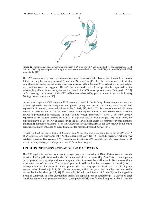
Figure 2: Comparison of three-dimensional structures of P. separata GBP and mouse EGF. Ribbon diagrams of GBP<br />
(left) and EGF (right) were generated using the atomic coordinates obtained from the PDB (entry sets 1BQF and 1EPG,<br />
respectively) [10].<br />
The ENF peptide gene is expressed in many stages and tissues of moths. Transcripts of multiple sizes were<br />
detected during the embryogenesis of B. mori and M. brassicae [15, 18]. The mRNAs were not detected<br />
immediately following the oviposition, but were detected within the next 24 h, indicating that their origins<br />
were not maternal but zygotic. The M. brassicae GBP mRNA is specifically expressed in the<br />
suboesophageal body in the embryo under the control of a HOX transcriptional factor, Deformed [15, 35].<br />
In M. sexta eggs, expression of the PP2 mRNA was enhanced by parasitization of the parasitoid wasp<br />
Trichogramma evanescens [36].<br />
In the larval stage, the ENF peptide mRNAs were expressed in the fat body, hemocytes, central nervous<br />
system, epidermis, muscle, wing disc, and gonads (ovary and testis), and among these tissues their<br />
expressions, in general, were predominant in the fat body [12, 16-18, 37]. In contrast, these mRNAs were<br />
detected in small amounts in the silk gland, midgut or Malpighian tubules. While a 0.6-0.8 kb ENF peptide<br />
mRNA is predominantly expressed in many tissues, longer transcripts of sizes >2 kb were strongly<br />
expressed in the central nervous systems of P. separata and P. includens [13, 16]. In B. mori, the<br />
expression level of PP mRNA changed during the last larval instar under the control of juvenile hormone<br />
and molting hormone ecdysone [18]. In the P. separata larvae, expression of the GBP mRNA in the central<br />
nervous system was enhanced by parasitization of the parasitoid wasp A. kariyai [38].<br />
Recently, it has been shown that a 1.5 kb embryonic PP mRNA of B. mori and a 2.5 kb larval GBP mRNA<br />
of P. separata are tricistronic mRNAs that encode not only the ENF peptide precursor but also two<br />
cytokine precursor-like proteins [19]. Orthologous tricistronic ENF peptide mRNAs were found in M.<br />
brassicae, S. cynthia pryeri, T. japonica, and N. himachala sangaica.<br />
4. PROTEIN EXPRESSION, ACTIVATION, AND INACTIVATION<br />
C<br />
The ENF peptide is translated as an inactive larger precursor, consisting of 120 to 150 amino acids, and the<br />
bioactive ENF peptide is located at the C-terminal end of the precursor (Fig. 3A). This precursor protein<br />
(preproprotein) has a signal peptide containing a number of hydrophobic residues at the N-terminus end and<br />
is secreted out of the cells. The secreted protein is still an inactive precursor (proprotein) and is<br />
subsequently cleaved to form the active peptide after receiving special stimuli, such as bleeding and<br />
microorganism infection [17-18, 39]. Serine proteases, which are yet to be identified, are presumably<br />
responsible for this cleavage [17, 39]. For example, following an infection of B. mori by a microorganism,<br />
a cellular component of the microorganism, such as the peptidoglycan of bacteria or -1, 3-glucan of fungi,<br />
stimulates hemocytes to generate reactive oxygen species (ROS) (see for detail <strong>chapter</strong> number 8), and the<br />
N<br />
N


