Molecular characterisation of odontoblast during primary, secondary ...
Molecular characterisation of odontoblast during primary, secondary ...
Molecular characterisation of odontoblast during primary, secondary ...

You also want an ePaper? Increase the reach of your titles
YUMPU automatically turns print PDFs into web optimized ePapers that Google loves.
Introduction<br />
superficial cells move to the centre <strong>of</strong> the injury regenerating and repairing the<br />
tissue and allowing its reorganisation.<br />
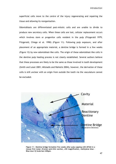
Odontoblasts are differentiated post-mitotic cells and are unable to divide to<br />
produce new secretory cells. When these cells are lost, cellular replacement occurs<br />
which involves stem or progenitor cells resident in the pulp (Fitzgerald 1979;<br />
Fitzgerald, Chiego et al. 1990) (Figure 11). Following pulp exposure, and after<br />
placement <strong>of</strong> an appropriate material, a dentine bridge is formed in a few weeks<br />
(Figure 12) by new <strong>odontoblast</strong>-like cells. The origin <strong>of</strong> these <strong>odontoblast</strong>-like cells in<br />
the dentine pulp healing process is not clearly established. Several authors believe<br />
that these processes are likely to be the same as those involved in tooth development<br />
(Smith and Lesot 2001; Mitsiadis and Rahiotis 2004), however, the derivation <strong>of</strong> these<br />
cells is still unclear with an origin from outside the tooth via the vasculature cannot<br />
be excluded.<br />
Figure 11 : Dentine bridge formation five weeks after pulp capping with MTA® in a<br />
mouse first molar (frontal semi-thin section, x50 magnification, methylene blue –<br />
Blue Azur II) Scale bar=200µm.<br />
47