View Annual Report 2012 - University of Illinois College of Medicine ...
View Annual Report 2012 - University of Illinois College of Medicine ...
View Annual Report 2012 - University of Illinois College of Medicine ...

You also want an ePaper? Increase the reach of your titles
YUMPU automatically turns print PDFs into web optimized ePapers that Google loves.
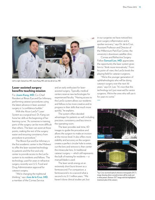
Left to right: Samuel Lee, MD, Joann Kang, MD, and Jose de la Cruz, MD<br />
Laser-assisted surgery<br />
benefits teaching mission<br />
For Joann Kang, MD, Co-Chief<br />
Resident at <strong>Illinois</strong> Eye and Ear Infirmary,<br />
performing cataract procedures using<br />
the latest advance in laser-assisted<br />
surgery is “a confidence builder.”<br />
With the Alcon LenSx® Laser<br />
System as a surgical tool, Dr. Kang can<br />
hone her skills at the beginning <strong>of</strong> her<br />
learning curve. “As someone in training,<br />
parts <strong>of</strong> the surgery can be more difficult<br />
than others. The laser can assist at those<br />
points, making the rest <strong>of</strong> the surgery<br />
easier and ensuring consistency from<br />
procedure to procedure.”<br />
The <strong>Illinois</strong> Eye and Ear Infirmary is<br />
the first academic center in the Midwest<br />
to <strong>of</strong>fer the laser-assisted technology<br />
to patients and the first and only<br />
center to provide access to the LenSx<br />
system to its residents and fellows. The<br />
technology, used for years in refractive<br />
surgery, recently won U.S. Food and<br />
Drug Administration approval for<br />
cataract surgery.<br />
“We’re changing the traditional<br />
thinking,” says Jose de la Cruz, MD,<br />
a member <strong>of</strong> the Cornea Service<br />
and an early enthusiast for laserassisted<br />
surgery. Typically, medical<br />
centers reserve new technologies for<br />
experienced faculty. “Having access to<br />
the LenSx system allows our residents<br />
and fellows to be more creative and to<br />
progress in their skills that much more<br />
quickly,” he explains.<br />
The system <strong>of</strong>fers decided<br />
advantages for patients as well, including<br />
precision, consistency and less time in<br />
the operating room.<br />
The laser provides real-time, 3D<br />
images to guide the procedure and<br />
allows the surgeon to make an incision<br />
at the micron level. It also <strong>of</strong>fers more<br />
stability and accuracy as the surgeon<br />
creates a perfect circular hole to enter,<br />
cut the lens and remove it, then center<br />
the intraocular lens. In traditional<br />
cataract surgery — which still represents<br />
the bulk <strong>of</strong> training for residents — a<br />
manual blade is used.<br />
The laser sends energy at an<br />
extremely short burst known as a<br />
femtosecond. For comparison, a<br />
femtosecond is to a second what a<br />
second is to 31.7 million years. “We<br />
haven’t done clinical studies yet, but<br />
One Vision <strong>2012</strong> 11<br />
in our surgeries we have noticed less<br />
post-surgery inflammation and a<br />
quicker recovery,” says Dr. de la Cruz,<br />
Assistant Pr<strong>of</strong>essor and Director <strong>of</strong><br />
the Millennium Park Eye Center, the<br />
university’s downtown satellite clinic.<br />
Cornea and Refractive Surgery<br />
Fellow Samuel Lee, MD, appreciates<br />
the opportunity the laser system gives<br />
him to “think more innovatively.” From<br />
his point-<strong>of</strong>-view, the LenSx levels the<br />
playing field for cataract surgeons.<br />
“We’re the younger generation <strong>of</strong><br />
ophthalmologists who will be doing<br />
cataract surgery over the next 30<br />
years,” says Dr. Lee. “It’s nice that this<br />
technology isn’t just reserved for senior<br />
surgeons. We’re the ones who will use it<br />
for years to come.”<br />
Top: Cross-sectional optical coherence tomography (OCT)<br />
image showing where corneal incisions will be made and<br />
in what pattern. Bottom: OCT image with the highlighted<br />
treatment plan <strong>of</strong> the cataract.

![CV Joan [W51] - University of Illinois College of Medicine at Chicago ...](https://img.yumpu.com/17336863/1/190x245/cv-joan-w51-university-of-illinois-college-of-medicine-at-chicago-.jpg?quality=85)