Barotrauma and Hypotension Resulting from Jet Ventilation in ...
Barotrauma and Hypotension Resulting from Jet Ventilation in ...
Barotrauma and Hypotension Resulting from Jet Ventilation in ...

You also want an ePaper? Increase the reach of your titles
YUMPU automatically turns print PDFs into web optimized ePapers that Google loves.
<strong>Barotrauma</strong> <strong>and</strong> <strong>Hypotension</strong> <strong>Result<strong>in</strong>g</strong><br />
<strong>from</strong> <strong>Jet</strong> <strong>Ventilation</strong> <strong>in</strong> Critically Ill Patients*<br />
Andrew Egol, D.O.; Judith A Culpepper, M. D.; <strong>and</strong> James V Snyder, M. D.<br />
We present the first reports of pneumoperitoneum secon- decrease the risk of such complications. We cannot, at<br />
dary to jet ventilation, bamtrauma secondary to jet ventila- present, recommend the use of h<strong>and</strong>-held jet ventilators<br />
tion thtough the suction port of a fiberoptic laryngoscope, unless both adequate exhalation space is guaranteed <strong>and</strong><br />
<strong>and</strong> hypotension due to jet ventilation via nasotracheal <strong>and</strong> direct imp<strong>in</strong>gement of the catheter's tip on the mucosal<br />
omtracheal catheters. We suggest that m<strong>in</strong>imiz<strong>in</strong>g airway surface is avoided.<br />
pressum <strong>and</strong> us<strong>in</strong>g jet catheters with side holes may help<br />
variety of techniques for jet ventilation have been<br />
A used <strong>in</strong> recent years as a means of deliver<strong>in</strong>g<br />
positive-pressure ventilation to patients dur<strong>in</strong>g such<br />
procedures as bronchoscopy, laryngoscopy, tracheal<br />
suction<strong>in</strong>g, endotracheal tube change, <strong>and</strong> <strong>in</strong> other<br />
circumstances where positive-pressure ventilation is<br />
required dur<strong>in</strong>g manipulation of the upper airway.14<br />
Several methods for h<strong>and</strong>-held jet ventilation dur<strong>in</strong>g<br />
these procedures have been described previou~ly.~.~"<br />
Although jet ventilation can appear simple <strong>and</strong> effective<br />
<strong>in</strong> provid<strong>in</strong>g oxygenation <strong>and</strong> ventilation for patients,<br />
potentially life-threaten<strong>in</strong>g complications can<br />
occur. We report the f<strong>in</strong>d<strong>in</strong>gs <strong>in</strong> three patients who<br />
suffered significant complications dur<strong>in</strong>g manually <strong>and</strong><br />
mechanically regulated jet ventilation <strong>and</strong> suggest<br />
alterations <strong>in</strong> technique that may prevent such problems.<br />
A 66-year-old white woman was admitted to the <strong>in</strong>tensive care unit<br />
(ICU) after fusion of the thoracic sp<strong>in</strong>e for osteoporotic vertebral<br />
compression fractures. The patient's postoperative problems <strong>in</strong>cluded<br />
retmperitoneal hematoma (which required reexploration of<br />
the surgical site), pneumonia, sepsis with Pseudomonas, renal<br />
failure, <strong>and</strong> the need for prolonged mechanical ventilation.<br />
On the Uth postoperative day, laryngoscopic exam<strong>in</strong>ation of the<br />
patienti upper airway was undertaken to evaluate whether any<br />
laryngeal <strong>in</strong>jury was present due to prolonged orotracheal <strong>in</strong>tubation.<br />
S<strong>in</strong>ce it seemed unlikely that the patient could soon be weaned<br />
<strong>from</strong> mechanical ventilation <strong>and</strong> then extubated, such damage, if<br />
present, would have been considered an <strong>in</strong>dication for tracheostomy.<br />
It was planned to temporarily remove the orotracheal tube over a<br />
hollow flexible stylet, over which the orotracheal tube would be<br />
replaced at the end of the exam<strong>in</strong>ation. The same stylet served to<br />
deliver jet ventilation dur<strong>in</strong>g exam<strong>in</strong>ation of the larynx. The h<strong>and</strong>held<br />
device for jet ventilation that was used (Instrumentation<br />
Industries) delivers oxygen at the available driv<strong>in</strong>g pressure. This is<br />
commonly 50 psi or 2,620 mm Hg. A ~ressure-reduc<strong>in</strong>g valve can be<br />
<strong>in</strong>corporated to lower the driv<strong>in</strong>gpressure, but it was not used <strong>in</strong> this<br />
*From the Critical Care Medic<strong>in</strong>e Tra<strong>in</strong><strong>in</strong>g Program, University<br />
Health Center, Pittsburgh.<br />
Manuscript received January 26; revision accepted December 18.<br />
98<br />
Downloaded From: http://journal.publications.chestnet.org/ on 07/13/2013<br />
case. The device is manually operated by a spr<strong>in</strong>g-loaded valve <strong>and</strong><br />
delivers gas via noncompliant tub<strong>in</strong>g to a jett<strong>in</strong>g stylet. The jett<strong>in</strong>g<br />
stylet <strong>in</strong> this case was an 18 French tracheal suction<strong>in</strong>g catheter with<br />
two end holes.<br />
After the conventional mechanical ventilator was disconnected,<br />
the stylet was passed down the endotracheal tube <strong>in</strong>to the trachea,<br />
<strong>and</strong> jet ventilation was <strong>in</strong>itiated. The patient almost immediately<br />
became hypotensive <strong>and</strong> cyanotic, with obvious abdom<strong>in</strong>al disten-<br />
tion. The jet stylet was withdrawn, <strong>and</strong> the patient was manually<br />
ventilated through the conventional endotracheal tube (which had<br />
not been removed) with aself-<strong>in</strong>flat<strong>in</strong>g bag us<strong>in</strong>g 100 percent oxygen.<br />
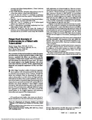
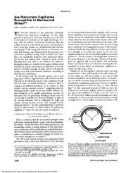
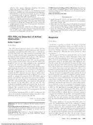
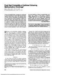
A chest roentgenogram confirmed the presence of a right-sided<br />
tension pneumothorax <strong>and</strong> pneumoperitoneum (Fig 1). A chest tube<br />
was immediately <strong>in</strong>serted, <strong>and</strong> the patient was returned to conven-<br />
tional mechanical ventilation. Her vital signs <strong>and</strong> arterial blood gas<br />
levels improved to their basel<strong>in</strong>e levels. The pneumothorax <strong>and</strong><br />
pneumoperitoneum resolved uneventfully. The patient survived this<br />
event <strong>and</strong>, after a prolonged stay <strong>in</strong> the hospital, was able to return to<br />
her home, ambulat<strong>in</strong>g with a walker.<br />
FIGURE 1. Chest roentgenogram show<strong>in</strong>g tension pneumothorax<br />
<strong>and</strong> pneurnoperitoneum which occurred with jet ventilation dur<strong>in</strong>g<br />
laryngoscopy (case 1).<br />
Barmuma <strong>and</strong> Hypdension hum <strong>Jet</strong> <strong>Ventilation</strong> (Em Culpepper, Snyder)
A man had his jaws wired closed follow<strong>in</strong>g surgical duction of<br />
prognathism. Because of the risk of airway compromise secondary to<br />
postoperative swell<strong>in</strong>g of soft tissue, he was extubated over a jett<strong>in</strong>g<br />
stylet.= This stylet was a double-lumen nasogastric sump tube<br />
<strong>in</strong>tended to be left <strong>in</strong> the trachea while the patient was observed for<br />
respiratory problems. Were ventilatory difficulty to occur, this stylet<br />
could deliver low-flow oxygen or jet ventilation; it could also function<br />
as a stylet over which a nasotracheal tube could aga<strong>in</strong> be <strong>in</strong>serted.<br />
Immediately after extubation, the patient breathed around the<br />
endotracheal stylet without difficulty. Upon <strong>in</strong>itiation of a trial<br />
demonstration ofjet ventilation with aventilator set at approximately<br />
20 psi <strong>and</strong> 30 percent <strong>in</strong>jection time (Instrument Development Corp<br />
vs 600), the patient rema<strong>in</strong>ed comfortable until he tried to talk. At<br />
that time, his mean arterial blood pressure fell rapidly to 30 mm Hg,<br />
presumably due to closure of his vocal cords around the catheter.<br />
Blood pressure returned to normal promptly after the jet ventilator<br />
was turned off <strong>and</strong> the patient was positioned horizontally. No<br />
evidence ofbarotraumawas apparent on achest roentgenogram. The<br />
patient was stable with the stylet left <strong>in</strong> the trachea for the rema<strong>in</strong><strong>in</strong>g<br />
period of observation after the <strong>in</strong>itial extubation; remwal of the<br />
stylet resulted <strong>in</strong> no further problem.<br />
A 62-yearold white man presented to the ICU with the superior<br />
vena cava syndrome <strong>and</strong> respiratory failure secondary to atelectasis<br />
of the right upper <strong>and</strong> middle lobes <strong>from</strong> an endobronchial small cell<br />
carc<strong>in</strong>oma. Several days later, when he was stabilizd <strong>and</strong> ready for<br />
extubation, an exam<strong>in</strong>ation of the upper airway with a flexible<br />
fiberoptic laryngompe was planned <strong>in</strong>order to assess damage to the<br />
- -<br />
uDwr airway <strong>from</strong> the <strong>in</strong>itial <strong>in</strong>tubation. which had been traumatic.<br />
The fiberoptic laryngompe was passed through the nasotracheal<br />
tube without difficulty, after the patient was given a 100-mg<br />
<strong>in</strong>travenous bolus of lidoca<strong>in</strong>e to dim<strong>in</strong>ish his cough reflex. The<br />
endotracheal tube was withdrawn <strong>from</strong> the larynx, over the<br />
laryngoscope, to facilitate the exam<strong>in</strong>ation of the larynx. Dur<strong>in</strong>g the<br />
entire procedure, jetted oxygen at 40 psi via the suction port of the<br />
fiberoptic laryngompe provided ventilation. The jet ventilator was<br />
the same as that described <strong>in</strong> case 1.<br />
Three m<strong>in</strong>utes <strong>in</strong>to the exam<strong>in</strong>ation, the patient developed<br />
significant abdom<strong>in</strong>al distention <strong>and</strong> rigidity, with progressive<br />
cyanosis. Attempts to pass a nasogastric tube for decompression of<br />
the stomach were unsuccessful. Arterial blood gas analysis showed<br />
comb<strong>in</strong>ed respiratory <strong>and</strong> metabolic acidosis with severe hypox-<br />
emia. Replacement of the endotracheal tube failed to impmve the<br />
movement of air, cyanosis persisted, <strong>and</strong> arterial hypotension<br />
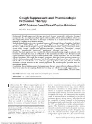
ensued. Roentgenograms of the abdomen <strong>and</strong> chest revealed<br />
marked pneumoperitoneum, elevated hemidiaphragms, distention<br />
of the gastro<strong>in</strong>test<strong>in</strong>al tract, <strong>and</strong> result<strong>in</strong>g significant volume reduc<br />
tion of both lungs (Fig 2) without apparent pneumomediast<strong>in</strong>um or<br />
pneumothorax. Rapidly adm<strong>in</strong>istered <strong>in</strong>travenous fluids partially<br />
corrected this patient's hypotension. Insertion of a 19-gauge catheter<br />
(Cook) <strong>in</strong>to the peritoneal space caused prompt decompression of<br />
the pneurnoperitoneum <strong>and</strong> resolution of the hypotension, hypox-<br />
emia, <strong>and</strong> respiratory acidosis. A nasogastric tube was then success-<br />
fully passed to facilitate gastric decompression.<br />
Subsequent roentgenograms showed a resolv<strong>in</strong>g pneumoperito-<br />
neum but still no evidence of pneumomediast<strong>in</strong>um or pneu-<br />
mothorax. Instillation of water-soluble contrast material <strong>in</strong>to the<br />
stomach through the patient's nasogastric tube failed to demonstrate<br />
perbration as the cause of the pneurnoperitoneum. The patient's<br />
family rehsed to grant permission for an exploratory laparotomy to<br />
exclude a perforated viscus. The peritoneal catheter was removed<br />
with<strong>in</strong> five hours. The pneurnoperitoneum resolved completely over<br />
several days.<br />
The patient's subsequent course was complicated by severe<br />
Downloaded From: http://journal.publications.chestnet.org/ on 07/13/2013<br />
pneumonia due to Cytomegalovirus <strong>and</strong> Pseudomonas aerog<strong>in</strong>osa,<br />
as well as renal failure <strong>and</strong> seizures. The patient died on the 57th day<br />
of hospitalization (51 days after develop<strong>in</strong>g the pneurnoperitoneum).<br />
Postmortem exam<strong>in</strong>ation revealed small cell carc<strong>in</strong>oma of the right<br />
upper lobe with superior vena caval obstruction. Metastases were<br />
present <strong>in</strong> the cerebral cortex, cerebellum, para-aortic <strong>and</strong> para-<br />
tracheal lymph nodes, liver, <strong>and</strong> adrenal gl<strong>and</strong>s. No evidence was<br />
present for an esophageal, gastric, tracheal, or pleural perforation.<br />
Mechanism of Injuries<br />
Theoretically, barotrauma <strong>from</strong> jet ventilation may<br />
be due to <strong>in</strong>jection <strong>in</strong>jury (analagous to grease-gun<br />
<strong>in</strong>jury) or to overdistention of lung. Injection <strong>in</strong>jury<br />
can occur when a high-pressure stream of gas is<br />
directed aga<strong>in</strong>st mucosa <strong>and</strong> penetrates <strong>in</strong>to sub-<br />
mucosal tissue or even perforates the wall altogether.<br />
This <strong>in</strong>jury is more likely when the jett<strong>in</strong>g catheter has<br />
only a term<strong>in</strong>al port, ensur<strong>in</strong>g excessive pressure (as<br />
high as the pressure of the gas source used) when that<br />
port is pressed aga<strong>in</strong>st mucosa. This <strong>in</strong>jury is unrelated<br />
to airway pressure <strong>and</strong> does not require a high volume<br />
of gas. The second form of jet-<strong>in</strong>duced barotrauma,<br />
that which is due to overdistention, may also occur<br />
without complete obstruction of the airway. If there is<br />
<strong>in</strong>sufficient time or space for jetted gas to be exhaled,<br />
pressure will build up with<strong>in</strong> the tracheobronchial<br />
tree, result<strong>in</strong>g <strong>in</strong> rupture at some po<strong>in</strong>t not necessarily<br />
close to the jet catheter. Oliverio et ale described one<br />
such case of pneumothorax dur<strong>in</strong>g laryngoscopic jet<br />
FIGURE 2. Film document<strong>in</strong>g pneurnoperitoneum at time of jet<br />
ventilation through fiberoptic laryngoscope (case 3).<br />
CHEST 1 88 I 1 I JULY. 11985
ventilation. In this case, prolapse of a large vocal cord<br />
papilloma apparently prevented an adequate outlet for<br />
exhalation, caus<strong>in</strong>g high <strong>in</strong>tratracheal pressures <strong>and</strong><br />
the result<strong>in</strong>g pneumothorax.<br />
Chang et all0 reported the f<strong>in</strong>d<strong>in</strong>gs <strong>in</strong> a patient who<br />
developed left-sided pneumothorax dur<strong>in</strong>g jet venti-<br />
lation for laryngoscopy <strong>in</strong> the operat<strong>in</strong>g room; the<br />
pneumothorax was probably secondary to the jet<br />
catheter hav<strong>in</strong>g been <strong>in</strong>advertently pushed beyond the<br />
, car<strong>in</strong>a <strong>and</strong> possibly wedged <strong>in</strong> a bronchus when the<br />
patient was moved. In our first patient, we th<strong>in</strong>k that<br />
the pneumothorax was similarly due to accidental en-<br />
dobronchial position<strong>in</strong>g of the jet catheter. Such mal-<br />
position<strong>in</strong>g could have resulted <strong>in</strong> either an <strong>in</strong>jection<br />
<strong>in</strong>jury or local pulmonary distention <strong>and</strong> rupture.<br />
S<strong>in</strong>ce the suction catheter that was used has holes only<br />
at its end, gas exits <strong>from</strong> the catheter tip under high<br />
pressure. When the tip is wedged <strong>in</strong>to a small airway,<br />
the distal pulmonary tissue may be quickly subjected<br />
to high pressure equal to the driv<strong>in</strong>g pressure (10 to 50<br />
psi) of the system. Dissection of gas through soft tissue<br />
planes, as discussed subsequently, presumbly resulted<br />
<strong>in</strong> pneumoperitoneum.<br />
Inadequate space <strong>and</strong> time for exhalation is probably<br />
also the etiologic mechanism when a patient be<strong>in</strong>g<br />
ventilated with a jet catheter through the larynx closes<br />
his cords around the catheter <strong>in</strong> laryngospasm or to<br />
talk. We believe the hypotension <strong>in</strong> our second patient<br />
to be due to high pleural pressure <strong>from</strong> this mecha-<br />
nism, with subsequent drop <strong>in</strong> venous return <strong>and</strong><br />
cardiac output.<br />
Pneumoperitoneum as a barotraumatic complication<br />
of ventilatory support usually occurs through disrup-<br />
tion of either alveoli or small airways. Air, once out of<br />
the bronchial tree, dissects along the perivascular<br />
space back to the mediast<strong>in</strong>um <strong>and</strong>, <strong>from</strong> there,<br />
through soft tissue planes to result <strong>in</strong> pneumomediasti-<br />
num, retroperitoneal air, <strong>and</strong> subcutaneous emphy-<br />
sema." Subsequent rupture of pleura or peritoneum<br />
will lead to pneumothorax <strong>and</strong> pneumoperitoneum,<br />
respectively. In conventional mechanical ventilation,<br />
these complications are related to peak <strong>in</strong>spiratory<br />
pressures, end-expiratory pressures, duration of <strong>in</strong>-<br />
spiratory time, <strong>and</strong> the presence of underly<strong>in</strong>g pulmo-<br />
nary disease."" Although pneumoperitoneum is a<br />
recognized complication of conventional mechanical<br />
ventilation, it has not been described previously as a<br />
complication of jet ventilation.<br />
In our first patient, pneumoperitoneum was pre-<br />
sumably secondary to pneumomediast<strong>in</strong>um, with fur-<br />
ther dissection of respiratory gas through soft tissue<br />
planes. In our third patient the mechanism is not so<br />
clear. Pneumoperitoneum associated with mechanical<br />
ventilation is usually associated with either pneu-<br />
mothorax or pneumomediast<strong>in</strong>um, <strong>and</strong> its occurrence<br />
<strong>in</strong> their absence suggests possible perforation of an<br />
Downloaded From: http://journal.publications.chestnet.org/ on 07/13/2013<br />
abdom<strong>in</strong>al viscus.14 In our third patient, pneumothorax<br />
or pneumomediast<strong>in</strong>um never became cl<strong>in</strong>ically<br />
or roentgenographically apparent. There was<br />
also no radiographic evidence of gas <strong>in</strong> the wall of the<br />
large bowel. Although the patient did not have a<br />
readily apparent gastric perforation on contrast exam<strong>in</strong>ation,<br />
a lower esophageal perforation was not<br />
ruled out, as this area was not exam<strong>in</strong>ed dur<strong>in</strong>g the<br />
contrast study, <strong>and</strong> no esophagoscopy was performed.<br />
We speculate that he may have had <strong>in</strong>advertent direction<br />
of gas flow under high pressure <strong>in</strong>to the esophagus,<br />
result<strong>in</strong>g <strong>in</strong> either a high esophagogastric pressure<br />
with distension <strong>and</strong> rupture or a local <strong>in</strong>jection <strong>in</strong>jury<br />
to the wall of the esophagus. The gas distention seen <strong>in</strong><br />
his gastro<strong>in</strong>test<strong>in</strong>al tract on roentgenograms supports<br />
either hypothesis. Either of these two mechanisms<br />
could result <strong>in</strong> tension pneumoperitoneum." Chang et<br />
alW described a case of severe abdom<strong>in</strong>al distension<br />
dur<strong>in</strong>g jet ventilation which was believed to be secondary<br />
to displacement of the jet catheter, allow<strong>in</strong>g part<br />
of the jet ventilator stream to be <strong>in</strong>troduced <strong>in</strong>to the<br />
patient's digestive tract. While this did not result <strong>in</strong><br />
pneumoperitoneum, the gastro<strong>in</strong>test<strong>in</strong>al distension,<br />
noted on abdom<strong>in</strong>al roentgenograms, impaired spontaneous<br />
breath<strong>in</strong>g to such a degree that the patient<br />
required re<strong>in</strong>tubation for mechanical ventilation.<br />
Another possible explanation for our third patient's<br />
barotrauma is that rupture occurred at the catheter tip<br />
with<strong>in</strong> the trachea due to <strong>in</strong>jection <strong>in</strong>jury, with air<br />
dissection <strong>in</strong>to the esophagus <strong>and</strong> through the lower<br />
mediast<strong>in</strong>um <strong>in</strong>to the peritoneal cavity. An additional<br />
possible mechanism is that gas jetted <strong>in</strong>to the trachea<br />
had <strong>in</strong>adequate room for exhalation, with <strong>in</strong>creased<br />
overall airway pressure result<strong>in</strong>g <strong>in</strong> rupture of the<br />
tracheobronchial tree at a site distant <strong>from</strong> the catheter<br />
tip, with dissection of gas through soft tissue planes to<br />
the peritoneum; however, there was no roentgenographically<br />
demonstrated free mediast<strong>in</strong>al or pleural<br />
air to support this hypothesis.<br />
Several authors have described the use ofjet ventilation<br />
dur<strong>in</strong>g fiberoptic bronchoscopy <strong>and</strong> laryngos-<br />
A variety of methods have been used which<br />
<strong>in</strong>clude ventilation through the bronchoscope (via the<br />
suction port of a flexible bronchoscope or an attached<br />
channel of a rigid bronchoscope) or through a 14-gauge<br />
catheter placed <strong>in</strong>side an endotracheal tube with an<br />
<strong>in</strong>ner diameter of 3 to 5 mm that was placed along the<br />
outside of the bronchoscope. While complications<br />
have been described with translaryngeal <strong>and</strong> percuta-<br />
neous transtracheal catheter jet ventilati~n,'~~~~~~" no<br />
complications previously have been reported second-<br />
ary to jet ventilation through the suction port of a<br />
fiberoptic bronchoscope or laryngoscope. We believe<br />
that both mechanisms of trauma discussed previously<br />
are <strong>in</strong>herent with this maneuver; however, direct visu-<br />
alization throughout the procedure should m<strong>in</strong>imize<br />
mutrauma <strong>and</strong> W(HISiOn trOm <strong>Jet</strong> Ver~uiatbr~ (€go/, cupepper; snyw
the risk of <strong>in</strong>jection <strong>in</strong>jury.<br />
Preuentwn of Znju y<br />
With either of the two methods of damage (<strong>in</strong>jection<br />
<strong>in</strong>jury <strong>and</strong> impaired exhalation), trauma is related to<br />
pressure. Pressure can be limited by avoid<strong>in</strong>g the<br />
factors which <strong>in</strong>crease it <strong>and</strong> by development <strong>and</strong> use<br />
of pressure-sens<strong>in</strong>g <strong>and</strong> cutoff devices. The amount of<br />
pressure <strong>in</strong> the airway is determ<strong>in</strong>ed by multiple<br />
<strong>in</strong>terrelat<strong>in</strong>g factors. Resistance to exhalation, time of<br />
exhalation, <strong>and</strong> m<strong>in</strong>ute ventilation determ<strong>in</strong>e positive<br />
end-expiratory pressure (PEEP). Peak airway pressure<br />
is determ<strong>in</strong>ed by PEEP, compliance of the lungs, <strong>and</strong><br />
tidal volume (VT) (ie, gas flow <strong>and</strong> <strong>in</strong>spiratory time).<br />
Gas flow is determ<strong>in</strong>ed by driv<strong>in</strong>g pressure <strong>and</strong> the<br />
radius <strong>and</strong> length of the jet catheter. Manipulat<strong>in</strong>g<br />
these factors may decrease the risk of <strong>in</strong>jury, as shown<br />
<strong>in</strong> the follow<strong>in</strong>g tabulation giv<strong>in</strong>g suggestions for<br />
decreas<strong>in</strong>g the risk with a h<strong>and</strong>-held jet:<br />
1. Be cont<strong>in</strong>ually aware of location of catheter tips:<br />
A. Avoid direct pressure aga<strong>in</strong>st mucosa.<br />
B. Avoid <strong>in</strong>sertion <strong>in</strong>to orifices <strong>from</strong> which exhalation<br />
may be restricted.<br />
C. Secure catheter to m<strong>in</strong>imize risk of migration.<br />
D. Use tracheal tubes with fixed-position jet catheter.<br />
2. Use catheters with side holes.<br />
3. M<strong>in</strong>imize resistance to exhalation:<br />
A. Use a smaller-diameter jet catheter <strong>and</strong> a larger<br />
endotracheal tube. Deflate the endotracheal tube<br />
cuff.<br />
4. Use suction: apply<strong>in</strong>g a negative pressure to the ex-<br />
piratory port will dim<strong>in</strong>ish entra<strong>in</strong>ment <strong>and</strong> VT, but<br />
also lower peak <strong>and</strong> mean pressure <strong>and</strong> PEEP.<br />
5. Use m<strong>in</strong>imal effective driv<strong>in</strong>g pressure.<br />
6. Encourage development <strong>and</strong> use of effective pressure-<br />
sensor <strong>and</strong> pressure-cutoff device.<br />
For example, the use of more side holes, a lower<br />
driv<strong>in</strong>g pressure, or a smaller catheter, or not advanc-<br />
<strong>in</strong>g the catheter as far, all might have prevented<br />
pulmonary rupture <strong>in</strong> the first case. Avoid<strong>in</strong>g contact<br />
with the mucosal surfice might have prevented <strong>in</strong>jury<br />
<strong>in</strong> the third case. When a jet catheter is <strong>in</strong>serted<br />
through the larynx, safe use requires that the patient<br />
not close his or her cords or lips <strong>and</strong> that expiratory<br />
obstruction is absent. Development of an effective<br />
pressure sensor <strong>and</strong> pressure cutoff would enhance<br />
safety <strong>in</strong> all applications. We recognize that these<br />
recommendations are contradictory-one must hope<br />
to achieve a balance among them to m<strong>in</strong>imize risk <strong>and</strong><br />
maximize benefit to the patient.<br />
These cases demonstrate the necessity of cont<strong>in</strong>ued<br />
awareness of the position of the jet catheter tip dur<strong>in</strong>g<br />
the entire procedure of jet ventilation. If a jet catheter<br />
is used that is long enough to slip past the car<strong>in</strong>a <strong>in</strong>to<br />
smaller airways, it may be safer to use a catheter with<br />
large proximal side holes so that gas can escape above<br />
the potentially wedged tip of the catheter. Such side<br />
Downloaded From: http://journal.publications.chestnet.org/ on 07/13/2013<br />
Table 1-Effect qfcatheter Vatidled on Preusum<br />
Delioetad by the <strong>Jet</strong>*<br />
Pressure at<br />
Catheter's<br />
<strong>Jet</strong> Catheter Tip, psi?<br />
3%-<strong>in</strong> 14gauge Deseret catheter No. 012-010<br />
(with open tip)<br />
With 4 side holes open 30<br />
With 2 side holes open 42<br />
With 0 side holes open 52<br />
16 French Argyle double-lumen Salem sump tube<br />
All holes are side holes; tip is closed<br />
With 11 holes open 0<br />
If tip is cut to open it<br />
With 11 side holes open 2<br />
With 9 side holes open 2<br />
With 7 side holes open 4<br />
With 5 side holes open 4<br />
With 3 side holes open 4<br />
With 1 side hole open 5<br />
With 0 side holes open 52<br />
14 French Superior suction catheter (with<br />
open tip)<br />
With 2 side holes open<br />
With 1 side hole open<br />
With 0 side holes open ---<br />
*H<strong>and</strong>-held jet ventilator (Instrumentation Laboratories).<br />
tAs measured by pressure gauge (US Gauge 13400-1).<br />
holes can markedly decrease pressure delivered at the<br />
tip but still function smoothly when used cl<strong>in</strong>ically for<br />
jet ventilation. A catheter with a closed tip <strong>and</strong> side<br />
holes appears to be even safer (Table 1). Inadvertent<br />
wedg<strong>in</strong>g of the catheter can be prevented by measur<strong>in</strong>g<br />
its length <strong>and</strong> mark<strong>in</strong>g the appropriate depth of<br />
<strong>in</strong>sertion before beg<strong>in</strong>n<strong>in</strong>g the procedure <strong>and</strong> secur<strong>in</strong>g<br />
the catheter h m further advance once it is properly<br />
positioned <strong>in</strong> the patient's trachea. Despite these<br />
precautions, the use of bl<strong>in</strong>dly placed jet catheters may<br />
result <strong>in</strong> a higher <strong>in</strong>cidence of direct <strong>in</strong>jury than if the<br />
procedure were performed under constant direct visualization.<br />
A catheter appropriate <strong>in</strong> some cl<strong>in</strong>icd<br />
situations is the HiLo <strong>Jet</strong> tracheal tube (No. 86613;<br />
Mall<strong>in</strong>ckrodt, Inc.). This tube consists of a conventional<br />
endotracheal tube with a separate lumen for the<br />
jet which is one-tenth the lumen provided for exhalation,<br />
thus ensur<strong>in</strong>g adequate space for exhalation.<br />
Furthermore, this catheter is short enough that <strong>in</strong>advertent<br />
wedg<strong>in</strong>g <strong>in</strong>to small airways is unlikely.<br />
Pressure-cutoff devices for jet ventilation rema<strong>in</strong> a<br />
problem. The use of a separate catheter to sense<br />
pressure <strong>and</strong> activate a cutoff entails risk of occlusion of<br />
the sens<strong>in</strong>g catheter (eg, by condensed water) <strong>and</strong> is<br />
thus <strong>in</strong>herently <strong>in</strong>adequate to protect the patient <strong>from</strong><br />
<strong>in</strong>jection <strong>in</strong>juries. Incorporation of the sens<strong>in</strong>g device<br />
with<strong>in</strong> the jet catheter is desirable but requires tolerance<br />
of the high pressures dur<strong>in</strong>g <strong>in</strong>spiration (eg, 40 to<br />
50 psi) while reta<strong>in</strong><strong>in</strong>g sensitivity to significant changes<br />
<strong>in</strong> expiratory pressure. The system must also be easily<br />
CHEST 1 88 I 1 1 JULY, 1198 101
adjustable. No system for jet ventilation that is com-<br />
mercially available at this time has all of these features.<br />
In summary, although jet ventilation is potentially<br />
useful <strong>in</strong> many circumstances <strong>in</strong> the <strong>in</strong>tensive care<br />
unit, it carries sigdcant risks of complications, <strong>in</strong>-<br />
clud<strong>in</strong>g barotrauma <strong>and</strong> hypotension. We believe that<br />
such complications can be m<strong>in</strong>imized with appropriate<br />
attention to details of technique, as noted previously.<br />
Development <strong>and</strong> use of additional safety devices is<br />
crucial to the safety of this procedure. The rout<strong>in</strong>e use<br />
of techniques for h<strong>and</strong>-held jet ventilation cannot, at<br />
the present time, be recommended unless adequate<br />
space hr exhalation is guaranteed <strong>and</strong> imp<strong>in</strong>gement of<br />
the catheter on mucosal surfaces is avoided.<br />
ACKNOWLEDGMENT: We thank Ms. Michele Macom for secre-<br />
tarial assistance <strong>and</strong> ME James Roth, Mr. Charles Kern, <strong>and</strong> Mr<br />
James O'Leary for their technical assistance.<br />
1 Borg U, Eriksson I, Sjostr<strong>and</strong> U. High-frequency positive-<br />
pressure ventilation (HFPPV): a review based upon its use<br />
dur<strong>in</strong>g bronchoscopy <strong>and</strong> for laryngoscopy <strong>and</strong> microlaryngeal<br />
surgery under general anesthesia. Anesth Analg [Cleve] 1980;<br />
59:594-603<br />
2 Satyanarayanax Capan L, Ramanathan S, Chalon J, Turndorf H.<br />
Bronchofiberscopic jet ventilation. Anesth Analg [Cleve] 1980;<br />
59:350-54<br />
3 Guntupalli K, Kla<strong>in</strong> M, Sladen A. High frequency jet ventilation<br />
<strong>and</strong> tracheal tube re<strong>in</strong>tubation. Crit Care Med 1981; 9:lW<br />
4 Guntupalli K, Kla<strong>in</strong> M, Sladen A. High frequency jet ventilation<br />
with endotracheal suction<strong>in</strong>g. Crit Care Med 1981; 9:lW<br />
5 Jaoobs HB, Smyth NPD, Witorsch I? Transtracheal catheter<br />
ventilation: cl<strong>in</strong>ical experience <strong>in</strong> 36 patients. Chest 1974;<br />
65:w<br />
6 Jaoobs HB. Emergency percutaneous transtracheal catheter <strong>and</strong><br />
Downloaded From: http://journal.publications.chestnet.org/ on 07/13/2013<br />
ventilator. J Trauma 1972; 12:50-5<br />
7 Smith RB, Schaer WB, Pfaeffle H. Percutaneous transtracheal<br />
ventilation for anaesthesia <strong>and</strong> resuscitation: a review <strong>and</strong> report<br />
of complications. Can Anaesth Soc J 1975; 22:607-12<br />
8 Smith RB, L<strong>in</strong>dholm CE, Kla<strong>in</strong> M. <strong>Jet</strong> ventilation for fiberoptic<br />
bronchoscopy under general anesthesia. Acta Anaesth Sc<strong>and</strong><br />
1976; U):lll-16<br />
9 Oliverio R Jr, Ruder CB, Fermon C, Cura A. Pneumothorax<br />
secondary to ball-valve obstruction dur<strong>in</strong>g jet ventilation. Anes-<br />
thesiology 1979; 51:255-56<br />
10 Chang JL, Bleyaert A, Bedger R. Unilateral pneumothorax<br />
follow<strong>in</strong>g jet ventilation dur<strong>in</strong>g general anesthesia. Anesthe-<br />
siology 1980; 53:244-46<br />
11 Mackl<strong>in</strong> CC. Transport of air along sheaths of pulmonic blood<br />
vessels <strong>from</strong> alveoli to mediast<strong>in</strong>um. Arch Intern Med 1939; 64:<br />
913-26<br />
12 Glauser FL, Bartlett RH. Pneumoperitoneum <strong>in</strong> association<br />
with pneumothorax. Chest 1974; 66:536-40<br />
13 Powner DJ, Snyder JV, Morris CW, Grenvik A. Retroperitoneal<br />
air dissection associated with mechanical ventilation. Chest<br />
1976; 69:739-42<br />
14 Gantt CB, Daniel WW, Hallenbeck GA. Nonsurgical pneu-<br />
moperitoneum. Am J Surg 1977; 134411-14<br />
15 Richard C, Guiochon A, Rimailho A, Ricome JL, Auzepy I?<br />
Pneumoperitoneum complicat<strong>in</strong>g status epilepticus. N Engl J<br />
Med 1981; 305:1651-52<br />
16 Chang JL, Meeuwis H, Bleyaert A, Bab<strong>in</strong>ski M, Petruscak J.<br />
Severe abdom<strong>in</strong>al distention follow<strong>in</strong>g jet ventilation dur<strong>in</strong>g<br />
general anesthesia. Anesthesiology 1978; 49:216<br />
17 S<strong>and</strong>ers RD. Two ventilat<strong>in</strong>g attachments for bronchoscopes.<br />
Del Med J 1967; 39:170-75<br />
18 Morales GA, Epste<strong>in</strong> BS, C<strong>in</strong>w B, Mk<strong>in</strong>s PA, Coakley CS.<br />
<strong>Ventilation</strong> dur<strong>in</strong>g general anesthesia for bronchoscopy. J Thorac<br />
Cardiovasc Surg 1969; 57:873-78<br />
19 Poon YK. A life threaten<strong>in</strong>g complication of cricothyroid mem-<br />
brane puncture. Anesth Analg [Cleve] 1976; 55:298301<br />
20 Spencer CD, Beaty HN. Complications of transtracheal aspira-<br />
tion. N Engl J Med 1972; 286:304-06