Bronchoalveolar Lavage in the Normal Volunteer Subject* - Chest
Bronchoalveolar Lavage in the Normal Volunteer Subject* - Chest
Bronchoalveolar Lavage in the Normal Volunteer Subject* - Chest

Create successful ePaper yourself
Turn your PDF publications into a flip-book with our unique Google optimized e-Paper software.
<strong>Bronchoalveolar</strong> <strong>Lavage</strong> <strong>in</strong> <strong>the</strong> <strong>Normal</strong><br />
<strong>Volunteer</strong> <strong>Subject*</strong><br />
1. Technical Aspects and Intersubject Variability<br />
David B. Ettensohn, M.D., F.C. C.P; Marilee f Jankowski, B.A., B. S.;<br />
Pamela G. Duncan: and Peggy A. Lalor, B. S.<br />
To <strong>in</strong>vestigate subject-to-subject variability <strong>in</strong> commonly<br />
used parameters from bronchoalveolar lavage (BAL), we<br />
analyzed <strong>the</strong> results of BAL from 78 consecutive normal<br />
volunteer subjects. The BAL was performed us<strong>in</strong>g three,<br />
40 ml aliquots of normal sal<strong>in</strong>e solution, <strong>in</strong> a l<strong>in</strong>gular<br />
subsegment. The same diameter bronchoscope (4.8 mm)<br />
was used and BAL was performed and analyzed identically<br />
<strong>in</strong> all subjects. While <strong>the</strong> percentage of lavageate returned<br />
was relatively consistent from subject to subject (63.4 + 10.8<br />
percent, mean ± SD), <strong>the</strong>re was marked variability <strong>in</strong> <strong>the</strong><br />
number of cells obta<strong>in</strong>ed (7.3 ± 3.9 x 10⁶). While low returns<br />
were generally associated with a dim<strong>in</strong>ished cell recovery,<br />
<strong>in</strong> <strong>the</strong> range of normal percentage returned, <strong>the</strong>re was no<br />
correlation with <strong>the</strong> number recovered. There was no<br />
correlation of cell number or percent lavageate returned<br />
ronchoalveolar lavage (BAL) is a well established,<br />
valuable research tool with which to exam<strong>in</strong>e<br />
alveolar l<strong>in</strong><strong>in</strong>g cells and fluid phase constituents of <strong>the</strong><br />
human lung."2 In contrast, <strong>the</strong>re have been mixed<br />
op<strong>in</strong>ions as to its cl<strong>in</strong>ical value3'4 except for <strong>the</strong> obvious<br />
advantages BAL affords <strong>in</strong> <strong>the</strong> diagnosis of <strong>in</strong>fectious<br />
diseases <strong>in</strong> immunocompromised patients.56 While<br />
<strong>the</strong>re is some prelim<strong>in</strong>ary <strong>in</strong>dication that BAL may be<br />
useful for stag<strong>in</strong>g, prognosticat<strong>in</strong>g and follow<strong>in</strong>g response<br />
to <strong>the</strong>rapy <strong>in</strong> some patients with <strong>in</strong>terstitial<br />
lung diseases,1-3'7'8 o<strong>the</strong>rs have found it to be of little<br />
cl<strong>in</strong>ical utility <strong>in</strong> <strong>the</strong> management of this group of<br />
pulmonary diseases.4'9 Cl<strong>in</strong>ical application of BAL is<br />
primarily hampered by variability <strong>in</strong> results. While<br />
this may be due to disease processes <strong>in</strong> patient<br />
subjects, methodologic differences employed by <strong>in</strong>vestigators<br />
to obta<strong>in</strong> and process <strong>the</strong> lavage specimens<br />
may also contribute to variability <strong>in</strong> results. Parameters<br />
known to <strong>in</strong>fluence <strong>the</strong> composition of <strong>the</strong> lavageate<br />
<strong>in</strong>clude <strong>the</strong> type and temperature of physiologic<br />
fluid <strong>in</strong>stilled,'° volume of <strong>the</strong> aliquot,"1 number of<br />
*From <strong>the</strong> Department of Medic<strong>in</strong>e, Division of Pulmonary Medic<strong>in</strong>e,<br />
Brown University Program <strong>in</strong> Medic<strong>in</strong>e, Memorial Hospital<br />
of Rhode Island, Pawtucket, and Brown University, Providence,<br />
RI.<br />
This study was supported <strong>in</strong> part by NIH grants HL-34116 and<br />
RR05862.<br />
Manuscript received November 12; revision accepted February 9.<br />
Repr<strong>in</strong>t requests: Dr Ettensohn, Division of Pulmonary Medic<strong>in</strong>e,<br />
The Memorial Hospital, Pawtucket, Rhode Island 02860<br />
Downloaded From: http://journal.publications.chestnet.org/ on 07/13/2013<br />
with gender, height, or any lung volume (as determ<strong>in</strong>ed on<br />
pulmonary function tests). There was relatively little variability<br />
<strong>in</strong> differential analysis of recovered cells. The BAL<br />
was well tolerated by all subjects; <strong>the</strong> chest roentgenogram<br />
was found to be of little utility <strong>in</strong> screen<strong>in</strong>g normal subjects,<br />
but pulmonary function test<strong>in</strong>g detected obstructive lung<br />
disease <strong>in</strong> three potential subjects lead<strong>in</strong>g to <strong>the</strong>ir disqualification<br />
as "normal." Our experience confirms <strong>the</strong> safety<br />
of bronchoalveolar lavage <strong>in</strong> normal volunteer subjects. We<br />
report <strong>the</strong> BAL results that can be expected for research<br />
or cl<strong>in</strong>ical comparison us<strong>in</strong>g a protocol described <strong>in</strong> detail.<br />
The variability of cell numbers from BAL, even under<br />
<strong>the</strong>se controlled conditions, precludes <strong>the</strong> rout<strong>in</strong>e use of<br />
this measurement from BAL performed for cl<strong>in</strong>ical purposes.<br />
aliquots,11, 2 and <strong>in</strong> which bronchopulmonary segment<br />
<strong>the</strong> lavage is performed.'0 Ano<strong>the</strong>r parameter which<br />
may <strong>in</strong>fluence BAL results, which has not been<br />
systematically <strong>in</strong>vestigated, is <strong>the</strong> variability <strong>in</strong>herent<br />
<strong>in</strong> human subjects.<br />
Data compiled from normal volunteer subjects serve<br />
as <strong>the</strong> standard for BAL analysis <strong>in</strong> many laboratories.<br />
Several groups have performed BAL on normal subjects<br />
and have published values for percentage of<br />
lavageate returned, cell numbers recovered, and differential<br />
analysis.13 While <strong>the</strong> results of most studies<br />
are similar, <strong>the</strong>re have also been widely disparate<br />
values reported for <strong>the</strong>se BAL parameters.4"13 Much<br />
of this variability may relate to <strong>the</strong> small number of<br />
normal subjects studied by different groups and technical<br />
differences <strong>in</strong> ei<strong>the</strong>r <strong>the</strong> performance of BAL or<br />
preparation of <strong>the</strong> cells for differential analysis. Incomplete<br />
understand<strong>in</strong>g of <strong>the</strong> extent of <strong>the</strong> variability<br />
that is found <strong>in</strong> normal subjects limits <strong>the</strong> extrapolation<br />
of f<strong>in</strong>d<strong>in</strong>gs to patients. Moreover, for <strong>in</strong>vestigational<br />
purposes, it is difficult to know whe<strong>the</strong>r lavage results<br />
from one group of normal volunteers is comparable to<br />
those described by o<strong>the</strong>rs. In <strong>the</strong> current study, we<br />
describe our BAL technique <strong>in</strong> detail and report our<br />
experience <strong>in</strong> <strong>the</strong> performance ofBAL and <strong>the</strong> analysis<br />
of <strong>the</strong> specimens us<strong>in</strong>g identical methodology <strong>in</strong> 78<br />
normal volunteer subjects.<br />
CHEST / 94 / 2 / AUGUST, 1988 275
MATERIALS AND METHODS<br />
Subjects<br />
The study was completed over an <strong>in</strong>terval of approximately five<br />
years. <strong>Normal</strong> volunteer subjects were solicited for <strong>the</strong> study by<br />
word of mouth and posted notices and were reimbursed for <strong>the</strong>ir<br />
participation. Rigid criteria for <strong>in</strong>clusion <strong>in</strong> <strong>the</strong> study <strong>in</strong>cluded <strong>the</strong><br />
follow<strong>in</strong>g: (1) lack of history of pulmonary disease; (2) nonsmokers;<br />
(3) normal posterior-anterior and lateral chest roentgenograms (if<br />
none <strong>in</strong> <strong>the</strong> previous year); (4) normal, complete pulmonary function<br />
tests; (5) normal heart and lung physical exam<strong>in</strong>ation; (6) tak<strong>in</strong>g no<br />
medication; and (7) no history of viral or o<strong>the</strong>r illness for several<br />
weeks prior to <strong>the</strong> study Performance of BAL <strong>in</strong> volunteer subjects<br />
was approved by <strong>the</strong> Committee for <strong>the</strong> Use of Human Subjects at<br />
Memorial Hospital of Rhode Island and <strong>in</strong>formed consent was<br />
obta<strong>in</strong>ed from all subjects prior to study Seventy-eight subjects (44<br />
men and 34 women), aged 20 to 36 years, participated <strong>in</strong> <strong>the</strong> study.<br />
The average age of <strong>the</strong> subjects was 26.3. Several potential subjects<br />
were excluded prior to pulmonary function test<strong>in</strong>g on <strong>the</strong> basis of<br />
historic evidence of lung disease, usually reactive airways disease.<br />
One potential subject had an abnormal chest roentgenogram show<strong>in</strong>g<br />
bilateral hilar adenopathy and was thus disqualified from<br />
participation. Three o<strong>the</strong>r subjects were disqualified for participation<br />
<strong>in</strong> <strong>the</strong> study by virtue of evidence of obstructive lung disease<br />
on pulmonary function test<strong>in</strong>g despite giv<strong>in</strong>g no history of airways<br />
disease. Appropriate local anes<strong>the</strong>sia could not be achieved <strong>in</strong> two<br />
o<strong>the</strong>r subjects and consequently, bronchoscopy was term<strong>in</strong>ated<br />
prior to enter<strong>in</strong>g <strong>the</strong> trachea.<br />
Bronchoscopy and BAL<br />
Subjects were <strong>in</strong>structed to take noth<strong>in</strong>g by mouth after midnight<br />
prior to <strong>the</strong> bronchoscopy All bronchoscopies were performed<br />
between 7:30 and 8:30 AM, by, or under <strong>the</strong> direct supervision of<br />
one of <strong>the</strong> authors, (D. B. E.). An <strong>in</strong>travenous l<strong>in</strong>e with 5 percent<br />
dextrose <strong>in</strong> water was established <strong>in</strong> an antecubital ve<strong>in</strong>. Atrop<strong>in</strong>e,<br />
0.6 mg, was adm<strong>in</strong>istered immediately via this l<strong>in</strong>e. Cardiac rate<br />
and rhythm were monitored electrocardiographically us<strong>in</strong>g precordial<br />
chest leads. Oxygen, 2 liters per m<strong>in</strong>ute, per nasal cannulae<br />
was adm<strong>in</strong>istered for <strong>the</strong> first 25 bronchoscopies. However, use of<br />
oxygen dur<strong>in</strong>g subsequent bronchoscopies was discont<strong>in</strong>ued s<strong>in</strong>ce<br />
ear oximetry showed no decrease of oxygen desaturation at any<br />
po<strong>in</strong>t dur<strong>in</strong>g <strong>the</strong> bronchoalveolar lavage procedure even when no<br />
supplemental 02 was used, and <strong>the</strong> nasal cannulae were a major<br />
source of discomfort for <strong>the</strong> subjects. There was no evidence that<br />
<strong>the</strong> adm<strong>in</strong>istration of 02 changed BAL results. Subjects were<br />
ma<strong>in</strong>ta<strong>in</strong>ed <strong>in</strong> a semirecumbent position throughout <strong>the</strong> procedure<br />
<strong>in</strong> an adjustable, dental chair. Local anes<strong>the</strong>sia of <strong>the</strong> posterior<br />
pharynx was achieved by an aerosolized spray of 2 percent xyloca<strong>in</strong>e<br />
delivered through an air driven atomizer. Subjects were <strong>in</strong>structed<br />
to pant and <strong>the</strong>n deeply <strong>in</strong>hale <strong>the</strong> aerosolized anes<strong>the</strong>tic. A<br />
bronchoscope (external diameter of 4.8 mm) was <strong>the</strong>n <strong>in</strong>troduced<br />
perorally or occasionally pernasally (three subjects required pernasal<br />
route due to excessive gag us<strong>in</strong>g <strong>the</strong> peroral route). Once <strong>the</strong><br />
posterior portion of <strong>the</strong> tongue and epiglottis were visualized, 2<br />
percent xyloca<strong>in</strong>e was adm<strong>in</strong>istered directly via <strong>the</strong> bronchoscope.<br />
Adequate anes<strong>the</strong>sia was usually achieved with two, 2 ml aliquots<br />
of xyloca<strong>in</strong>e. The bronchoscope was <strong>the</strong>n advanced and <strong>the</strong> larynx<br />
and vocal cords were similarly anes<strong>the</strong>tized and <strong>the</strong>n passed. One<br />
milliliter of 2 percent xyloca<strong>in</strong>e was <strong>the</strong>n applied to <strong>the</strong> ma<strong>in</strong> car<strong>in</strong>a,<br />
<strong>the</strong> left ma<strong>in</strong> stem bronchus, <strong>the</strong> secondary car<strong>in</strong>a, and at <strong>the</strong><br />
l<strong>in</strong>gular orifice. The bronchoscope was <strong>the</strong>n advanced <strong>in</strong>to a B4 or<br />
B5 l<strong>in</strong>gular bronchus until it was completely wedged <strong>in</strong> such a<br />
position as to allow visualization of <strong>the</strong> distal bronchus. Three<br />
aliquots of40 ml of sterile normal sal<strong>in</strong>e solution at room temperature<br />
were <strong>in</strong>stilled through <strong>the</strong> bronchoscope and <strong>the</strong>n suctioned back<br />
(immediately after each <strong>in</strong>stillation) <strong>in</strong>to a sterile, 250 ml side-arm<br />
276<br />
Downloaded From: http://journal.publications.chestnet.org/ on 07/13/2013<br />
2, L<br />
-15"10 TO BRONCHOSCOPE<br />
-250<br />
TO WALL SUCTION<br />
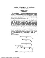
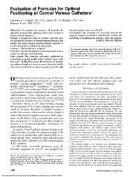
FIGURE 1. Diagram of modification of a 250 ml filtration flask for<br />
collection of fluid returned by suction of BAL. The entire unit can<br />
withstand repeated steam autoclav<strong>in</strong>g.<br />
filtration flask adapted to function as a suction trap (see Fig 1). This<br />
entire unit is steam autoclavable to provide sterility should <strong>the</strong> cells<br />
be used later for tissue culture. Wall suction pressure was used but<br />
was applied <strong>in</strong> variable strength, controlled by partial occlusion of<br />
<strong>the</strong> suction port with <strong>the</strong> f<strong>in</strong>ger of <strong>the</strong> operator to permit sufficient<br />
suction without collaps<strong>in</strong>g <strong>the</strong> airway. Subjects were <strong>in</strong>structed to<br />
take slow deep breaths follow<strong>in</strong>g <strong>in</strong>stillation and removal of <strong>the</strong> third<br />
(and f<strong>in</strong>al) aliquot of sal<strong>in</strong>e solution. The bronchoscope was withdrawn<br />
and <strong>the</strong> subject was observed for possible adverse reactions<br />
(20 to 30 m<strong>in</strong>utes) before be<strong>in</strong>g allowed to depart. Each was<br />
encouraged to cough and to deep brea<strong>the</strong> for <strong>the</strong> rema<strong>in</strong>der of <strong>the</strong><br />
day and to report any adverse effects.<br />
Process<strong>in</strong>g of <strong>the</strong> BAL Specimen<br />
A total of IOOU penicill<strong>in</strong> g/ml and 100 ,ug streptomyc<strong>in</strong>/ml were<br />
added to <strong>the</strong> lavageate which was immediately decanted through<br />
four layers of sterile cotton gauze <strong>in</strong>to one or two polystyrene, 50<br />
ml centrifuge tubes. The specimen was <strong>the</strong>n centrifuged at 5OOg for<br />
20 m<strong>in</strong>utes at room temperature. The supernatant fluid was<br />
decanted, aliquoted, and stored at - 700 C. The cell pellets were<br />
resuspended with gentle pipett<strong>in</strong>g with a plugged Pasteur pipette<br />
<strong>in</strong> 3 ml phosphate buffered sal<strong>in</strong>e solution (PBS), pooled and brought<br />
up to 50 ml by addition of PBS, and <strong>the</strong>n re-centrifuged at 500 g<br />
for 20 m<strong>in</strong>utes. This wash<strong>in</strong>g step was repeated a total of three times<br />
and <strong>the</strong> cells were <strong>the</strong>n resuspended <strong>in</strong> 3 to 5 ml of culture medium.<br />
A tenfold dilution of a 50 pl aliquot of this suspension was used to<br />
count <strong>the</strong> cells on a hemocytometer. Viability of <strong>the</strong> cells was<br />
determ<strong>in</strong>ed by trypan blue dye exclusion. Cytocentrifuge preparations<br />
were made by plac<strong>in</strong>g 100 to 200 pd of a twofold to threefold<br />
dilution (ie, an approximate total of 1-2 x 10⁵ cells) of <strong>the</strong> orig<strong>in</strong>al<br />
cell suspension <strong>in</strong> <strong>the</strong> cytocentrifuge cones and centrifug<strong>in</strong>g (at 250<br />
BAL <strong>in</strong> <strong>Normal</strong> <strong>Volunteer</strong> Subjects. 1. Technical Aspects (Ettensohn et al)
Table 1-BAL Analysis of<strong>Normal</strong> <strong>Volunteer</strong> Subjects<br />
No. %Return Cell No./<br />
Group of of 120 ml Cell No. ml BAL<br />
Studied Subj Age* BAL X 106 Fluid x 103 %ALVM0t %LY %PMN %EOS<br />
All 78 26.3±3.6 63.4±10.8 7.28±3.9 93.7±45.1 95.1±2.9 3.9±2.4 0.7±0.8 0.17±.9<br />
Subjects (20-36) (23.3-79.2) (0.69-23) (9.9-267) (84.2-99.3) (0.7-14.4) (0-3.2) (0-7.1)<br />
Men 44 26.0+3.6 61.7+12.3 7.29+4.3 94.2±49.3 94.9+3.2 4.2+2.7 0.8±0.8 0.2±1<br />
(20-36) (23.3-67.2) (0.69-23) (9.9-267) (84.2-99.3) (0. 7-14.4) (0-3) (0-7.1)<br />
Women 34 26.6±3.6 64.5±7.3 7.25±3.4 93.5±40.5 95.5±2.4 3.7±1.9 0.7±0.8 0.1±0.5<br />
(22-35) (50-79.2) (3.1-15.5) (3.7-200) (89.9-99.2) (0.8-9) (0-3.2) (0-2.6)<br />
*All data presented as mean ± SD of <strong>the</strong> mean (range).<br />
tAbbreviations used: AlvM0, alveolar macrophages; Ly, lymphocytes; PMN, neutrophils; EOS, eosionophils.<br />
rpm for five m<strong>in</strong>utes) <strong>the</strong> cells onto clean glass slides us<strong>in</strong>g filter<br />
papers. These preparations were allowed to air dry and were <strong>the</strong>n<br />
sta<strong>in</strong>ed by a modified Wright-Giemsa technique. A total of 500 to<br />
1,000 cells were counted <strong>in</strong> <strong>the</strong> random field count<strong>in</strong>g technique<br />
and results reported as percentage of each cell type present. The<br />
dist<strong>in</strong>ction between macrophages and lymphocytes was confirmed<br />
by nonspecific esterase sta<strong>in</strong><strong>in</strong>g. Over <strong>the</strong> time course of <strong>the</strong> study,<br />
<strong>the</strong> vast majority of differential analysis was performed by ei<strong>the</strong>r<br />
one of <strong>the</strong> authors (M.J.J. or PA.L.) each tra<strong>in</strong>ed <strong>in</strong> <strong>the</strong> technique<br />
of differential analysis by D. B. E.<br />
Statistics<br />
Student's t-test was used to assess for significant differences<br />
between groups for <strong>the</strong> same parameter, and l<strong>in</strong>ear regression<br />
analysis was used to determ<strong>in</strong>e correlation between two parameters.<br />
A p55 percent) and when <strong>the</strong> entire group was analyzed,<br />
<strong>the</strong>re was no correlation between cell number<br />
recovered and percentage lavageate returned<br />
(r =0.27). Male subjects, as expected, had a greater<br />
total lung capacity than female subjects (6.9 ± 0. 9L vs<br />
5.2 ± . 8L, p
z<br />
w<br />
CC<br />
w<br />
a-<br />
-j<br />
"Cm<br />
lm<br />
cLw<br />
w<br />
0<br />
w<br />
C)<br />
-j<br />
-j<br />
w<br />
U.<br />
0 -j<br />
z<br />
0w<br />
w<br />
LL.<br />
U-<br />
100<br />
98<br />
A. ALVEOLAR MACROPHAGES<br />
96<br />
94<br />
92<br />
90<br />
88<br />
86<br />
84<br />
____ 4~~~~~~4<br />
* *<br />
16 B. LYMPHOCYTES<br />
14<br />
12<br />
10<br />
8 * U s<br />
6- a. * * U +sd<br />
4 n 6r<br />
2 *u U<br />
*~~~* i *<br />
0O%U.<br />
8<br />
5-<br />
4,<br />
3<br />
2.<br />
3,<br />
C. NEUTROPHILS<br />
A<br />
A<br />
A<br />
A<br />
A&<br />
A<br />
A<br />
d<br />
s<br />
-sd<br />
sO<br />
A AA<br />
AM<br />
-A--*AA Ah e d.<br />
A<br />
ui _,A--A ___ .L -, over, for research purposes, idiosyncrasies oftechnique<br />
_<br />
D. EOSINOPHILS<br />
0<br />
* 0- 0<br />
OBSERVATIONS<br />
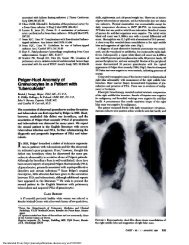
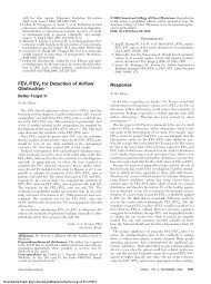
FIGURE 3. Scattergrams of <strong>the</strong> percentage of A, alveolar macrophages;<br />
B, lymphocytes; C, neutrophils; and D, eos<strong>in</strong>ophils (as<br />
determ<strong>in</strong>ed by analysis of cytocentrifuge preparation of BAL cells<br />
from 78 consecutive normal volunteer subjects).<br />
extremely rare and were similarly not <strong>in</strong>cluded <strong>in</strong> <strong>the</strong><br />
differential analysis.<br />
Viability of BAL Cells<br />
The viability (mean +±SD) of BAL cells was<br />
91.4 8.6 percent with a range from 66 to 100 percent.<br />
Viability of cells from seven subjects (9 percent) was<br />
less than 80 percent, but <strong>the</strong>re was no evident<br />
explanation for <strong>the</strong> lower than normal cell viability<br />
Safety and ComplicationslAdverse Reaction<br />
There were no complications of any consequence<br />
related to <strong>the</strong> performance of BAL <strong>in</strong> any subject.<br />
Virtually all subjects noted mild transient light headedness<br />
of 15 to 20 m<strong>in</strong>ute duration immediately<br />
278<br />
2<br />
Uo<br />
Downloaded From: http://journal.publications.chestnet.org/ on 07/13/2013<br />
0<br />
±sd<br />
-sd<br />
follow<strong>in</strong>g term<strong>in</strong>ation of <strong>the</strong> BAL. Approximately 50<br />
percent of <strong>the</strong> subjects reported be<strong>in</strong>g more tired than<br />
usual for up to 24 hours follow<strong>in</strong>g <strong>the</strong> BAL. Subjects<br />
were specifically requested to monitor for fever, but<br />
only seven reported any temperature elevation and <strong>in</strong><br />
only one subject was <strong>the</strong> elevation >37.7°C. This<br />
isolated subject noted onset offever (39.1C) and chills<br />
approximately four hours after <strong>the</strong> procedure which<br />
subsided over <strong>the</strong> next 24 hours without morbid<br />
sequelae. No cardiac arrhythmias were noted.<br />
DISCUSSION<br />
There is considerable variation <strong>in</strong> <strong>the</strong> manner <strong>in</strong><br />
which BAL is performed and <strong>the</strong> specimen processed<br />
<strong>in</strong> both cl<strong>in</strong>ical and research applications of BAL.<br />
There is little evidence to support <strong>the</strong> use of one<br />
technique over <strong>the</strong> o<strong>the</strong>r, and <strong>in</strong> all likelihood, if BAL<br />
specimens are processed similarly <strong>in</strong> an <strong>in</strong>dividual<br />
laboratory, results may be compared to <strong>the</strong> locally<br />
established norms. However, <strong>the</strong>re may be some<br />
danger <strong>in</strong> compar<strong>in</strong>g results obta<strong>in</strong>ed and published<br />
by o<strong>the</strong>r <strong>in</strong>vestigators who may have used a different<br />
size bronchoscope or process<strong>in</strong>g techniques. More-<br />
may also limit comparison of results of <strong>in</strong> vitro<br />
functional assays <strong>in</strong> which BAL cells are used. The<br />
purpose of <strong>the</strong> current study was not to promote or<br />
espouse a particular method, but ra<strong>the</strong>r to report on<br />
<strong>the</strong> subject-to-subject variability <strong>in</strong> BAL results found<br />
<strong>in</strong> a relatively large population of normal subjects<br />
when us<strong>in</strong>g rigidly controlled conditions.<br />
All bronchoscopies and process<strong>in</strong>g of <strong>the</strong> BAL<br />
specimens were performed by or under <strong>the</strong> direct<br />
supervision of one experienced <strong>in</strong>dividual, and all<br />
BAL was performed us<strong>in</strong>g <strong>the</strong> same diameter bronchoscope,<br />
<strong>the</strong> same type of fluid, <strong>the</strong> same relative<br />
location (l<strong>in</strong>gula), at <strong>the</strong> same time of day. The subject<br />
pool was also controlled by several criteria. Despite<br />
<strong>the</strong> uniformity of <strong>the</strong> BAL protocol, <strong>the</strong>re was considerable<br />
variability <strong>in</strong> both <strong>the</strong> percentage of return and<br />
cell number obta<strong>in</strong>ed (Table 1) and with <strong>the</strong> exception<br />
of BALs with low percentage return, <strong>the</strong>re was no<br />
correlation between cell number and percentage of<br />
return. There was also no correlation of ei<strong>the</strong>r value<br />
with any lung volume. Similarly, <strong>the</strong>re was no correlation<br />
ofnumbers of lavage cells per milliliter lavageate<br />
with any lung volume. Not surpris<strong>in</strong>gly <strong>the</strong>n, even<br />
though female subjects had significantly smaller lung<br />
volumes than males, <strong>the</strong>re was no correlation between<br />
cell numbers or percentage of return and <strong>the</strong> gender<br />
of <strong>the</strong> lavage subject and no significant gender-related<br />
differences <strong>in</strong> any lavage parameter. A possible explanation<br />
of this observation may be that similar surface<br />
area of lung was lavaged <strong>in</strong> all subjects irrespective of<br />
lung size. S<strong>in</strong>ce <strong>the</strong> same diameter bronchoscope was<br />
used for all subjects, it is likely that bronchi of nearly<br />
BAL <strong>in</strong> <strong>Normal</strong> <strong>Volunteer</strong> Subjects. 1. Technical Aspects (Ettensohn et al)
identical diameter were lavaged. In animal studies,<br />
analysis of bronchial casts <strong>in</strong>dicate that <strong>the</strong> cross<br />
sectional area of <strong>the</strong> bronchus is related to <strong>the</strong> number<br />
of term<strong>in</strong>al units served by that bronchus. 14 However,<br />
<strong>the</strong> wide variation <strong>in</strong> <strong>the</strong> number of cells recovered<br />
from subject to stubject <strong>in</strong>dicates that ei<strong>the</strong>r this is a<br />
subject-dependent parameter, or <strong>the</strong>re are o<strong>the</strong>r,<br />
noncontrollable factors that contribute to this variability<br />
As illustrated <strong>in</strong> <strong>the</strong> companion study to <strong>the</strong> current<br />
manuscript, <strong>the</strong>re is limited support for <strong>the</strong> former<br />
possibility 15 but no way to discount <strong>the</strong> latter. S<strong>in</strong>ce<br />
greater than 25 percent of <strong>the</strong> cells may be lost by<br />
wash<strong>in</strong>g <strong>the</strong> cells three times as <strong>in</strong> <strong>the</strong> current study'2<br />
(necessary to remove <strong>the</strong> xyloca<strong>in</strong>e to reduce <strong>in</strong>terference<br />
<strong>in</strong> functional, <strong>in</strong> vitro assays'6), it is possible that<br />
greater or lesser loss of cells from subject to subject<br />
may have contributed to <strong>the</strong> variability However,<br />
similar BAL analyses by o<strong>the</strong>rs" <strong>in</strong> which <strong>the</strong> cells<br />
were not washed have yielded results similar to those<br />
described here<strong>in</strong>.<br />
In contrast to <strong>the</strong> variability noted <strong>in</strong> <strong>the</strong> percentage<br />
of lavageate and cell number recovered, <strong>the</strong>re was<br />
marked uniformity <strong>in</strong> <strong>the</strong> differential analysis of <strong>the</strong><br />
recovered cells. This suggests consistency <strong>in</strong> both <strong>the</strong><br />
subject pool and observers perform<strong>in</strong>g <strong>the</strong> differential<br />
analysis. On <strong>the</strong> basis of this present study, we have<br />
established normal values for differential analysis of<br />
lavage cells as del<strong>in</strong>eated by <strong>the</strong> mean ±2 standard<br />
deviations (ie, 95 percent confidence limits). These<br />
are as follow: alveolar macrophages, >89 percent;<br />
lymphocytes,
D. <strong>Bronchoalveolar</strong> lavage <strong>in</strong> <strong>the</strong> diagnosis of diffuse pulmonary<br />
<strong>in</strong>filtrates <strong>in</strong> <strong>the</strong> immunocompromised host. Ann Intern Med<br />
1984; 101:1-7<br />
6 Mart<strong>in</strong> WJ II, Smith TF, Brut<strong>in</strong>el WM, Cocherill FR III,<br />
Douglas WW Role of bronchoalveolar lavage <strong>in</strong> <strong>the</strong> assessment<br />
of opportunistic pulmonary <strong>in</strong>fections: utility and complications.<br />
Mayo Cl<strong>in</strong> Proc 1987; 62:549-57<br />
7 Keough BA, Hunn<strong>in</strong>ghake GW L<strong>in</strong>e BR, Crystal RG. The<br />
alveolitis of pulmonary sarcoidosis: evaluation of natural history<br />
and alveolitis-dependent changes <strong>in</strong> lung function. Am Rev<br />
Respir Dis 1983; 128:256-65<br />
8 O'Donnell K, Keough BA, Cart<strong>in</strong> A, Crystal RG. Pharmacologic<br />
suppression of <strong>the</strong> neutrophil component of <strong>the</strong> alveolitis <strong>in</strong><br />
idiopathic pulmonary fibrosis. Am Rev Respir Dis 1987; 136:288-<br />
92<br />
9 Watters LC, Schwarz MI, Cherniack RM, Waldron JA, Dunn<br />
TL, Stanford RE, et al. Idiopathic pulmonary fibrosis: pretreatment<br />
bronchoalveolar lavage cellular constituents and <strong>the</strong>ir<br />
relationship with lung histopathology and cl<strong>in</strong>ical response to<br />
<strong>the</strong>rapy. Am Rev Respir Dis 1987; 135:696-704<br />
10 P<strong>in</strong>gleton SK, Harrison GF, Stechschulte DJ, Wesselius LJ,<br />
Kirby GR, Ruth WE. Effect of location, pH and temperature of<br />
<strong>in</strong>stillate <strong>in</strong> bronchoalveolar lavage <strong>in</strong> normal volunteers. Am<br />
Rev Respir Dis 1983; 128:1035-37<br />
11 Davis GS, Giancola MS, Costanza MC, Low RB. Analysis of<br />
sequential bronchoalveolar lavage samples from healthy human<br />
2800<br />
volunteers. Am Rev Respir Dis 1982; 126:611-16<br />
12 Merrill W, O'Hearn E, Rank<strong>in</strong> J, Naegel G, Matthay RA,<br />
Reynolds HY K<strong>in</strong>etic analysis of respiratory tract prote<strong>in</strong>s<br />
recovered dur<strong>in</strong>g a sequential lavage protocol. Am Rev Respir<br />
Dis 1982; 126:617-19<br />
13 Reynolds HY, Chretien J. Respiratory tract fluids: analysis of<br />
content and contemporary use <strong>in</strong> understand<strong>in</strong>g lung disease.<br />
DM 1984; 5:1-103<br />
14 McBride JT, Chuang C. A technique for quantitat<strong>in</strong>g airway size<br />
from bronchial casts. J Appl Physiol 1985; 58:1015-27<br />
15 Ettensohn DB, Redondo A, Jankowski MJ, Duncan PG. <strong>Bronchoalveolar</strong><br />
lavage <strong>in</strong> <strong>the</strong> normal volunteer subject: 2. Safety<br />
and results of repeated BAL and its use <strong>in</strong> <strong>the</strong> assessment of<br />
<strong>in</strong>trasubject variability <strong>Chest</strong> 1988; 94:281-85<br />
16 Cullen BF, Chretien PB, Leventhal BG. The effect of lignoca<strong>in</strong>e<br />
on PHA-stimulated human lymphocyte transformation. Br J<br />
Anesth 1972; 44:1247-51<br />
17 Salt<strong>in</strong>i C, Hance AJ, Ferrans VJ, Basart F, Bitterman PB, Crystal<br />
RG. Accurate quantification of cells recovered by bronchoalveolar<br />
lavage. Am Rev Respir Dis 1984; 130:650-58<br />
18 Rank<strong>in</strong> JA, Snyder PE, Schacter EN, Martag RE. <strong>Bronchoalveolar</strong><br />
lavage: its safety <strong>in</strong> subjects with mild asthma. <strong>Chest</strong><br />
1984; 85:723-28<br />
19 Burns DM, Shure D, Francoz R, Kalafer M, Harrell J, Witztun<br />
M, Moser K. The physiological consequences of sal<strong>in</strong>e lobar<br />
lavage <strong>in</strong> healthy human adults. Am Rev Respir Dis 1983;<br />
127:695-70<br />
Course <strong>in</strong> Allergy and Immunology<br />
The Pacific Presbyterian Medical Center will present <strong>the</strong> 18th Course on Allergy and<br />
Immunology on October 14 <strong>in</strong> San Francisco. For <strong>in</strong>formation, contact Herbert S. Kaufman,<br />
M.D., 2340 Clay Street, Room 313, San Francisco 94115 (415:923-3441).<br />
Downloaded From: http://journal.publications.chestnet.org/ on 07/13/2013<br />
BAL <strong>in</strong> <strong>Normal</strong> <strong>Volunteer</strong> Subjects. 1. Technical Aspects (Ettensohn et a/)