The Axillary Vein: An Alternative Approach for Percutaneous ... - Chest
The Axillary Vein: An Alternative Approach for Percutaneous ... - Chest
The Axillary Vein: An Alternative Approach for Percutaneous ... - Chest

You also want an ePaper? Increase the reach of your titles
YUMPU automatically turns print PDFs into web optimized ePapers that Google loves.
<strong>The</strong> <strong>Axillary</strong> <strong>Vein</strong>: <strong>An</strong> <strong>Alternative</strong> <strong>Approach</strong><br />
<strong>for</strong> <strong>Percutaneous</strong> Pulmonary Artery<br />
Catheterization*<br />
Claude Martin, M.D.;Jean Pierre Auffray, M.D.; Pierre Saux, M.D.;<br />
Jacques Albanese, M.D.; and Fran#{231}oLsGouin, M.D.<br />
<strong>The</strong> adllary vein route was investigated prospectively <strong>for</strong><br />
percutaneous pulmonary artery catheterization in 79 patients<br />
who underwent 83 attempts. Forty nine of these<br />
patients were tracheostomized and under mechanical yentilation<br />
and eight had hemostasis disorders. Successful<br />
catheterization was achieved 74 times in the 79 patients.<br />
Pulmonary artery was reached within 6 ± 2.1 mm after the<br />
catheter was set in place in the axillary vein. Less than 1 mm<br />
was needed in 53 cases. Puncture of the axillary artery was<br />
noted in II patients without complication. No other significant<br />
complication was noted following the punctures. Mean<br />
duration ofcatheterizations was 3.6 ± 2 days. No septicemia<br />
was related to the catheterization procedure. One thrombosis<br />
of the axillary vein was noted, Catheterization of the<br />
pulmonary artery via the axillary vein is safe, simple and<br />
reliable and can represent an alternative method should the<br />
use of other routes be unsuccessful.<br />
theterization of the pulmonary artery (PA) with a<br />
flow-directed, balloon-tipped catheter is a common<br />
procedure be<strong>for</strong>e anesthesia <strong>for</strong> patients with<br />
cardiac disease who undergo major surgery, or <strong>for</strong><br />
patients under intensive care.’ Successful placement<br />
ofthe catheter requires reliable and safe entry into the<br />
intrathoracic circulation. <strong>The</strong> vessels usually selected<br />
include veins of the external jugular veins<br />
(EJV),5’6 internal jugular veins (IJV),257’0 subclavian<br />
veins (SCV)5” and femoral veins.su Some difficulties<br />
can arise with each of these routes . <strong>The</strong> arm veins are<br />
often in poor condition after several days of treatment<br />
in the intensive care unit (ICU). Catheterization of the<br />
PA via the femoral vein can be difficult without the<br />
help of fluoroscopic guidance,’ a technique which is<br />
not always available at patient’s bedside in the ICU.<br />
Cannulation of the 1W or SCV is associated with a<br />
number ofminor and some more serious complications<br />
including arterial puncture and pneumothorax.’’4<br />
Entry into the intrathoracic circulation via the axillary<br />
vein (AV) is an alternative Clinical practice<br />
fur several years supports the impression that PA catheterization<br />
via the axillary vein is safe, reliable and<br />
simp1e. In this study, the feasibility ofPA catheterization<br />
using the axillary vein approach was investigated<br />
prospectively in unanesthetized patients and in patients<br />
under mechanical ventilation <strong>for</strong> treatment of<br />
adult respiratory distress syndrome (ARDS).<br />
MATERIAL AND METHODS<br />
Seventy nine patients, 18 women and 61 men ranging in age from<br />
6D#{233}partement d’<strong>An</strong>esth#{233}sie-R#{233}animation Marseille-Sud, HOpital<br />
Sainte-Marguerite, Marseille, France.<br />
Reptint requests: Di Martin, Hopital St. Marguerite, 13099 Marseille,<br />
France<br />
18 to 75 years, were included in the study. <strong>The</strong>y underwent 83<br />
cannulations ofthe axillary vein and were carefully followed up in a<br />
prospective manner during a 17-month period. In<strong>for</strong>med consent<br />
was obtained from the 34 patients with angina who were scheduled<br />
<strong>for</strong> elective surgical procedures.<br />
<strong>The</strong>re were ten women and 24 men ranging in age from 42 to 75<br />
years, mean age 62 ± 2 years (mean ± SEM). Twenty seven of these<br />
patients underwent abdominal aortic aneurysmectoiy. <strong>The</strong> other<br />
seven patients underwent hepatic resection <strong>for</strong> malignant tumors.<br />
All patients were catheterized in the operating room. Catheterizalion<br />
ofthe PA was also per<strong>for</strong>med in 45 patients with ARDS. Eight<br />
women and 37 men ranging in age from 18 to 67 years, mean age<br />
50 ± 3 years). For these sedated patients, in<strong>for</strong>med consent was obtamed<br />
from the family. All patients were tracheostomized and under<br />
controlled mechanical ventilation with positive end-expiratory pressure<br />
(PEEP) above 10 cmH2O. Ventilation with PEEP was used to<br />
treat hypoxemia following bacterial pneumonia in 23 patients,<br />
aspiration pneumoniain six, peritonitis in eight, fat embolism in five,<br />
and viral pneumonitis in three. Eight patients had blood platelet<br />
counts below 70,000 mm3 and four patients below 20,000 mm3.<br />
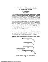
<strong>The</strong> axillary vein was cannulated following an original approach.’<br />
<strong>The</strong> patient lay supine, the arm to be cannulated was abducted away<br />
from the patient side, and the hand placed behind the occiput. All<br />
catheters were set in place using sterile technique including masks,<br />
gowns, gloves and sterile draping. <strong>The</strong> axilla was shaved, cleansed<br />
with an iodine solution (Betadine) and towelled. <strong>The</strong> operator stood<br />
on the same side as the puncture site. <strong>The</strong> axillary artery was<br />
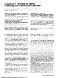
palpated and its course was noted(Fig 1). After alocalanesthetic was<br />
injected into the skin and subcutaneous tissues, the azillaryvein was<br />
cannulated initially with a 20-gauge, 51 mm (2 inch) needle. <strong>The</strong><br />
needle was inserted high in the axilla at an angle of3O#{176} to the skin<br />
parallel to the course ofthe artery, toward the chest wall and 1 cm<br />
medial to the artery(Fig 1). Puncture ofthe axillaryvelnwas realized<br />
higher in the axilla than with Spracklen’s method.’7 After the needle<br />
was inserted into the vein, a 40-cm long, 0.64-mm (0.025 inch)<br />
diameter straight guide wire was threaded with subsequent 8.0-<br />
French dilator/sheath unit (Desilet, Vygon Laboratories) guided<br />
over the wire, and 7.0-French PA catheter (93A-131 7 F, Edwards<br />
Laboratories) inserted into the sheath. Fluoroscopic guidance was<br />
never used in this study. Once the tipofthe PAcatheterwas setin the<br />
superior vena cave., the balloon was inflated with 1.2 ml of air and<br />
694 <strong>The</strong> AxiiIary Win <strong>Approach</strong> <strong>for</strong> Pulmonary Artery Cathetedzation (Martin eta!)<br />
Downloaded From: http://journal.publications.chestnet.org/ on 08/30/2013
FIGURE 1. Position of patient <strong>for</strong> axillary vein<br />
catheterization. <strong>The</strong> lateral border of the pectoralis<br />
majo muscle and the course of the<br />
axillary vein are marked with a skin peflcil. <strong>The</strong><br />
axillary artery is palpated with the left hand.<br />
pushed while the vascular pressure and the ECG were carefully<br />
monitored.’3 Once the wedge pressure was obtained, the balloon<br />
was deflated. Be<strong>for</strong>e the PA catheter was secured to the skin with a<br />
suture, the arm was stretched out and put at an angle of3O#{176} with the<br />
thorax. <strong>The</strong> pressure tracing was then carefully observed. If wedge<br />
pressure appeared with an inflation volume of less than 1 ml, the<br />
balloon was deflated and the catheter withdrawn centrally. With this<br />
technique we insured that the catheter tip was located as centrally as<br />
possible. Daily chest x-ray films were reviewed to check the position<br />
of the catheter tip.<br />
During placement of the PA catheters, careful monitoring of the<br />
oscilloscope tracing was completed <strong>for</strong> signs of myocardial ischeinia<br />
(angina or depression ofST-segment on the EGG ofi mm or greater),<br />
premature ventricular contractions (PVCs), runs of ventricular<br />
tachycardia (three of more successive PVCs) or any other arrhythmia.<br />
Following catheter placement, puncture sites were<br />
cleansed with Betadine solution; then a sterile gauze impregnated<br />
with Betadine was applied to the site. Finally, the catheter and<br />
sterile gauze were covered with a sterile occlusive dressing. <strong>An</strong><br />
adhesive, transparent, semipermeable film was used (Op-Site,<br />
Smith and Nephew, mc). <strong>The</strong> 10-cm segment ofcatheter extending<br />
from the puncture site was coiled within the dressing. Sterile<br />
dressing changes were carried out every day, using the same<br />
technique. Stopcocks and tubing were changed daily, using sterile<br />
technique. <strong>The</strong>se procedures were eventually per<strong>for</strong>med several<br />
times a day ifdressings were soiled or unstuck. Patients were visited<br />
every day and puncture sites were inspected <strong>for</strong> hematoma and<br />
evidence of infection (redness, swelling, heat). Cultures of the<br />
catheters were obtained only when catheter-related infection, and<br />
catheter-related bacteremia were suspected, or when puncture sites<br />
became infected. Catheter-related infection was defined as follows:<br />
temperatures38.5#{176}C, white blood cell count12,500/mm3, no<br />
other apparent source ofinfection. Catheter related bacteremia was<br />
defined as follows: species from catheter tip culture and from<br />
separate blood culture obtained by venipuncture identical, no other<br />
source of bacteremia, temperature38.5#{176}C, white blood cell<br />
count12,500/mm3. In all patients, blood cultures were obtained<br />
when body temperature was38.5#{176}C. Body temperature was<br />
checked every four hours. <strong>The</strong> catheters were aseptically removed<br />
by intensive care nurses. <strong>The</strong> insertion site was cleaned with an<br />
iodine solution and the distal 3 cm of the catheter cut off with a<br />
scalpel blade. In the laboratory, catheter segment was transferred<br />
from a transport tube (Culturette, Marion Corp) onto the surface of<br />
blood agar plates. Growth of 15 or more colonies on a semiquan-<br />
titative plate was regarded as positive, indicating catheter infection.<br />
In the other patients, when no catheter-related infection or bacteremia<br />
were suspected, the cultures of catheter tip were not<br />
obtained.<br />
RESULTS<br />
<strong>Axillary</strong> vein catheterization <strong>for</strong> placement of a PA<br />
catheter was attempted 83 times in the 79 patients. A<br />
total of 74 catheterizations were successful (89.2 percent).<br />
<strong>The</strong> right axillary vein was utilized 36 times and<br />
the left axillary vein 47 times. In six cases (7.2 percent),<br />
the operator failed to catheterize either axillary vein<br />
and in three cases (3.6 percent) the guide wire could<br />
not be successfully introduced into the vein. <strong>The</strong>re<strong>for</strong>e,<br />
of the 74 cases in whom the PA catheter could be<br />
introduced into the axillary vein, 74 (100 percent)<br />
underwent successful PA catheterization. This required<br />
less than three punctures in 53 cases (64 percent).<br />
<strong>The</strong> time necessary to per<strong>for</strong>m the cannulations<br />
was 13 ± 2 mm (range: 25 sec to 45 mm). <strong>The</strong> time required<br />
to reach PA was 6 ± 2. 1 mm after the tip of the<br />
catheter was inserted into the vein. In 53 cases (64 percent)<br />
the PA was reached within 1 mm or less. <strong>The</strong><br />
entire PA catheterization procedure was 18 ± 3 mm in<br />
the 74 cases in whom PA was catheterized successfully.<br />
In five cases (6 percent) ventricular extrasystoles were<br />
observed, but none ofthe patients exhibited ventricular<br />
tachycardia. No patient experienced angina or<br />
EGG changes. Arterial punctures occurred 11 times (13<br />
percent) and large hematomas were always avoided by<br />
prolonged pressure (3 mm or more) very easily<br />
achieved because of the superficial position of the<br />
axillary artery. No other complication was noted. <strong>The</strong><br />
catheters were in place <strong>for</strong> 2. 1 ± 1. 5 days in patients<br />
undergoing surgery and <strong>for</strong> 4. 6 ± 1. 5 days in patients<br />
with ARDS. No septicemia or bacteremia was related<br />
to the PA catheterization. In the ARDS group, five<br />
cases of catheter-related infection were suspected.<br />
CHEST I 90 I 5 I NOVEMBER, 1986 695<br />
Downloaded From: http://journal.publications.chestnet.org/ on 08/30/2013
Catheters were withdrawn and cultures of the tips<br />
showed Staphylococcus aureus in three cases, Proteus<br />
mirabilis in one case, and Pseudomonas maltophilia in<br />
one case. Blood cultures obtained from these patients<br />
did not grow any organism. One thrombosis (1.2<br />
percent) of the axillary and subclavian vein was<br />
clinically diagnosed and successfully treated with<br />
heparin. No late neurologic sequel and no damage of<br />
the axillary plexus were noted.<br />
DISCUSSION<br />
Placement of a PA catheter requires entry into the<br />
intrathoracic circulation. <strong>The</strong> central vessels can be<br />
reached percutaneously from several sites. Catheterization<br />
of the deeper venous route (IJV or SCV) is<br />
associated with serious complications such as arterial<br />
puncture, pneumothorax or accidental entry ofthe PA<br />
catheter into the mediastinum.’’4 <strong>An</strong> approach via<br />
the antecubital vein is safer,3 but catheterization of this<br />
vein is often impossible in patients after several days of<br />
hospitalization, especially in the ICU. Puncture of the<br />
EJV is also simple and safe,6#{176}but the vein is not present<br />
in all patients. Catheterization ofthe AV can be an<br />
alternative<br />
method.<br />
In this study, PA catheterization was achieved in 89.2<br />
percent of attempts. This success rate is lower than<br />
with the IVJ which is the vessel most often selected fbr<br />
entry into the intrathoracic circulation.29”#{176} Cannulation<br />
ofthe AV is proposed as an alternative route <strong>for</strong> PA<br />
catheterization and must be compared with other alternative<br />
routes. <strong>The</strong> 89.2 percent success rate of AV<br />
compares favorably with the 77 percent success rate<br />
reported <strong>for</strong> EJV by Schwartz et al,6 with 48 percent of<br />
the patients catheterized after two or less cannulation<br />
attempts. This 89. 2 percent success rate of AV also<br />
compares favorably with the 75 percent reported <strong>for</strong> PA<br />
catheterization via the antecubital vein.3 <strong>The</strong> time of<br />
catheter insertion via the AV seems acceptable. Cannulation<br />
of the AV is perfurmed within a few minutes<br />
and needs less than three cannulation attempts in 64<br />
percent. In a few cases, more time was needed <strong>for</strong> AV<br />
cannulation. This was related in most cases to procedures<br />
per<strong>for</strong>med by residents during their first year of<br />
training. As with other vein routes, procedures perfurmed<br />
with more skilled and experienced operators<br />
are considerably more rapid. Malposition of the arm<br />
can lead to some difficulties in AV cannulation. Should<br />
this occur, it is of great importance to place the hand<br />
correctly behind the occiput be<strong>for</strong>e a new puncture is<br />
attempted. <strong>The</strong>re are no controlled studies to compare<br />
the time required to achieve the entire procedure of PA<br />
catheterization via AV and other routes. However, this<br />
time averages 15 mm using IJV and 21 mm using<br />
basilic vein.3 In this study, the time needed was 18 ± 3<br />
mm, which is acceptable. Arterial puncture occurred<br />
at a similar frequency as during IJV puncture.7’#{176}’4#{176} No<br />
complication was noted fullowing these arterial punctures.<br />
<strong>The</strong>y were all easily treated with finger pressure<br />
enhanced by the superficial position of the artery. No<br />
pneumothorax followed the PA catheterization. <strong>The</strong><br />
risk ofneurologic sequel after puncture ofthe nerves of<br />
the arm or <strong>for</strong>earm seems more theoretic than real. No<br />
complication ofthat kind was noted. This is confirmed<br />
by Spracklen et al’7 and by a study based on over 300<br />
cannulations over several years.’<br />
A low incidence of ventricular dysrhythmias was<br />
observed in this study (6 percent). Frequency of this<br />
complication is highly variable, ranging from 72 percents<br />
to 10 percent.3 In our study, cardiac monitoring<br />
was done using oscilloscope tracing and not paper recording.<br />
Should the latter technique be used, the<br />
incidence of dysrythmias could have been higher. In<br />
six cases, chest x-ray films obtained daily allowed<br />
diagnosis of spontaneous inward migration of the<br />
catheter which was withdrawn to a proper position. No<br />
kinking in the AV was noted. One thrombosis ofthe AV<br />
was diagnosed. In this study, only clinical signs were<br />
used to investigate this complication and we cannot<br />
exclude that routine use of phlebography might have<br />
shown a greater incidence of thrombosis of the AV.<br />
Catheter-related sepsis can cause serious nosocomial<br />
infections. No case ofcatheter-induced bateremia was<br />
noted in our patients (all of them had blood cultures<br />
drawn when body temperature was38.5#{176}C). Catheter-related<br />
infection was suspected in five cases (6<br />
percent). Resolution ofthe infection was obtained in all<br />
cases after catheter removal. Catheter tip cultures<br />
were not obtained in all the patients, but only when<br />
clinical features were consistent with local or general<br />
catheter-related sepsis. For this reason, we cannot<br />
exclude that positive-catheter cultures (growth on the<br />
blood agar), without local or general infection, might<br />
have been present in some of our patients. This<br />
incidence ofcatheter-related sepsis was comparable in<br />
a previous study of more than 300 AV cannulations<br />
where septicemia occurred in 2.7 percent of the<br />
patients.’8 <strong>The</strong>se patients were at risk of catheterrelated<br />
infection since most of them were tracheostomized<br />
and average duration ofcatheterization was 18.6<br />
days.’8<br />
As a practical point, the administration of medications<br />
through a distal vein in the catheterized arm<br />
should be discouraged. This study does not provide<br />
controlled data about this point, but venous stagnation<br />
might occur following AV cannulation and cause elevated<br />
drug concentrations and tissue slough.<br />
It is concluded that PA catheterization is per<strong>for</strong>med<br />
easily, reliably and relatively free from complications<br />
via the AV. This technique represents an alternative<br />
method ofPA catheterization which may have merit in<br />
certain patients in case of inability to use the other<br />
sites.<br />
696 <strong>The</strong> Axiliary Win <strong>Approach</strong> <strong>for</strong> Pulmonary Artery CatheterizatiOn (Martin et a!)<br />
Downloaded From: http://journal.publications.chestnet.org/ on 08/30/2013
REFERENCES<br />
1 Swan HJC, Ganz W, Forrester J, Marcus M, Diamond G,<br />
Chonette D. Catheterization of the heart in man with use of a<br />
flow-directed balloon-lipped catheter. N Engi J Med 1970;<br />
283:447-51<br />
2 Shah KB, Ran UK, Laughlin 5, El-Etr AA. A review of<br />
pulmonary artery catheterization in 6.245 patients. <strong>An</strong>esthesiology<br />
1984; 61:271-75<br />
3 Dc Lange 55, Boscoe MJ, Stanley TH. <strong>Percutaneous</strong> pulmonary<br />
artery catheterization via the arm be<strong>for</strong>e anaesthesia: success<br />
rate, frequency of complications and arterial and heart rate<br />
responses. Br J <strong>An</strong>aesth 1981; 53:1167-71<br />
4 Mandel S, Barash P. <strong>The</strong> proximalbasilicvein: anewapproach <strong>for</strong><br />
introduction ofa flow guided catheter into the pulmonary artery.<br />
J Thorac Cardiovasc Surg 1976; 71:376-77<br />
5 Boyd KD, Thomas SJ, Gold J, Boyd AD. A prospective study of<br />
complications of pulmonary artery catheterizations in 500 consecutive<br />
patients. <strong>Chest</strong> 1983; 84:245-49<br />
6 Schwartz AJ, Jobes DR, Levy WJ, Palermo L, Ellison N.<br />
Intrathoracic vascular catheterization via the external jugular<br />
vein. <strong>An</strong>esthesiology 1982; 56:400-02<br />
7 KatzJD, Cronan LH, Barash PB, Mandel SD. Pulmonary artery<br />
flow-guided catheters in the perioperative period: indications<br />
and complications. JAMA 1977; 237:2832-35<br />
8 Luau JK, Stanley TH, Webster LR, Bidway AV. Arterial blood<br />
pressure and pulse rate responses to pulmonary and radial<br />
arterial catheterization prior to cardiac and major vascular<br />
operations. <strong>An</strong>esthesiology 1979; 51:265-69<br />
9 ZadianJ, Lowenstein E, Hallowell P. Lappas DG. Routine use of<br />
the Swan-Ganz flow-directed pulmonary artery catheter in adult<br />
cardiac surgical patients: time <strong>for</strong> insertions, success rate, and<br />
incidence ofcomplications in 76 consecutive attempts (abstract).<br />
<strong>An</strong>esthesiology 1975; 43 5: 237<br />
10 Zerr C, Levrot A, Fauchon G, Lebreton P, QuesnelJ, Khayat A.<br />
Le cath#{233}t#{233}risme de l’art#{233}repulmonaire. Int#{233}rfitde la voie<br />
jugulaire interne, 75 cas. <strong>An</strong>esth <strong>An</strong>alg R#{233}anim1981; 38:15-9<br />
11 McMichan JC, Michel L. Guide wire-sheath technique <strong>for</strong> pulmonary<br />
artery catheterization and central vein cannulation.<br />
Intensive Care Med 1979; 5:37-9<br />
12 Rosen H, Latto IP, Ng WS. Handbook ofpercutaneous central<br />
venous catheterization. London: WB Saunders, 1981<br />
13 Christensen KH, Nerstrom B, Baden H. Complications of<br />
percutaneous catheterization ofthe subclavian vein in 129 cases.<br />
Acta Chir Scand 1967; 133:615-19<br />
14 Schwartz AJ, Jobes DR. Grewhow DE, Stephenson LW, Ellison<br />
N. Carotid artery puncture with internal jugular cannulation<br />
using the Seldinger technique: incidence, recognition, treatment,<br />
and prevention (abstract). <strong>An</strong>esthesiology 1979; 51S:160<br />
15 Auffray JR Martin C, Houvenaeghel M, Rocca B, Chevalier A,<br />
Gouin F. Cath#{233}t#{233}risme de Ia veine axillaire en reanimation.<br />
<strong>An</strong>alyse d’une s#{233}rie de 151 observations. <strong>An</strong>n Fr <strong>An</strong>esth RCanim<br />
1983; 2:266-69<br />
16 Ayim EN. <strong>Percutaneous</strong> catheterization ofthe axillary vein and<br />
proximal basilic vein. <strong>An</strong>aesthesia 1977; 32:753-59<br />
17 Spracklen FA, Niesche F, Lord PW, Beterman M. <strong>Percutaneous</strong><br />
catheterization of the axillary vein. Cardiovas Res 1967;<br />
1:297-300<br />
18 Gouin F, Martin C, Saux P. Central venous and pulmonary artery<br />
catheterizations via the axillary vein. Acta <strong>An</strong>aesthesiol Scand<br />
1985; Suppl. 81:27-9<br />
19 Thomas F, Burke JP, ParkerJ, Orme JF, Gardner RM, Clemmer<br />
TP, et al <strong>The</strong> risk ofinfection related to radial vs femoral sites <strong>for</strong><br />
arterial catheterization. Crit Care Med 1983; 11:807-12<br />
20 Belani KG, Buckley JJ, Gordon JR, Castaneda W. <strong>Percutaneous</strong><br />
cervical central venous placement: a comparison ofthe internal<br />
and external jugular vein mutes. <strong>An</strong>esth <strong>An</strong>alg (Cleve) 1980;<br />
59:40-4<br />
21 Albertson TE, Fisher CJ, Vera Z. Accidental mediastinal entry<br />
via left internal jugular vein cannulation. Intensive Care Med<br />
1985; 11:154-57<br />
22 Defalque RJ. Subclavian venipuncture: a review. <strong>An</strong>esth <strong>An</strong>alg<br />
(Cleve) 1968; 47:677-82<br />
23 Wilson JN, Grow JB, Demong CV, Prevedel AE, Owens JC.<br />
Central venous pressure in optimal blood volume maintenance.<br />
Arch Surg 1962; 85:563-68<br />
24 Kaplan JA. Hemodynamic monitoring. In: Cardiac anesthesia.<br />
New York: Crane and Stratton, 1979; 86<br />
CHEST I 90 1 5 I NOVEMBER, 1986 697<br />
Downloaded From: http://journal.publications.chestnet.org/ on 08/30/2013