Bronchoalveolar Lavage in the Normal Volunteer Subject* - Chest
Bronchoalveolar Lavage in the Normal Volunteer Subject* - Chest
Bronchoalveolar Lavage in the Normal Volunteer Subject* - Chest

You also want an ePaper? Increase the reach of your titles
YUMPU automatically turns print PDFs into web optimized ePapers that Google loves.
MATERIALS AND METHODS<br />
Subjects<br />
The study was completed over an <strong>in</strong>terval of approximately five<br />
years. <strong>Normal</strong> volunteer subjects were solicited for <strong>the</strong> study by<br />
word of mouth and posted notices and were reimbursed for <strong>the</strong>ir<br />
participation. Rigid criteria for <strong>in</strong>clusion <strong>in</strong> <strong>the</strong> study <strong>in</strong>cluded <strong>the</strong><br />
follow<strong>in</strong>g: (1) lack of history of pulmonary disease; (2) nonsmokers;<br />
(3) normal posterior-anterior and lateral chest roentgenograms (if<br />
none <strong>in</strong> <strong>the</strong> previous year); (4) normal, complete pulmonary function<br />
tests; (5) normal heart and lung physical exam<strong>in</strong>ation; (6) tak<strong>in</strong>g no<br />
medication; and (7) no history of viral or o<strong>the</strong>r illness for several<br />
weeks prior to <strong>the</strong> study Performance of BAL <strong>in</strong> volunteer subjects<br />
was approved by <strong>the</strong> Committee for <strong>the</strong> Use of Human Subjects at<br />
Memorial Hospital of Rhode Island and <strong>in</strong>formed consent was<br />
obta<strong>in</strong>ed from all subjects prior to study Seventy-eight subjects (44<br />
men and 34 women), aged 20 to 36 years, participated <strong>in</strong> <strong>the</strong> study.<br />
The average age of <strong>the</strong> subjects was 26.3. Several potential subjects<br />
were excluded prior to pulmonary function test<strong>in</strong>g on <strong>the</strong> basis of<br />
historic evidence of lung disease, usually reactive airways disease.<br />
One potential subject had an abnormal chest roentgenogram show<strong>in</strong>g<br />
bilateral hilar adenopathy and was thus disqualified from<br />
participation. Three o<strong>the</strong>r subjects were disqualified for participation<br />
<strong>in</strong> <strong>the</strong> study by virtue of evidence of obstructive lung disease<br />
on pulmonary function test<strong>in</strong>g despite giv<strong>in</strong>g no history of airways<br />
disease. Appropriate local anes<strong>the</strong>sia could not be achieved <strong>in</strong> two<br />
o<strong>the</strong>r subjects and consequently, bronchoscopy was term<strong>in</strong>ated<br />
prior to enter<strong>in</strong>g <strong>the</strong> trachea.<br />
Bronchoscopy and BAL<br />
Subjects were <strong>in</strong>structed to take noth<strong>in</strong>g by mouth after midnight<br />
prior to <strong>the</strong> bronchoscopy All bronchoscopies were performed<br />
between 7:30 and 8:30 AM, by, or under <strong>the</strong> direct supervision of<br />
one of <strong>the</strong> authors, (D. B. E.). An <strong>in</strong>travenous l<strong>in</strong>e with 5 percent<br />
dextrose <strong>in</strong> water was established <strong>in</strong> an antecubital ve<strong>in</strong>. Atrop<strong>in</strong>e,<br />
0.6 mg, was adm<strong>in</strong>istered immediately via this l<strong>in</strong>e. Cardiac rate<br />
and rhythm were monitored electrocardiographically us<strong>in</strong>g precordial<br />
chest leads. Oxygen, 2 liters per m<strong>in</strong>ute, per nasal cannulae<br />
was adm<strong>in</strong>istered for <strong>the</strong> first 25 bronchoscopies. However, use of<br />
oxygen dur<strong>in</strong>g subsequent bronchoscopies was discont<strong>in</strong>ued s<strong>in</strong>ce<br />
ear oximetry showed no decrease of oxygen desaturation at any<br />
po<strong>in</strong>t dur<strong>in</strong>g <strong>the</strong> bronchoalveolar lavage procedure even when no<br />
supplemental 02 was used, and <strong>the</strong> nasal cannulae were a major<br />
source of discomfort for <strong>the</strong> subjects. There was no evidence that<br />
<strong>the</strong> adm<strong>in</strong>istration of 02 changed BAL results. Subjects were<br />
ma<strong>in</strong>ta<strong>in</strong>ed <strong>in</strong> a semirecumbent position throughout <strong>the</strong> procedure<br />
<strong>in</strong> an adjustable, dental chair. Local anes<strong>the</strong>sia of <strong>the</strong> posterior<br />
pharynx was achieved by an aerosolized spray of 2 percent xyloca<strong>in</strong>e<br />
delivered through an air driven atomizer. Subjects were <strong>in</strong>structed<br />
to pant and <strong>the</strong>n deeply <strong>in</strong>hale <strong>the</strong> aerosolized anes<strong>the</strong>tic. A<br />
bronchoscope (external diameter of 4.8 mm) was <strong>the</strong>n <strong>in</strong>troduced<br />
perorally or occasionally pernasally (three subjects required pernasal<br />
route due to excessive gag us<strong>in</strong>g <strong>the</strong> peroral route). Once <strong>the</strong><br />
posterior portion of <strong>the</strong> tongue and epiglottis were visualized, 2<br />
percent xyloca<strong>in</strong>e was adm<strong>in</strong>istered directly via <strong>the</strong> bronchoscope.<br />
Adequate anes<strong>the</strong>sia was usually achieved with two, 2 ml aliquots<br />
of xyloca<strong>in</strong>e. The bronchoscope was <strong>the</strong>n advanced and <strong>the</strong> larynx<br />
and vocal cords were similarly anes<strong>the</strong>tized and <strong>the</strong>n passed. One<br />
milliliter of 2 percent xyloca<strong>in</strong>e was <strong>the</strong>n applied to <strong>the</strong> ma<strong>in</strong> car<strong>in</strong>a,<br />
<strong>the</strong> left ma<strong>in</strong> stem bronchus, <strong>the</strong> secondary car<strong>in</strong>a, and at <strong>the</strong><br />
l<strong>in</strong>gular orifice. The bronchoscope was <strong>the</strong>n advanced <strong>in</strong>to a B4 or<br />
B5 l<strong>in</strong>gular bronchus until it was completely wedged <strong>in</strong> such a<br />
position as to allow visualization of <strong>the</strong> distal bronchus. Three<br />
aliquots of40 ml of sterile normal sal<strong>in</strong>e solution at room temperature<br />
were <strong>in</strong>stilled through <strong>the</strong> bronchoscope and <strong>the</strong>n suctioned back<br />
(immediately after each <strong>in</strong>stillation) <strong>in</strong>to a sterile, 250 ml side-arm<br />
276<br />
Downloaded From: http://journal.publications.chestnet.org/ on 07/13/2013<br />
2, L<br />
-15"10 TO BRONCHOSCOPE<br />
-250<br />
TO WALL SUCTION<br />
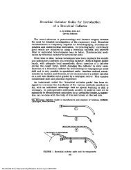
FIGURE 1. Diagram of modification of a 250 ml filtration flask for<br />
collection of fluid returned by suction of BAL. The entire unit can<br />
withstand repeated steam autoclav<strong>in</strong>g.<br />
filtration flask adapted to function as a suction trap (see Fig 1). This<br />
entire unit is steam autoclavable to provide sterility should <strong>the</strong> cells<br />
be used later for tissue culture. Wall suction pressure was used but<br />
was applied <strong>in</strong> variable strength, controlled by partial occlusion of<br />
<strong>the</strong> suction port with <strong>the</strong> f<strong>in</strong>ger of <strong>the</strong> operator to permit sufficient<br />
suction without collaps<strong>in</strong>g <strong>the</strong> airway. Subjects were <strong>in</strong>structed to<br />
take slow deep breaths follow<strong>in</strong>g <strong>in</strong>stillation and removal of <strong>the</strong> third<br />
(and f<strong>in</strong>al) aliquot of sal<strong>in</strong>e solution. The bronchoscope was withdrawn<br />
and <strong>the</strong> subject was observed for possible adverse reactions<br />
(20 to 30 m<strong>in</strong>utes) before be<strong>in</strong>g allowed to depart. Each was<br />
encouraged to cough and to deep brea<strong>the</strong> for <strong>the</strong> rema<strong>in</strong>der of <strong>the</strong><br />
day and to report any adverse effects.<br />
Process<strong>in</strong>g of <strong>the</strong> BAL Specimen<br />
A total of IOOU penicill<strong>in</strong> g/ml and 100 ,ug streptomyc<strong>in</strong>/ml were<br />
added to <strong>the</strong> lavageate which was immediately decanted through<br />
four layers of sterile cotton gauze <strong>in</strong>to one or two polystyrene, 50<br />
ml centrifuge tubes. The specimen was <strong>the</strong>n centrifuged at 5OOg for<br />
20 m<strong>in</strong>utes at room temperature. The supernatant fluid was<br />
decanted, aliquoted, and stored at - 700 C. The cell pellets were<br />
resuspended with gentle pipett<strong>in</strong>g with a plugged Pasteur pipette<br />
<strong>in</strong> 3 ml phosphate buffered sal<strong>in</strong>e solution (PBS), pooled and brought<br />
up to 50 ml by addition of PBS, and <strong>the</strong>n re-centrifuged at 500 g<br />
for 20 m<strong>in</strong>utes. This wash<strong>in</strong>g step was repeated a total of three times<br />
and <strong>the</strong> cells were <strong>the</strong>n resuspended <strong>in</strong> 3 to 5 ml of culture medium.<br />
A tenfold dilution of a 50 pl aliquot of this suspension was used to<br />
count <strong>the</strong> cells on a hemocytometer. Viability of <strong>the</strong> cells was<br />
determ<strong>in</strong>ed by trypan blue dye exclusion. Cytocentrifuge preparations<br />
were made by plac<strong>in</strong>g 100 to 200 pd of a twofold to threefold<br />
dilution (ie, an approximate total of 1-2 x 10⁵ cells) of <strong>the</strong> orig<strong>in</strong>al<br />
cell suspension <strong>in</strong> <strong>the</strong> cytocentrifuge cones and centrifug<strong>in</strong>g (at 250<br />
BAL <strong>in</strong> <strong>Normal</strong> <strong>Volunteer</strong> Subjects. 1. Technical Aspects (Ettensohn et al)