Spinal meningiomas: surgical management and outcome
Spinal meningiomas: surgical management and outcome
Spinal meningiomas: surgical management and outcome

You also want an ePaper? Increase the reach of your titles
YUMPU automatically turns print PDFs into web optimized ePapers that Google loves.
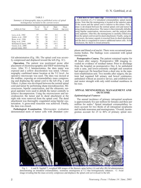
TABLE 1<br />
Summary of demographic data in published series of spinal<br />
<strong>meningiomas</strong> included in the current review<br />
No. of No. of Male/ Mean<br />
Authors & Year Cases Ops Female Age (yrs)<br />
Levy, et al., 1982 97 97 4:1 53<br />
Solero, et al., 1989 174 184 3:1 56<br />
Roux, et al., 1996 54 54 4:1 54<br />
King, et al., 1998 78 78 4.1:1 62<br />
Klekamp & Samii, 1999 117 130 3.9:1 57<br />
Gezen, et al., 2000 36 36 3:1 49<br />
present study, 2003 25 25 4.2:1 60<br />
Gd administration (Fig. 1B). The spinal cord was severely<br />
compressed <strong>and</strong> displaced toward the left (Fig. 1C).<br />
Operation. The patient was positioned prone after<br />
placement of electromyographic <strong>and</strong> SSEP monitoring devices.<br />
After T1–3 laminectomies, the dura mater was<br />
exposed <strong>and</strong> a yellow discoloration was noted. Ultrasonography<br />
confirmed tumor location at the T-2 level. An<br />
operative microscope was used. The dura was incised at<br />
the midline, exposing an extramedullary mass compressing<br />
<strong>and</strong> displacing the spinal cord to the left (Fig. 2 <strong>and</strong><br />
Video). A plane was developed between the normal spinal<br />
cord <strong>and</strong> tumor by performing sharp microdissection. Microscissors,<br />
bipolar cauterization, <strong>and</strong> the ultrasonic <strong>surgical</strong><br />
aspirator were used to debulk the tumor centrally to<br />
achieve decompression. Using the microscissors <strong>and</strong> microdissector,<br />
the tumor <strong>and</strong> its dural attachment at the<br />
right side were removed from the spinal cord. The dural<br />
attachment was thoroughly coagulated using bipolar cauterization.<br />
A gross-total resection was achieved. Finally,<br />
the dura was closed.<br />
Pathological Examination. Microscopic evaluation<br />
demonstrated nests of tumor cells with abundant cyto-<br />
O. N. Gottfried, et al.<br />
Click here to view video clip: Demonstrated in the video clip<br />
is the resection of a T-2 intradural extramedullary spinal meningioma.<br />
Note that the meningioma is located at the superior aspect<br />
of the screen <strong>and</strong> the spinal cord is inferior to this tumor. First, a<br />
plane is developed between the normal spinal cord <strong>and</strong> tumor by<br />
using sharp microdissection. The tumor is then internally debulked<br />
using bipolar cauterization, microscissors, <strong>and</strong> the <strong>surgical</strong> ultrasonic<br />
aspirator. After this, the meningioma is carefully rolled away<br />
from the spinal cord toward its dural attachment. Finally, using microscissors,<br />
the tumor capsule is resected from its dural attachment<br />
<strong>and</strong> the dura is coagulated to remove additional tumor cells. At the<br />
conclusion, the dural is closed primarily.<br />
plasm <strong>and</strong> bl<strong>and</strong> oval nuclei. There were occasional psammoma<br />
bodies. The findings were consistent with spinal<br />
meningioma.<br />
Postoperative Course. The patient remained supine for<br />
48 hours after surgery. Postoperative MR imaging revealed<br />
no evidence of residual tumor. Prior to discharge<br />
from the hospital on postoperative Day 4, he ambulated<br />
with a cane, <strong>and</strong> lower-extremity strength <strong>and</strong> sensation<br />
had improved. He benefited from a short stay in the inpatient<br />
rehabilitation unit. Two months after surgery, the patient<br />
had regained full urinary <strong>and</strong> bowel continence,<br />
ambulated without an assistive device, <strong>and</strong> exhibited normal<br />
motor strength with only mild residual numbness in<br />
his toes.<br />
SPINAL MENINGIOMAS: MANAGEMENT AND<br />
OUTCOME<br />
Epidemiological Features<br />
The annual incidence of primary intraspinal neoplasm<br />
is approximately five per million for females <strong>and</strong> three per<br />
million for males. 9 <strong>Spinal</strong> intradural extramedullary tumors<br />
account for two thirds of all intraspinal neoplasms<br />
<strong>and</strong> include neuromas <strong>and</strong> <strong>meningiomas</strong>. 1 Overall, <strong>meningiomas</strong><br />
account for 25 to 46% of primary spinal neo-<br />
Fig. 1. A <strong>and</strong> B: Sagittal T 1 -weighted MR images of the thoracic spine before (A) <strong>and</strong> after (B) Gd administration<br />
demonstrating an intradural extramedullary, isointense meningioma at T-2 that homogenously enhances. C: Axial<br />
image revealing that the mass severely compresses <strong>and</strong> displaces the spinal cord (arrowhead) to the left.<br />
2 Neurosurg. Focus / Volume 14 / June, 2003