Spinal meningiomas: surgical management and outcome
Spinal meningiomas: surgical management and outcome
Spinal meningiomas: surgical management and outcome

You also want an ePaper? Increase the reach of your titles
YUMPU automatically turns print PDFs into web optimized ePapers that Google loves.
in 64%, <strong>and</strong> 32% of the patients were nonambulatory at<br />
the time of presentation.<br />
Prior to the advent of MR imaging, spinal <strong>meningiomas</strong><br />
were often confused with multiple sclerosis, syringomyelia,<br />
pernicious anemia, <strong>and</strong> herniated disc. 8,16 Levy, et<br />
al., 16 reported that the rate of misdiagnosis in their pre–<br />
MR imaging series was 33% <strong>and</strong> that errors in diagnosis<br />
delayed treatment, sometimes leading to inappropriate<br />
surgery. Seven of their patients underwent inappropriate<br />
surgeries, including lumbar disc exploration <strong>and</strong> knee surgery.<br />
In our series, a misdiagnosis of multiple sclerosis<br />
was made in one case <strong>and</strong> arthritis in another.<br />
Magnetic resonance imaging is the best imaging technique<br />
for diagnosing spinal <strong>meningiomas</strong>. It clearly delineates<br />
the level of the tumor <strong>and</strong> its relation to the cord,<br />
which is useful in planning surgery. In the past, plain radiography<br />
was used to detect calcified <strong>meningiomas</strong>, but<br />
calcifications are only visible on 2 to 5% of plain x-ray<br />
films. 25 Prior to the advent of MR imaging, myelography<br />
was the radiological modality of choice. 16,25 Klekamp <strong>and</strong><br />
Samii 13 reported that MR imaging led to earlier diagnosis<br />
of spinal <strong>meningiomas</strong> by 6 months <strong>and</strong>, on average, patients<br />
suffered less severe neurological deficits at the time<br />
of surgery. Typically, spinal <strong>meningiomas</strong> were isointense<br />
to the normal spinal cord on T 1 - <strong>and</strong> T 2 -weighted images,<br />
<strong>and</strong> they displayed intense enhancement after gadolinium<br />
injection. 8<br />
Intraoperative ultrasonography has been useful in the<br />
identification <strong>and</strong> localization of <strong>meningiomas</strong>, 17 providing<br />
useful information about its size <strong>and</strong> the degree to<br />
which it displaces the spinal cord. Typical ultrasonographic<br />
features include echogenicity, irregular surfaces,<br />
<strong>and</strong> tight adherence to the dura. 17<br />
Surgical Approach<br />
In the reviewed series, all patients underwent classic<br />
posterior laminectomies. In cases in which <strong>meningiomas</strong><br />
were located anteriorly the laminectomy was extended laterally<br />
toward the articular process to provide sufficient<br />
exposure <strong>and</strong> cause minimal displacement of the spinal<br />
cord. Excluding those in the earlier series, patients underwent<br />
surgeries involving the operating microscope (Table<br />
4). Ultrasonography provided detailed localization of<br />
<strong>meningiomas</strong>, thereby avoiding unnecessarily large dural<br />
openings. The goal of surgery was to minimize displacement<br />
of the spinal cord by undertaking an appropriately<br />
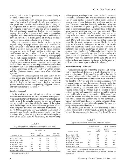
TABLE 4<br />
Surgical equipment <strong>and</strong> percent of complete resections*<br />
Op Device (%)<br />
Micro- US Total Re-<br />
Authors & Year scope Aspirator Laser section (%)<br />
Levy, et al., 1982 NA NA NA 82<br />
Solero, et al., 1989 17 NA NA 97<br />
Roux, et al., 1996 63 18 22 93<br />
King, et al., 1998 NA NA NA 99<br />
Klekamp & Samii, 1999 NA NA NA 89<br />
Gezen, et al., 2000 100 NA NA 97<br />
present study, 2003<br />
* US = ultrasonic <strong>surgical</strong>.<br />
100 63 4 92<br />
O. N. Gottfried, et al.<br />
wide exposure, making the tumor <strong>and</strong> its dural attachment<br />
accessible. Sometimes this was accomplished by cutting<br />
one or more dentate ligaments. After dural opening, a<br />
plane was developed between the arachnoid <strong>and</strong> the tumor.<br />
The tumor was then internally debulked using suction,<br />
an ultrasonic <strong>surgical</strong> aspirator, microscissors, or<br />
laser. In the more recent series, increased use of the ultrasonic<br />
<strong>surgical</strong> aspirator <strong>and</strong> laser was apparent. After<br />
debulking, in the majority of cases the tumor was rolled<br />
away from the spinal cord <strong>and</strong> toward its dural attachment.<br />
The tumor was then removed from its dural attachment.<br />
Dura with remaining tumor was either coagulated<br />
using bipolar cauterization or resected (Table 5). In the<br />
majority of cases, including our series, the dural attachment<br />
was cauterized rather than resected. The dural attachment<br />
was always cauterized in cases involving an<br />
anterior dural attachment. Additionally, in most cases the<br />
dura was closed primarily, compared with suturing in a<br />
graft, which was performed far less frequently (Table 5).<br />
Another option was separation of the dura into an outer<br />
<strong>and</strong> inner layer <strong>and</strong> to resect the tumor with the inner layer,<br />
leaving the outer layer available for closure. 23<br />
Neuromonitoring Techniques<br />
Neuromonitoring may reduce the likelihood of postoperative<br />
neurological deficits as a result of resecting spinal<br />
cord <strong>meningiomas</strong>. This modality provides data on the<br />
extent of tissue manipulation, does not compromise function,<br />
<strong>and</strong> may potentially lead to preservation of function;<br />
the benefits justify its use <strong>and</strong> the risks associated with<br />
extended operating time. Two commonly used modalities<br />
described in the reviewed series were muscle Tc-MEP <strong>and</strong><br />
SSEP monitoring. Transcranial-MEPs were obtained by<br />
placing stimulating electrodes over the patient’s motor<br />
cortical regions <strong>and</strong> recording electromyographic activity<br />
either epidurally or at the extremities. 14 This method was<br />
useful for tumors involving the anterior <strong>and</strong>/or anterolateral<br />
spinal canal <strong>and</strong> compressing of the corticospinal<br />
tracts. The SSEP recordings were useful in tumors located<br />
in the posterior or posterolateral aspect of the canal <strong>and</strong><br />
compressing the dorsal columns.<br />
The advantage of muscle Tc-MEP monitoring is that it<br />
allows assessment of motor function from the cortex to<br />
beyond the neuromuscular junction. The Tc-MEPs were<br />
TABLE 5<br />
Approaches to dural attachment <strong>and</strong> dural closure<br />
Resection (%) Dural Repair (%)<br />
Coagu- Primary<br />
Authors & Year Dural lation Closure Graft<br />
Levy, et al., 1982 57 15 15 8*<br />
Solero, et al., 1989 14 86 86 14<br />
Roux, et al., 1996 15 85 89 11<br />
King, et al., 1998 26 74 NA NA<br />
Klekamp & Samii, 1999 NA NA NA NA<br />
Gezen, et al., 2000 NA NA NA NA<br />
present study, 2003 16† 88 88 12<br />
* Performed in 77% of patients with the dura left open.<br />
† One patient underwent a split-thickness removal of dura followed by<br />
coagulation.<br />
4 Neurosurg. Focus / Volume 14 / June, 2003