Spinal meningiomas: surgical management and outcome
Spinal meningiomas: surgical management and outcome
Spinal meningiomas: surgical management and outcome

You also want an ePaper? Increase the reach of your titles
YUMPU automatically turns print PDFs into web optimized ePapers that Google loves.
<strong>Spinal</strong> <strong>meningiomas</strong><br />
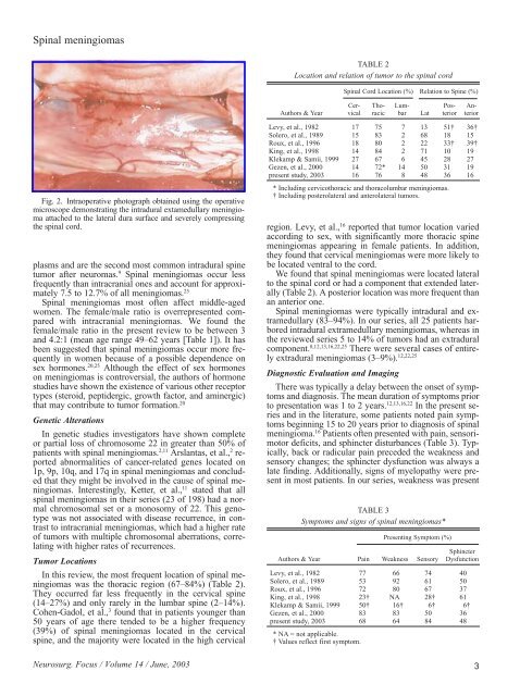
Fig. 2. Intraoperative photograph obtained using the operative<br />
microscope demonstrating the intradural extamedullary meningioma<br />
attached to the lateral dura surface <strong>and</strong> severely compressing<br />
the spinal cord.<br />
plasms <strong>and</strong> are the second most common intradural spine<br />
tumor after neuromas. 9 <strong>Spinal</strong> <strong>meningiomas</strong> occur less<br />
frequently than intracranial ones <strong>and</strong> account for approximately<br />
7.5 to 12.7% of all <strong>meningiomas</strong>. 25<br />
<strong>Spinal</strong> <strong>meningiomas</strong> most often affect middle-aged<br />
women. The female/male ratio is overrepresented compared<br />
with intracranial <strong>meningiomas</strong>. We found the<br />
female/male ratio in the present review to be between 3<br />
<strong>and</strong> 4.2:1 (mean age range 49–62 years [Table 1]). It has<br />
been suggested that spinal <strong>meningiomas</strong> occur more frequently<br />
in women because of a possible dependence on<br />
sex hormones. 20,25 Although the effect of sex hormones<br />
on <strong>meningiomas</strong> is controversial, the authors of hormone<br />
studies have shown the existence of various other receptor<br />
types (steroid, peptidergic, growth factor, <strong>and</strong> aminergic)<br />
that may contribute to tumor formation. 20<br />
Genetic Alterations<br />
In genetic studies investigators have shown complete<br />
or partial loss of chromosome 22 in greater than 50% of<br />
patients with spinal <strong>meningiomas</strong>. 2,11 Arslantas, et al., 2 reported<br />
abnormalities of cancer-related genes located on<br />
1p, 9p, 10q, <strong>and</strong> 17q in spinal <strong>meningiomas</strong> <strong>and</strong> concluded<br />
that they might be involved in the cause of spinal <strong>meningiomas</strong>.<br />
Interestingly, Ketter, et al., 11 stated that all<br />
spinal <strong>meningiomas</strong> in their series (23 of 198) had a normal<br />
chromosomal set or a monosomy of 22. This genotype<br />
was not associated with disease recurrence, in contrast<br />
to intracranial <strong>meningiomas</strong>, which had a higher rate<br />
of tumors with multiple chromosomal aberrations, correlating<br />
with higher rates of recurrences.<br />
Tumor Locations<br />
In this review, the most frequent location of spinal <strong>meningiomas</strong><br />
was the thoracic region (67–84%) (Table 2).<br />
They occurred far less frequently in the cervical spine<br />
(14–27%) <strong>and</strong> only rarely in the lumbar spine (2–14%).<br />
Cohen-Gadol, et al., 3 found that in patients younger than<br />
50 years of age there tended to be a higher frequency<br />
(39%) of spinal <strong>meningiomas</strong> located in the cervical<br />
spine, <strong>and</strong> the majority were located in the high cervical<br />
Neurosurg. Focus / Volume 14 / June, 2003<br />
TABLE 2<br />
Location <strong>and</strong> relation of tumor to the spinal cord<br />
<strong>Spinal</strong> Cord Location (%) Relation to Spine (%)<br />
Cer- Tho- Lum- Pos- An-<br />
Authors & Year vical racic bar Lat terior terior<br />
Levy, et al., 1982 17 75 7 13 51† 36†<br />
Solero, et al., 1989 15 83 2 68 18 15<br />
Roux, et al., 1996 18 80 2 22 33† 39†<br />
King, et al., 1998 14 84 2 71 10 19<br />
Klekamp & Samii, 1999 27 67 6 45 28 27<br />
Gezen, et al., 2000 14 72* 14 50 31 19<br />
present study, 2003 16 76 8 48 36 16<br />
* Including cervicothoracic <strong>and</strong> thoracolumbar <strong>meningiomas</strong>.<br />
† Including posterolateral <strong>and</strong> anterolateral tumors.<br />
region. Levy, et al., 16 reported that tumor location varied<br />
according to sex, with significantly more thoracic spine<br />
<strong>meningiomas</strong> appearing in female patients. In addition,<br />
they found that cervical <strong>meningiomas</strong> were more likely to<br />
be located ventral to the cord.<br />
We found that spinal <strong>meningiomas</strong> were located lateral<br />
to the spinal cord or had a component that extended laterally<br />
(Table 2). A posterior location was more frequent than<br />
an anterior one.<br />
<strong>Spinal</strong> <strong>meningiomas</strong> were typically intradural <strong>and</strong> extramedullary<br />
(83–94%). In our series, all 25 patients harbored<br />
intradural extramedullary <strong>meningiomas</strong>, whereas in<br />
the reviewed series 5 to 14% of tumors had an extradural<br />
component. 8,12,13,16,22,25 There were several cases of entirely<br />
extradural <strong>meningiomas</strong> (3–9%). 12,22,25<br />
Diagnostic Evaluation <strong>and</strong> Imaging<br />
There was typically a delay between the onset of symptoms<br />
<strong>and</strong> diagnosis. The mean duration of symptoms prior<br />
to presentation was 1 to 2 years. 12,13,16,22 In the present series<br />
<strong>and</strong> in the literature, some patients noted pain symptoms<br />
beginning 15 to 20 years prior to diagnosis of spinal<br />
meningioma. 16 Patients often presented with pain, sensorimotor<br />
deficits, <strong>and</strong> sphincter disturbances (Table 3). Typically,<br />
back or radicular pain preceded the weakness <strong>and</strong><br />
sensory changes; the sphincter dysfunction was always a<br />
late finding. Additionally, signs of myelopathy were present<br />
in most patients. In our series, weakness was present<br />
TABLE 3<br />
Symptoms <strong>and</strong> signs of spinal <strong>meningiomas</strong>*<br />
Presenting Symptom (%)<br />
Sphincter<br />
Authors & Year Pain Weakness Sensory Dysfunction<br />
Levy, et al., 1982 77 66 74 40<br />
Solero, et al., 1989 53 92 61 50<br />
Roux, et al., 1996 72 80 67 37<br />
King, et al., 1998 23† NA 28† 61<br />
Klekamp & Samii, 1999 50† 16† 6† 6†<br />
Gezen, et al., 2000 83 83 50 36<br />
present study, 2003 68 64 84 48<br />
* NA = not applicable.<br />
† Values reflect first symptom.<br />
3