Spinal meningiomas: surgical management and outcome
Spinal meningiomas: surgical management and outcome
Spinal meningiomas: surgical management and outcome

Create successful ePaper yourself
Turn your PDF publications into a flip-book with our unique Google optimized e-Paper software.
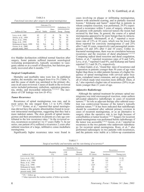
TABLE 6<br />
Functional <strong>outcome</strong>s after surgery for spinal <strong>meningiomas</strong><br />
Outcome (%)<br />
Ambulatory (%)<br />
Im- Dete-<br />
Authors & Year proved Stable riorated Preop Postop<br />
Levy, et al., 1982 83 17 0 70 76<br />
Solero, et al., 1989 53 37 10 70 92<br />
Roux, et al., 1996 85 13 2 67 94<br />
King, et al., 1998 95 1 4 74 97<br />
Klekamp & Samii, 1999 NA NA NA 74 94<br />
Gezen, et al., 2000 83 14 3 33 75<br />
present study, 2003 92 0 8 68 96<br />
tive bladder dysfunction exhibited normal function after<br />
surgery. Some patients suffered transient neurological<br />
worsening postoperatively, typically secondary to vasogenic<br />
edema or as a result of dissection, but function generally<br />
recovered after 6 months. 13,22,25<br />
Surgical Complications<br />
Mortality <strong>and</strong> morbidity rates were low. In published<br />
studies, the mortality rate ranged from 0 to 3% (Table 7),<br />
<strong>and</strong> the cause of death was unrelated to the primary disease<br />
in all cases reviewed. Causes of death in the reviewed<br />
series included pulmonary embolism, aspiration pneumonia,<br />
stroke, <strong>and</strong> myocardial infarction. 12,13,16,25 The incidence<br />
of CSF leakage was low (0–4%).<br />
Tumor Recurrence<br />
Recurrence of spinal <strong>meningiomas</strong> was rare, <strong>and</strong> in<br />
most series the rate ranged from 1.3 to 6.4% (Table<br />
7). 8,12,16,22,25 Ketter, et al., 11 reported that spinal <strong>meningiomas</strong><br />
did not have the genetic abnormalities found in recurrent<br />
intracranial <strong>meningiomas</strong>, suggesting that they had a<br />
more indolent nature. The slow growth of spinal <strong>meningiomas</strong><br />
<strong>and</strong> their presentation in patients at a late age contributed<br />
to the low recurrence rates. 12 In the reviewed series,<br />
recurrences occurred at 1 to 17 years (Table 7). In our<br />
series, one patient (4%) suffered a recurrence 2 years after<br />
subtotal resection of a large, infiltrative conus medullaris<br />
meningioma.<br />
Significantly higher recurrence rates were found in<br />
TABLE 7<br />
Surgical morbidity <strong>and</strong> mortality <strong>and</strong> the rate/timing of recurrence*<br />
O. N. Gottfried, et al.<br />
cases involving en plaque or infiltrating <strong>meningiomas</strong>,<br />
tumors with arachnoid scarring, <strong>and</strong> in partially resected<br />
lesions. 13 Klekamp <strong>and</strong> Samii 13 stated that in patients in<br />
whom complete resection, was performed, 29.5% experienced<br />
a recurrance within 5 years of surgery, whereas in<br />
all patients with partially removed tumors the lesion had<br />
recurred by that time. In general, the course of a spinal<br />
meningioma appeared to be more benign than its intracranial<br />
counterpart. Mirimanoff, et al., 18 reported a recurrence<br />
rate of 13% at 10 years, which was far lower than<br />
those reported for convexity <strong>meningiomas</strong> (3 <strong>and</strong> 25%<br />
after 5 <strong>and</strong> 10 years, respectively) <strong>and</strong> parasagittal <strong>meningiomas</strong><br />
(18 <strong>and</strong> 24% after 5 <strong>and</strong> 10 years). Unlike intracranial<br />
<strong>meningiomas</strong>, there was no correlation between<br />
recurrence <strong>and</strong> the resection of dural attachment. 8,12,13,25<br />
Comparing radical dural resection <strong>and</strong> dural coagulation,<br />
Solero, et al., 25 reported recurrence rates of 8 <strong>and</strong> 5.6%,<br />
Levy, et al., 16 reported 4 <strong>and</strong> 0%, <strong>and</strong> Klekamp <strong>and</strong> Samii<br />
reported 31.3 <strong>and</strong> 26.1%, respectively.<br />
Cohen-Gadol, et al., 3 found that rates of recurrence <strong>and</strong><br />
reoperation in patients younger than 50 years of age were<br />
higher than those in older patients because of a higher frequency<br />
of spinal <strong>meningiomas</strong> with cervical spine locations,<br />
extradural tumor extension, <strong>and</strong> en plaque growth,<br />
all of which made total resection more difficult. Deen, et<br />
al., 4 also reported a higher rate of recurrence (20%) in patients<br />
younger than 21 years of age.<br />
Adjunctive Radiotherapy<br />
Although the optimal treatment for primary spinal meningioma<br />
was total micro<strong>surgical</strong> resection, some authors<br />
advocated adjunctive radiotherapy in cases of recurrent<br />
tumors. 8,22 Its role as adjuvant therapy after subtotal resection<br />
was controversial because of the tumor’s typically<br />
indolent nature. 8,22 It has been indicated that radiotherapy<br />
should be considered after subtotal primary excision in<br />
cases of recurrent <strong>meningiomas</strong>, or as an alternative to<br />
surgery when the operative risk is too high because of<br />
comorbidities or tumor location. 8,18,22 Surgery for recurrent<br />
spinal <strong>meningiomas</strong> was performed before radiotherapy if<br />
the tumor was accessible. 8,18,22 Gezen, et al., 8 reported no<br />
recurrence in the two patients who underwent radiotherapy<br />
for recurrent spinal meningioma. Roux, et al., 22 also<br />
performed radiosurgery in two patients with recurrences,<br />
<strong>and</strong> the patients were stable at a follow-up examination<br />
Morbidity (%)<br />
Recurrence<br />
Mortality CSF Wound Mean<br />
Authors & Year (%) Leak Infection FU (mos) Incidence Timing (yrs)<br />
Levy, et al., 1982 1 3 1 84 3.1 8–16<br />
Solero, et al., 1989 1 1 1 180 6.4 1–17<br />
Roux, et al., 1996 0 0 1 28 3.7 2<br />
King, et al., 1998 1 4 0 132† 1.3 14<br />
Klekamp & Samii, 1999 2 4 1 20 14.7 NA<br />
Gezen, et al., 2000 3 3 6 108 5.6 5–8<br />
present study, 2003 0 0 0 23 4.0 2<br />
* FU = follow up.<br />
† Value represents the median number of follow-up months.<br />
6 Neurosurg. Focus / Volume 14 / June, 2003