Fatal tracheal rupture in an extremely preterm infant - Swiss Society ...
Fatal tracheal rupture in an extremely preterm infant - Swiss Society ...
Fatal tracheal rupture in an extremely preterm infant - Swiss Society ...

Create successful ePaper yourself
Turn your PDF publications into a flip-book with our unique Google optimized e-Paper software.
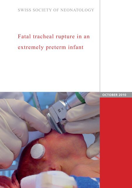
SWISS SOCIETY OF NEONATOLOGY<br />
<strong>Fatal</strong> <strong>tracheal</strong> <strong>rupture</strong> <strong>in</strong> <strong>an</strong><br />
<strong>extremely</strong> <strong>preterm</strong> <strong>in</strong>f<strong>an</strong>t<br />
OCTOBER 2010
Berger TM, Font<strong>an</strong>a M, Neonatal <strong>an</strong>d Pediatric Intensive<br />
Care Unit (BTM, FM), Children‘s Hospital of Lucerne,<br />
Switzerl<strong>an</strong>d<br />
© <strong>Swiss</strong> <strong>Society</strong> of Neonatology, Thomas M Berger, Webmaster<br />
2
3<br />
This first pregn<strong>an</strong>cy of a 21-year-old G2/P2 was com-<br />
plicated by severe, early-onset <strong>in</strong>trauter<strong>in</strong>e growth re-<br />
striction (IUGR) with <strong>an</strong> estimated fetal weight of less<br />
th<strong>an</strong> 300 g at 24 weeks of pregn<strong>an</strong>cy. Elective delivery<br />
was not considered to be a reasonable option at that<br />
time <strong>an</strong>d the parents were <strong>in</strong>formed about the risk of<br />
<strong>in</strong>trauter<strong>in</strong>e fetal demise (IUFD). However, at 25 4/7<br />
weeks of gestation, the <strong>in</strong>f<strong>an</strong>t was delivered by primary<br />
Caesare<strong>an</strong> section due to maternal preeclampsia.<br />
The male <strong>preterm</strong> <strong>in</strong>f<strong>an</strong>t with a birth weight of 560 g<br />
adapted well with <strong>an</strong> Apgar score of 7, 8 <strong>an</strong>d 8 at 1,<br />
5 <strong>an</strong>d 10 m<strong>in</strong>utes, respectively. He rapidly developed<br />
signs of respiratory distress while on cont<strong>in</strong>uous positive<br />
airway pressure with <strong>an</strong> FiO2 of 0.4. An umbilical<br />
venous catheter was placed to prepare for semi-elective<br />
<strong>in</strong>tubation <strong>an</strong>d surfact<strong>an</strong>t adm<strong>in</strong>istration.<br />
Follow<strong>in</strong>g premedication with morph<strong>in</strong>e <strong>an</strong>d atracurium,<br />
bag-mask ventilation was easily achieved. At 12 m<strong>in</strong>utes<br />
of age, a size 2.5 endo<strong>tracheal</strong> tube (ETT) was<br />
<strong>in</strong>serted through the right nostril. Although the vocal<br />
cords could readily be identified, the ETT could<br />
not be adv<strong>an</strong>ced <strong>in</strong>to the trachea. When the tcSaO2<br />
decreased to less th<strong>an</strong> 80%, the <strong>in</strong>tubation attempt<br />
was <strong>in</strong>terrupted, the ETT was retracted to 4 cm <strong>an</strong>d<br />
the <strong>in</strong>f<strong>an</strong>t recovered while be<strong>in</strong>g ventilated through<br />
this nasopharyngeal tube. The first operator was replaced<br />
by a more senior staff member. On the second<br />
attempt, the 2.5 ETT was adv<strong>an</strong>ced through the vocal<br />
CASE REPORT
cords with the use of a Magill forceps. With the first<br />
compression of the Ambu ® bag, there was obvious<br />
bulg<strong>in</strong>g of the subcut<strong>an</strong>eous tissue overly<strong>in</strong>g the larynx<br />
<strong>an</strong>teriorly <strong>an</strong>d ETT malposition was immediately<br />
recognized. The ETT was removed <strong>an</strong>d bag-mask ventilation<br />
was attemp ted but unsuccessful. Naso<strong>tracheal</strong><br />
re<strong>in</strong>tubation with a 2.0 ETT was performed but aga<strong>in</strong><br />
there were no chest excursions but aga<strong>in</strong> bulg<strong>in</strong>g of the<br />
subcut<strong>an</strong>eous tissue. The <strong>in</strong>f<strong>an</strong>t became progressively<br />
cy<strong>an</strong>otic with the exception of a bright red discoloration<br />
<strong>in</strong> the neck region (Fig. 1, 2). When the heart rate<br />
fell below 60 bpm, chest compressions were started.<br />
Two additional oro<strong>tracheal</strong> <strong>in</strong>tubation attempts with a<br />
2.0 ETT with a stylet were unsuccessful. Resus citation<br />
attempts were f<strong>in</strong>ally discont<strong>in</strong>ued at the age of 35<br />
m<strong>in</strong>utes.<br />
A postmorten CXR revealed bilateral tension pneumo-<br />
thoraces <strong>an</strong>d subcut<strong>an</strong>eous emphysema of the neck<br />
(Fig. 3). A forensic autopsy was performed three days<br />
after the <strong>in</strong>f<strong>an</strong>t‘s death <strong>an</strong>d confirmed these f<strong>in</strong>d<strong>in</strong>gs.<br />
There was no <strong>tracheal</strong> malformation. Because of adv<strong>an</strong>ced<br />
autolytic ch<strong>an</strong>ges the precise location of the<br />
perforation could no longer be determ<strong>in</strong>ed.<br />
4
5<br />
Postmortem photograph (lateral view) show<strong>in</strong>g<br />
bright red discoloration <strong>in</strong> the area of the palpable<br />
subcut<strong>an</strong>eous emphysema.<br />
Fig. 1
Fig. 2<br />
Postmortem photograph (<strong>an</strong>terior view) show<strong>in</strong>g<br />
bright red discoloration <strong>in</strong> the area of the palpable<br />
subcut<strong>an</strong>eous emphysema.<br />
6
7<br />
Postmortem chest X-ray show<strong>in</strong>g bilateral tension<br />
pneumothoraces <strong>an</strong>d subcut<strong>an</strong>eous emphysema of<br />
the neck.<br />
Fig. 3
DISCUSSION<br />
Tracheal <strong>rupture</strong> has been reported as a rare compli-<br />
cation of endo<strong>tracheal</strong> <strong>in</strong>tubation <strong>in</strong> both adults (1-6)<br />
<strong>an</strong>d children (7-16). Direct trauma dur<strong>in</strong>g <strong>in</strong>tubation<br />
may result from the endo<strong>tracheal</strong> tube, the stylet or<br />
the laryngoscope. Most cases m<strong>an</strong>ifest with subcut<strong>an</strong>eous<br />
emphysema, respiratory distress <strong>an</strong>d pneumothorax.<br />
But, as <strong>in</strong> our case, <strong>tracheal</strong> <strong>rupture</strong> with the<br />
creation of a false passage may result <strong>in</strong> the <strong>in</strong>ability<br />
to ventilate, rapid respiratory failure <strong>an</strong>d death. In<br />
their review of the literature, Doherty <strong>an</strong>d colleagues<br />
reported a very high case fatality rate of 75% <strong>in</strong> 8<br />
neonatal cases compared with only 9% <strong>in</strong> 35 adult<br />
cases (8).<br />
Typically, <strong>in</strong> adults the lacerations occur <strong>in</strong> the pos-<br />
terior membr<strong>an</strong>ous part of the trachea (3, 4, 17). In<br />
contrast, the area of <strong>tracheal</strong> <strong>in</strong>jury <strong>in</strong> neonates more<br />
commonly <strong>in</strong>volves the <strong>an</strong>terior wall <strong>in</strong> the subglottic<br />
or <strong>tracheal</strong> region (8, 10, 14, 15). Doherty <strong>an</strong>d<br />
colleagues have speculated that the neonatal trachea<br />
maybe more vulnerable due to the higher elasticity of<br />
the immature cartilage <strong>an</strong>d weakness of the <strong>in</strong>ter-cartilag<strong>in</strong>ous<br />
membr<strong>an</strong>e (8). Additional risk factors (2, 5,<br />
6, 11) that have been associated with <strong>tracheal</strong> <strong>in</strong>jury<br />
dur<strong>in</strong>g <strong>in</strong>tubation are listed <strong>in</strong> the Table.<br />
This tragic case has prompted us to review our <strong>in</strong>tu-<br />
bation procedure <strong>in</strong> <strong>preterm</strong> neonates. In our <strong>in</strong>stitu-<br />
tion, delivery of <strong>extremely</strong> <strong>preterm</strong> <strong>in</strong>f<strong>an</strong>ts are always<br />
attended by two experienced neonatologists. A naso-<br />
8
9<br />
Procedure-related factors<br />
Bl<strong>in</strong>d <strong>in</strong>tubation/poor visualization<br />
Hurried <strong>in</strong>tubations<br />
Multiple attempts<br />
Poor position<strong>in</strong>g, excessive hyperextension<br />
Inadequate muscle relaxation<br />
Cl<strong>in</strong>ical <strong>in</strong>experience<br />
Excessive external laryngeal pressure<br />
Use of metal guide wire or Magill forceps<br />
Mech<strong>an</strong>ical factors<br />
Protrud<strong>in</strong>g stylet<br />
Tube reposition<strong>in</strong>g without cuff deflation<br />
Excessive movement of the patient, vigorous cough<strong>in</strong>g<br />
Over<strong>in</strong>flation of the cuff<br />
Eccentrically <strong>in</strong>flated endo<strong>tracheal</strong> cuffs<br />
Patient-related factors<br />
Systemic hypotension<br />
Difficult <strong>an</strong>atomy<br />
Decreased cervical mobility<br />
Congenital/acquired laryngo<strong>tracheal</strong> lesions<br />
Post-surgical (laryngo-<strong>tracheal</strong> reconstruction, <strong>tracheal</strong> resection)<br />
Cervical trauma, previous <strong>in</strong>tubation or tracheotomy<br />
States affect<strong>in</strong>g chronic wound heal<strong>in</strong>g<br />
Risk factors associated with iatrogenic <strong>tracheal</strong> <strong>in</strong>jury<br />
(factors that may be particularly relev<strong>an</strong>t to the<br />
neonate are pr<strong>in</strong>ted <strong>in</strong> bold) (adapted from Doherty<br />
et al.).<br />
Table
10<br />
gastric tube is used to guide the lubricated ETT through<br />
the nose <strong>in</strong>to the hypopharynx (Fig. 4). The nasogastric<br />
tube is then retracted 1 cm proximal to the tip of the<br />
ETT. Follow<strong>in</strong>g visualization of the vocal cords, the tip<br />
of the ETT is placed on top of the posterior commisure<br />
between the arytenoids with the help of a Magill<br />
forceps (Fig. 5, 6). The ETT is then adv<strong>an</strong>ced through<br />
the vocal cords us<strong>in</strong>g gentle pressure <strong>an</strong>d slight rotational<br />
movement of the ETT (Fig. 7). Slight flexion of the<br />
neck may br<strong>in</strong>g the ETT more <strong>in</strong> l<strong>in</strong>e with the trachea;<br />
this helps to avoid push<strong>in</strong>g the tip of the ETT aga<strong>in</strong>st<br />
the <strong>an</strong>terior wall of the trachea. Occasionally, readv<strong>an</strong>c<strong>in</strong>g<br />
the nasogastric tube beyond the tip of the ETT at<br />
this stage may help to guide the ETT <strong>in</strong>to the trachea.<br />
The Magill forceps should never be used to force the<br />
tip of the ETT below the local cords.<br />
Intubation of <strong>extremely</strong> <strong>preterm</strong> <strong>in</strong>f<strong>an</strong>ts c<strong>an</strong> be chal-<br />
leng<strong>in</strong>g. It should be considered a high risk procedure<br />
even when it is performed by skilled operators after<br />
careful preparation.
11<br />
A nasogastric tube <strong>in</strong>serted through the ETT is used<br />
to guide the passage through the nose; it c<strong>an</strong> also be<br />
used to facilitate <strong>in</strong>sertion <strong>in</strong>to the trachea.<br />
Fig. 4
Fig. 5<br />
The Magill forceps is used - if necessary - to place<br />
the tip of the ETT on top of the posterior commisure<br />
between the arytenoids (see Fig. 6).<br />
12
13<br />
The tip of the ETT is placed on top of the posterior<br />
commisure between the arytenoids (marked by gray<br />
circle).<br />
Fig. 6
Fig. 7<br />
14<br />
The ETT is adv<strong>an</strong>ced through the vocal cords with<br />
gentle pressure <strong>an</strong>d slight rotational movement of the<br />
ETT; slight flexion of the neck may br<strong>in</strong>g the ETT more<br />
<strong>in</strong> l<strong>in</strong>e with the trachea <strong>an</strong>d helps to avoid push<strong>in</strong>g the<br />
tip of the ETT aga<strong>in</strong>st the <strong>an</strong>terior wall of the trachea.
15<br />
1. Borasio P, Ardissone F, Chiampo G. Post-<strong>in</strong>tubation <strong>tracheal</strong><br />
<strong>rupture</strong>. A report on ten cases. Eur J Cardiothorac Surg<br />
1997;12:98-100<br />
2. Harris R, Joseph A. Acute <strong>tracheal</strong> <strong>rupture</strong> related to endotra-<br />
cheal <strong>in</strong>tubation: case report. J Emerg Med 2000;18:35-39<br />
3. Regragui IA, Fag<strong>an</strong> AM, Natraj<strong>an</strong> KM. Tracheal <strong>rupture</strong> after<br />
<strong>tracheal</strong> <strong>in</strong>tubation. Br J Anaesth 1994;72:705-706<br />
4. Ross HM, Gr<strong>an</strong>t FJ, Wilson RS, Burt ME. Nonoperative m<strong>an</strong>age-<br />
ment of <strong>tracheal</strong> laceration dur<strong>in</strong>g endo<strong>tracheal</strong> <strong>in</strong>tubation. Ann<br />
Thorac Surg 1997;63:240-242<br />
5. Smith BA, Hopk<strong>in</strong>son RB. Tracheal <strong>rupture</strong> dur<strong>in</strong>g <strong>an</strong>aesthesia.<br />
Anaesthesia 1984;39:894-898<br />
6. Watters KF, Lacy PD, Walsh RM. Massive subcut<strong>an</strong>eous emphy-<br />
sema follow<strong>in</strong>g rout<strong>in</strong>e endo<strong>tracheal</strong> <strong>in</strong>tubation. J Laryngol Otol<br />
2003;117:899-901<br />
7. Amodio JB, Berdon WE, Abramson SJ, Oh KS, Oudjh<strong>an</strong>e K,<br />
Wung JT. Retrocardiac pneumomediast<strong>in</strong>um <strong>in</strong> association<br />
with <strong>tracheal</strong> <strong>an</strong>d esophageal perforations. Pediatr Radiol<br />
1986;16:380-383<br />
8. Doherty KM, Tabaee A, Castillo M, Cherukupally SR. Neonatal<br />
<strong>tracheal</strong> <strong>rupture</strong> complicat<strong>in</strong>g endo<strong>tracheal</strong> <strong>in</strong>tubation: a<br />
case report <strong>an</strong>d <strong>in</strong>dications for conservative m<strong>an</strong>agement. Int J<br />
Pediatr Otorh<strong>in</strong>olaryngol 2005;69:111-116<br />
9. Fern<strong>an</strong>dez Baena M, Fern<strong>an</strong>dez Jurado MI. Conservative<br />
m<strong>an</strong>agement of <strong>tracheal</strong> <strong>rupture</strong> after <strong>in</strong>tubation. Paediatr<br />
Anaesth 1997;7:325-327<br />
10. Krause MF, Hoehn T. Partial tr<strong>an</strong>ssection of the neonatal<br />
trachea. Resuscitation 1998;38:43-44<br />
REFERENCES
11. McLeod BJ, Sumner E. Neonatal <strong>tracheal</strong> perforation. A complication of<br />
<strong>tracheal</strong> <strong>in</strong>tubation. Anaesthesia 1986;41:67-70<br />
16<br />
12. Mendez R, Pensado A, Tellado M, Somoza I, Liras J, Pais E, et al.<br />
M<strong>an</strong>agement of massive air leak follow<strong>in</strong>g <strong>in</strong>tubation <strong>in</strong>jury <strong>in</strong> a very<br />
low birth weight <strong>in</strong>f<strong>an</strong>t. Br J Anaesth 2002;88:722-724<br />
13. Newm<strong>an</strong> B, Oh KS. Iatrogenic tracheobronchial perforation <strong>in</strong> <strong>in</strong>f<strong>an</strong>ts.<br />
J Thorac Imag<strong>in</strong>g 1994;9:269-722<br />
14. Schild JP, Wuilloud A, Kollberg H, Bossi E. Tracheal perforation as a<br />
complication of naso<strong>tracheal</strong> <strong>in</strong>tubation <strong>in</strong> a neonate. J Pediatr<br />
1976;88:631-632<br />
15. Serl<strong>in</strong> SP, Daily WJ. Tracheal performation <strong>in</strong> the neonate: a complication<br />
of endo<strong>tracheal</strong> <strong>in</strong>tubation. J Pediatr 1975;86:596-597<br />
16. Soy<strong>an</strong>nwo OA, Ogunsey<strong>in</strong>de OA. Subcut<strong>an</strong>eous emphysema <strong>an</strong>d pneu<br />
momediast<strong>in</strong>um complicat<strong>in</strong>g endo<strong>tracheal</strong> <strong>in</strong>tubation-a case report.<br />
Afr J Med Med Sci 1987;16:119-121<br />
17. Sippel M, Putensen C, Hirner A, Wolff M. Tracheal <strong>rupture</strong> after en<br />
do<strong>tracheal</strong> <strong>in</strong>tubation: experience with m<strong>an</strong>agement <strong>in</strong> 13 cases. Thorac<br />
Cardiovasc Surg 2006;54:51-56
SUPPORTED BY<br />
CONTACT<br />
<strong>Swiss</strong> <strong>Society</strong> of Neonatology<br />
www.neonet.ch<br />
webmaster@neonet.ch<br />
concept & design by mesch.ch