Cardiomegaly in a premature neonate after venous umbilical ...
Cardiomegaly in a premature neonate after venous umbilical ...
Cardiomegaly in a premature neonate after venous umbilical ...

Create successful ePaper yourself
Turn your PDF publications into a flip-book with our unique Google optimized e-Paper software.
SWISS SOCIETY OF NEONATOLOGY<br />
<strong>Cardiomegaly</strong> <strong>in</strong> a <strong>premature</strong><br />
<strong>neonate</strong> <strong>after</strong> <strong>venous</strong><br />
<strong>umbilical</strong> catheterization<br />
JULY 2008
Schlapbach LJ, Pfammatter J-P, Nelle M, McDougall<br />
FJ, Division of Neonatology (SLJ, NM, McDFJ), Divi-<br />
sion of Pediatric Cardiology (PJ-P), Department of<br />
Pediatrics, University of Berne, Switzerland<br />
© Swiss Society of Neonatology, Thomas M Berger, Webmaster
Umbilical <strong>venous</strong> catheters (UVC) are frequently used <strong>in</strong><br />
<strong>neonate</strong>s requir<strong>in</strong>g hyperosmolar parenteral nutrition,<br />
catecholam<strong>in</strong>es or when no peripheral <strong>venous</strong> access<br />
can be established. Catheterization of the <strong>umbilical</strong><br />
ve<strong>in</strong> allows rapid central access, but may be associated<br />
with various complications (1). Cl<strong>in</strong>icians are particular-<br />
ly aware of catheter-associated <strong>in</strong>fections and throm-<br />
bosis. Due to the widespread use of <strong>umbilical</strong> l<strong>in</strong>es,<br />
neonatologists should keep rare complications <strong>in</strong> m<strong>in</strong>d<br />
as well. We present a case of a newborn with pericar-<br />
dial effusion follow<strong>in</strong>g UVC placement.<br />
An extremely <strong>premature</strong> <strong>in</strong>fant weigh<strong>in</strong>g 590 grams<br />
was born at 5 weeks gestational age by caesarean<br />
section for severe pre-eclampsia and deteriorat<strong>in</strong>g fetal<br />
Doppler studies. The baby was <strong>in</strong>tubated for respiratory<br />
distress syndrome with<strong>in</strong> the first hour of life and um-<br />
bilical <strong>venous</strong> and arterial l<strong>in</strong>es were placed (UVC .5<br />
Charrière s<strong>in</strong>gle-lumen, UAC .5 Charrière, ArgyleTM<br />
polyurethane, Tyco Healthcare, Tullamore, Ireland). The<br />
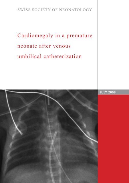
position of the catheters, both of which had been <strong>in</strong>-<br />
serted too far (Fig. 1), was corrected accord<strong>in</strong>g to the<br />
CXR by 1.5 cm (UVC) and cm (UAC). After receiv<strong>in</strong>g<br />
porc<strong>in</strong>e surfactant (Curosurf®), the <strong>in</strong>fant was success-<br />
fully weaned from mechanical ventilation. CXR before<br />
planned extubation on day three unexpectedly sho-<br />
wed cardiomegaly with a heart-to-lung ratio of 0.69<br />
(Fig. ). Echocardiography was performed immediate-<br />
ly and revealed a large echo-free pericardial effusion<br />
INTRODUCTION<br />
CASE REPORT
DISCUSSION<br />
measur<strong>in</strong>g 6 mm <strong>in</strong> diameter (Fig. ). Both atrial and<br />
ventricular function were adequate, without diastolic<br />
<strong>in</strong>dentation of the atrial wall. The UVC tip was float<strong>in</strong>g<br />
with<strong>in</strong> the right atrium. On the X-ray, the UVC was still<br />
positioned too high project<strong>in</strong>g <strong>in</strong>to the cardiac silhou-<br />
ette (Fig. ), although its <strong>in</strong>itial position had been ade-<br />
quately corrected. Aspiration through the UVC yielded<br />
bloody fluid, and blood gas and chemical analysis of the<br />
aspirate were consistent with blood and not parenteral<br />
nutrition. Ultrasound exam<strong>in</strong>ation ruled out pleural and<br />
abdom<strong>in</strong>al effusions. Stool cultures of the <strong>in</strong>fant and<br />
the mother were negative for enterovirus. Maternal se-<br />
rologies were negative for TORCHS, Parvovirus B19, and<br />
Coxiella burnetii.<br />
S<strong>in</strong>ce the <strong>in</strong>fant rema<strong>in</strong>ed hemodynamically stable with<br />
no signs of cardiovascular compromise, we decided<br />
aga<strong>in</strong>st emergency pericardiocentesis. After remov<strong>in</strong>g<br />
the UVC, the effusion gradually resolved with<strong>in</strong> a few<br />
days, and the <strong>in</strong>fant was successfully extubated. The<br />
later cl<strong>in</strong>ical course was complicated by bronchopul-<br />
monary dysplasia and osteopathy of prematurity. Later<br />
cardiac follow-up revealed no functional or anatomical<br />
pathology.<br />
Pericardial effusion is a well-known complication of<br />
peripherally <strong>in</strong>serted central catheters (PICC), with an<br />
estimated <strong>in</strong>cidence of 1.8/1000 catheters ( ). The ma-<br />
jority of <strong>in</strong>fants with reported pericardial effusion beca-
5<br />
me acutely symptomatic due to cardiac tamponade and<br />
deteriorated rapidly with signs of respiratory distress,<br />
cyanosis, tachycardia or bradycardia, mottled sk<strong>in</strong>, and<br />
arterial hypotension f<strong>in</strong>ally lead<strong>in</strong>g to cardiopulmonary<br />
arrest not responsive to standard <strong>in</strong>terventions ( ). No-<br />
tably, approximately a quarter of the cases were first<br />
diagnosed post-mortem at autopsy ( ). The mortality is<br />
very high ( 5-65%) ( , ) and those resuscitated success-<br />
fully improved only <strong>after</strong> emergency pericardiocentesis<br />
was performed. Analysis of the aspirated liquid usually<br />
reflected the composition of the parenteral nutrition.<br />
Follow<strong>in</strong>g a series of case reports on <strong>in</strong>fant deaths attri-<br />
buted to PICC-associated cardiac tamponade, guidel<strong>in</strong>es<br />
have been published aimed at reduc<strong>in</strong>g the risk of car-<br />
diac perforation ( -5).<br />
Contrary to PICC, the <strong>in</strong>cidence of pericardial effusion<br />
associated with UVC is unknown but case reports have<br />
documented sudden cardiovascular compromise <strong>in</strong> <strong>in</strong>-<br />
fants with UVC due to pericardial tamponade (6-8).<br />
Perforation and catheter migration are thought to occur<br />
as a result of both mechanical pressure by the catheter<br />
tip repeatedly push<strong>in</strong>g aga<strong>in</strong>st the contract<strong>in</strong>g heart<br />
wall and endocardial <strong>in</strong>jury caused by hyperosmotic pa-<br />
renteral nutrition fluids. Transmural diffusion of paren-<br />
teral nutrition fluids <strong>in</strong>to the pericardial space further<br />
contributes to the accumulation of fluid. Contrary to<br />
catheter-associated <strong>in</strong>fections and thrombosis which<br />
<strong>in</strong>crease over time, pericardial effusion may occur directly
Fig. 1<br />
UVC<br />
UAC<br />
Chest X-ray on day one <strong>after</strong> <strong>in</strong>tubation and <strong>in</strong>sertion<br />
of <strong>venous</strong> (UVC) and arterial (UAC) <strong>umbilical</strong> l<strong>in</strong>es.<br />
6
UVC<br />
UAC<br />
Incidental f<strong>in</strong>d<strong>in</strong>g of cardiomegaly on chest X-ray<br />
before extubation on day three. UVC: <strong>umbilical</strong><br />
<strong>venous</strong> catheter; UAC: <strong>umbilical</strong> arterial catheter.<br />
Fig. 2
<strong>after</strong> <strong>in</strong>sertion of catheters, or later, with a peak at<br />
three days follow<strong>in</strong>g catheter <strong>in</strong>sertion ( ). Malposition<br />
of central catheters is considered the ma<strong>in</strong> risk factor<br />
for pericardial effusion, particularly if the catheter tip<br />
projects <strong>in</strong>to the right atrium or shows angulation ( ,<br />
). The catheter tip should be positioned at the junction<br />
of the vena cava <strong>in</strong>ferior and right atrium with the tip<br />
ly<strong>in</strong>g outside the cardiac silhouette. However, catheter<br />
<strong>in</strong>ward migration, as experienced <strong>in</strong> the present case,<br />
has been described and is attributed to retraction of<br />
the mummify<strong>in</strong>g cord, changes <strong>in</strong> abdom<strong>in</strong>al girth and<br />
catheter dislocation dur<strong>in</strong>g manipulations (6, 9). The-<br />
refore, even <strong>after</strong> correct <strong>in</strong>itial placement, the UVC<br />
position should be checked regularly us<strong>in</strong>g X-ray or<br />
ultrasound.<br />
The differential diagnosis of neonatal pericardial effu-<br />
sion <strong>in</strong>cludes immune and non-immune hydrops fetalis,<br />
congenital <strong>in</strong>fections such as TORCHS and Parvovirus<br />
B19, and rarely myopericarditis caused by Enteroviridae,<br />
ma<strong>in</strong>ly Echovirus and Cocksackievirus, or Coxiella burnetii.<br />
The present case illustrates that pericardial effusion<br />
may progress asymptomatically before hemodynamic<br />
changes become evident. The <strong>in</strong>cidence of catheter-asso-<br />
ciated pericardial effusion may therefore be underesti-<br />
mated. Extremely low birth-weight <strong>in</strong>fants might be at<br />
particular risk due to the th<strong>in</strong> myocardial wall with rela-<br />
tively large catheters often - as <strong>in</strong> this case - be<strong>in</strong>g <strong>in</strong>i-<br />
tially <strong>in</strong>serted too far. Consider<strong>in</strong>g the potentially lethal<br />
8
9<br />
*<br />
*<br />
*<br />
Echocardiography demonstrat<strong>in</strong>g large pericardial<br />
effusion (asterisks) on day three.<br />
*<br />
Fig. 3
complications of UVC, neonatologists should carefully<br />
consider the <strong>in</strong>dication for plac<strong>in</strong>g UVCs and remove<br />
UVCs as soon as possible. Whether percutaneous long<br />
l<strong>in</strong>es represent a safer alternative rema<strong>in</strong>s unclear and<br />
further prospective studies compar<strong>in</strong>g UVC and PICC<br />
are needed (1). Mal-positioned UVCs should be cor-<br />
rected immediately and the position should be verified<br />
<strong>after</strong>wards. F<strong>in</strong>ally, neonatologists should ma<strong>in</strong>ta<strong>in</strong> a<br />
high <strong>in</strong>dex of suspicion for pericardial tamponade and<br />
readily perform echocardiography <strong>in</strong> acutely ill <strong>in</strong>fants<br />
with UVCs.<br />
In conclusion, pericardial effusion may occur asympto-<br />
matically <strong>after</strong> <strong>umbilical</strong> <strong>venous</strong> catheterization and<br />
should be suspected <strong>in</strong> <strong>in</strong>fants with central catheters<br />
and progressive cardiomegaly. Prompt removal of ca-<br />
theters and, if signs of pericardial tamponade are pre-<br />
sent, emergency pericardiocentesis, may prove life-sav<strong>in</strong>g.<br />
For related cases, see also COTM 10/ 001 and COTM<br />
1 / 00 .<br />
10
11<br />
1. Butler-O‘Hara M, Buzzard CJ, Reubens L, McDermott MP,<br />
DiGrazio W, D‘Angio CT. A randomized trial compar<strong>in</strong>g<br />
long-term and short-term use of <strong>umbilical</strong> <strong>venous</strong> catheters<br />
<strong>in</strong> <strong>premature</strong> <strong>in</strong>fants with birth weights of less than<br />
1 51 grams. Pediatrics 006;118:e 5- 5<br />
. Beardsall K, White DK, P<strong>in</strong>to EM, Kelsall AW. Pericardial ef<br />
fusion and cardiac tamponade as complications of neonatal<br />
long l<strong>in</strong>es: are they really a problem? Arch Dis Child Fetal<br />
Neonatal Ed 00 ;88:F 9 - 95<br />
. Darl<strong>in</strong>g JC, Newell SJ, Mohamdee O, Uzun O, Cull<strong>in</strong>ane CJ,<br />
Dear PR. Central <strong>venous</strong> catheter tip <strong>in</strong> the right atrium: a risk<br />
factor for neonatal cardiac tamponade. J Per<strong>in</strong>atol 001;<br />
1: 61- 6<br />
. Nowlen TT, Rosenthal GL, Johnson GL, Tom DJ, Vargo TA.<br />
Pericardial effusion and tamponade <strong>in</strong> <strong>in</strong>fants with central<br />
catheters. Pediatrics 00 ;110:1 -1<br />
5. Pezzati M, Filippi L, Chiti G, Dani C, Rossi S, Bert<strong>in</strong>i G,<br />
Rubaltelli FF. Central <strong>venous</strong> catheters and cardiac tamponade<br />
<strong>in</strong> preterm <strong>in</strong>fants. Intensive Care Med 00 ; 0: 5 - 56<br />
6. Traen M, Schepens E, Laroche S, van Overmeire B. Cardiac<br />
tamponade and pericardial effusion due to <strong>venous</strong> <strong>umbilical</strong><br />
catheterization. Acta Paediatr 005;9 :6 6-6 8<br />
. Sehgal A, Cook V, Dunn M. Pericardial effusion associated<br />
with an appropriately placed <strong>umbilical</strong> <strong>venous</strong> catheter.<br />
J Per<strong>in</strong>atol 00 ; : 1 - 19<br />
8. Onal EE, Saygili A, Koc E, Turkyilmaz C, Okumus N, Atalay Y.<br />
Cardiac tamponade <strong>in</strong> a newborn because of <strong>umbilical</strong> <strong>venous</strong><br />
catheterization: is correct position safe? Paediatr Anaesth<br />
00 ;1 :95 -956<br />
9. Salvadori S, Piva D, Filippone M. Umbilical <strong>venous</strong> l<strong>in</strong>e<br />
displacement as a consequence of abdom<strong>in</strong>al girth variation.<br />
J Pediatr 00 ;1 1:<br />
REFERENCES