Caudal regression sequence: a forgotten disorder? - Swiss Society ...
Caudal regression sequence: a forgotten disorder? - Swiss Society ...
Caudal regression sequence: a forgotten disorder? - Swiss Society ...

Create successful ePaper yourself
Turn your PDF publications into a flip-book with our unique Google optimized e-Paper software.
SWISS SOCIETY OF NEONATOLOGY<br />
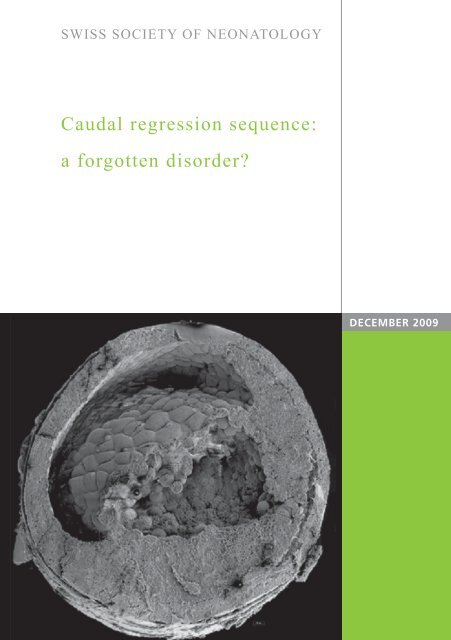
<strong>Caudal</strong> <strong>regression</strong> <strong>sequence</strong>:<br />
a <strong>forgotten</strong> <strong>disorder</strong>?<br />
DECEMBER 2009
Zoubir SA, Newman CJ, Vial Y, Meyrat B , Zambelli PY,<br />
Truttmann AC, Division of Neonatology (ZSA, TAC),<br />
Neurorehabilitation Unit (NCJ), Department of<br />
Pediatrics, Department Pediatric Surgery (VY),<br />
Department of Obstetrics and Gynaecology (MB),<br />
Orthopaedic Hospital (ZPY), University Center Hospital<br />
and University of Lausanne, Switzerland<br />
© <strong>Swiss</strong> <strong>Society</strong> of Neonatology, Thomas M Berger, Webmaster<br />
2
3<br />
We present a classical case of caudal <strong>regression</strong> se-<br />
quence (CRS) in a term newborn of a diabetic mother.<br />
This sporadic <strong>disorder</strong> is more frequent in diabetic<br />
mothers but can also be seen in other circumstances.<br />
Because of early antenatal diagnosis which is often<br />
followed by termination of pregnancy, the clinical<br />
syndrome is nowadays rarely seen postnatally.<br />
The mother is a 38-year-old Mauritian G5/P3 who was<br />
diagnosed with type I diabetes at the age of 16. She<br />
had first been managed by diet and after one year<br />
by insulin. Compliance was inconsistent, and, consequently,<br />
she developed severe bilateral retinopathy<br />
several years later.<br />
Her previous pregnancies resulted in two normal term<br />
deliveries in 1998 and 1999, a miscarriage in 2004<br />
and a voluntary interruption of pregnancy in 2005.<br />
At 4 weeks of this current pregnancy, there was an<br />
episode of severe hyperglycemia (blood glucose 20<br />
mmol/l) without ketoacidosis. At that time, the glycosylated<br />
hemoglobin (HbA1C) was normal and, incidentally,<br />
the patient was found to be pregnant. Because<br />
of poor diabetic control, she was hospitalized at<br />
25 weeks gestation for better diabetic management<br />
(HbA1C of 6.2%, normal < 6.0%). Additional risk factors<br />
included significant overweight with a BMI of 28,<br />
moderate nicotine consumption (
gynaecologist‘s office was difficult to perform because<br />
of the patient‘s overweight but was described as nor-<br />
mal. The lower part of the legs were not visible, but<br />
the length of the femur was between P25-50 percentile<br />
while other parameters were at P50-75. The nonvisualization<br />
of the lower parts of the legs was ascribed<br />
to the fact that the fetus was in a low position.<br />
Two further ultrasound examinations were performed<br />
with identical findings. The baby was delivered by<br />
emergency caesarean section at 38 weeks gestation<br />
because of non-reassuring fetal heart rate tracings. No<br />
resuscitation was required at birth, Apgar scores were<br />
4, 9, and 9 at 1, 5 and 10 minutes, respectively, her<br />
weight was 2970 g (P10-50) with a length of 43.5 cm<br />
(< P10) and a head circumference of 35.5 cm (P50-90).<br />
Immediately following delivery, obvious malformations<br />
of the lower extremities were noticed: the buttocks<br />
and lower limbs appeared atrophic and no bony structures<br />
were palpable in the lumbar and sacral regions.<br />
The limbs were immobilized in a frog-like position,<br />
fixed in flexion with webbing between the thigh and<br />
the leg, and a notch on each side was noted anterior<br />
to the great trochanters. The feet were also hypotrophic<br />
and in equinovarus position (Fig. 1, 2). Otherwise,<br />
the exam was normal with a normal female genitalia,<br />
an anteposed and patent anus with a weak anal reflex.<br />
Further investigations included a babygram, a MRI, and<br />
a voiding cystoureterogram (VCUG). On the babygram,<br />
4
5<br />
Frog-like position of the hypotrophic lower extre-<br />
mities with contractures of hip and knee joints.<br />
Fig. 1
Fig. 2<br />
Anteposed anus, small buttocks and dimples in the<br />
trochanteric region.<br />
6
7<br />
no lumbar or sacral vertebral bodies were visible, and<br />
the iliac bone was hypoplastic with fused wings (Fig. 3).<br />
The MRI definitely confirmed lumbar and sacral agenesis<br />
with the spinal cord fixed at T12 (tethered cord)<br />
(Fig. 4). The brain was normal. The abdominal MRI<br />
confirmed agenesis of the right kidney. There was<br />
no vesicoureteral reflux (Fig. 5). The baby was managed<br />
with nasal CPAP for transient tachypnea of the<br />
newborn. Feedings were started at 48 hours of life.<br />
Because of frequent regurgitation an upper gastrointestinal<br />
contrast study was done which confirmed<br />
gastroesophageal reflux. The baby was discharged<br />
after 10 days with antibiotic prophylaxis to prevent<br />
urinary tract infections and metoclopramide and omeprazole<br />
for gastroesophageal reflux.<br />
The baby was seen at 2, 4 and 12 months in the out-<br />
patient clinic. She is growing between P10-50 for<br />
weight and P50-90 for head circumference. She has<br />
had no febrile episodes nor urinary tract infections.<br />
She has anal incontinence and episodes of constipation<br />
have been managed with paraffin oil. She has had<br />
persistent moderate gastroesophageal regurgitations<br />
without clinical evidence of esophagitis. Cystomanometry<br />
performed at one and four months revealed<br />
high pressure in the bladder with normal voiding, reduced<br />
bladder volume and compliance. The left kidney<br />
shows hypertrophic compensation on ultrasonography<br />
with compensatory hyperactivity on renal scintigraphy.<br />
She is managed by a multidisciplinary team
of pediatric surgeons, ortho pedics, occupational the-<br />
rapists, social workers and neurorehabilitation specia-<br />
lists. Except for limitations due to the malformation of<br />
her lower extremities, her psychomotor development<br />
is normal.<br />
8
9<br />
Babygram with absence of lumbar and sacral<br />
vertebral bodies, fused iliac and ischial bones.<br />
Fig. 3
Fig. 4<br />
T2-weigthed MR image: sagittal view of spine and<br />
brain demonstrating an interruption of the spine at<br />
the 11th thoracic vertebral body.<br />
10
11<br />
VCUG showing no reflux; note the abnormal pelvic<br />
bony structures.<br />
Fig. 5
DISCUSSION The caudal <strong>regression</strong> <strong>sequence</strong> (CRS) is a rare malfor-<br />
mation with a broad spectrum of manifestations that<br />
ranges from sacral agenesis to the most severe form<br />
of the <strong>sequence</strong> as seen in our case. The cause of CRS<br />
is unknown, but maternal diabetes, genetic predisposition,<br />
and vascular hypoperfusion have been suggested<br />
as possible causative factors.<br />
There is a strong association with maternal diabetes,<br />
either type 1 or type 2 (1,2). The incidence of CRS is<br />
estimated to be 1:60‘000 births with a male:female<br />
ratio of 2.7:1 (3) and it is 200-250 times higher when<br />
the mother is diabetic. A high level of glucose at 6-8<br />
weeks of gestation is known to induce renal defects<br />
in mammals (4).<br />
A defect in the induction of caudal elements be-<br />
fore the 7th week of gestation leads to the CRS by<br />
compro mising cellular migration, neurulation and/or<br />
differentiation (5). It seems that a faulty gastrulation<br />
with subsequent abnormal development of the notochord<br />
leads to the CRS (6). The structures that are developmentally<br />
separated from these caudal elements<br />
are spared. A vascular origin with an anomaly of the<br />
unpaired vessels originating from the aorta was described<br />
as a possible etiology, operating like in sirenomelia<br />
where the persistent vitelline artery “steals”<br />
blood from the lower part of the body (7).<br />
12
13<br />
The association of CRS with other malformations such<br />
as Chiari I malformation and Pierre Robin <strong>sequence</strong><br />
have been reported (8). CRS is usually a sporadic <strong>disorder</strong>,<br />
but an association with chromosome 18p deletion<br />
has been described, with a dysmorphic and hypomimic<br />
face, a short neck, hypotonia with motor retardation,<br />
and an MRI showing a partial sacral and coccygeal<br />
agenesis and non progressive periventricular<br />
white matter lesions (9).<br />
The diagnosis can be made antenatally by ultrasono-<br />
graphy and fetal MRI. A short crown-rump length on<br />
the ultrasound in the first trimester is associated with<br />
CRS (10), and the diagnosis is also possible around 22<br />
weeks gestation by demonstrating the frog-like position<br />
and immobility of the lower limbs, the interruption<br />
of the spine and the defect of vertebrae in typical<br />
cases. The clinical spectrum is wide, comprising developmental<br />
anomalies of the caudal vertebrae, neural<br />
tube, urogenital and digestive organs.<br />
Major complications are the orthopedic deformities<br />
related to the syndrome and scoliosis commonly seen<br />
in the lumbosacral agenesis. Bladder and bowel incontinence,<br />
a subsequent high risk of urinary tract<br />
infections and renal impairment are further complications<br />
and are associated with high morbidity rates in<br />
these patients.
14<br />
Supportive treatment aims at preservation of renal<br />
function by prevention and treatment of urinary infections.<br />
Rehabilitative efforts focus on increasing independence<br />
and preventing secondary orthopedic complications;<br />
they include physiotherapy, occupational<br />
therapy and the provision of supportive devices (orthotics,<br />
wheelchair). Support of a psychologist and a<br />
social worker is also needed.<br />
Because of tight diabetes control prenatally and during<br />
pregnancy, CRS is rarely seen nowadays. Abnormal fetal<br />
leg position and low position of the fetus specially<br />
in a diabetic mother should raise the possibility of a<br />
CRS. When suspected antenatally, termination of pregnancy<br />
is currently offered.
15<br />
1. Twining P, McHugo J, Pilling D. Textbook of foetal<br />
abnormalities. Philadelphia, Pa: Saunders, 2000; 158-160<br />
2. Jones K L. <strong>Caudal</strong> dysplasia <strong>sequence</strong>. In : Smith’s Recognizable<br />
Patterns of Human Malformation, 6th edition, Philadelphia,<br />
Saunders, 2006, 730-731<br />
3. Singh S K, Singh R D, Sharma A. <strong>Caudal</strong> <strong>regression</strong> syndrome-<br />
case report and review of literature. Pediatr Surg Int<br />
2005;21:578-581<br />
4. Kanwar Y S, Nayak B, Lin S et al. Hyperglycemia: its<br />
imminent effects on mammalian nephrogenesis. Pediatric<br />
nephrology 2005;20:858-866<br />
5. Sadler TW. Langman’s Medical Embroyology. 8th edition,<br />
Philadelphia, Lippincott Williams & Wilkins, 2000, 61-110<br />
6. Dias MS, Walker ML. The embryogenesis of complex dysraphic<br />
malformations: a <strong>disorder</strong> of gastrulation? Pediatr Neurosurg<br />
1992;18:229-253<br />
7. Hentschel J, Stierkorb E, Schneider G et al. <strong>Caudal</strong> <strong>regression</strong><br />
<strong>sequence</strong>: vascular origin? J Perinatol. 2006;26:445-447<br />
8. Tubbs R S, Oakes W J. Chiari I malformation, caudal <strong>regression</strong><br />
syndrome and Pierre Robin syndrome: a previously unreported<br />
combination. Childs Nerv Syst 2006;22:1507-1508<br />
‚<br />
9. Kacinski M, Jaworek M, Skowronek-Bała B. <strong>Caudal</strong> <strong>regression</strong><br />
syndrome associated with white matter lesions and<br />
chromosome 18p11.2 deletion. Brain Dev 2007;29:164-166<br />
10. Smith AS, Grable AB I, Levine D. <strong>Caudal</strong> <strong>regression</strong><br />
syndrome in the foetus of diabetic mother. Radiology<br />
2004;230:229-233<br />
REFERENCES
SUPPORTED BY<br />
CONTACT<br />
<strong>Swiss</strong> <strong>Society</strong> of Neonatology<br />
www.neonet.ch<br />
webmaster@neonet.ch<br />
concept & design by mesch.ch