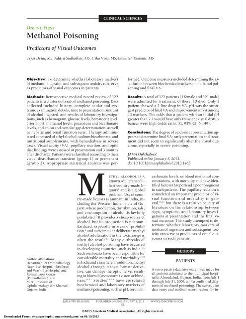
Methanol Poisoning
Methanol Poisoning
Methanol Poisoning

Create successful ePaper yourself
Turn your PDF publications into a flip-book with our unique Google optimized e-Paper software.
ONLINE FIRST<br />
<strong>Methanol</strong> <strong>Poisoning</strong><br />
Predictors of Visual Outcomes<br />
CLINICAL SCIENCES<br />
Tejas Desai, MS; Aditya Sudhalkar, MS; Usha Vyas, MS; Bakulesh Khamar, MS<br />
Objective: To determine whether laboratory markers<br />
of methanol ingestion and subsequent toxicity can serve<br />
as predictors of visual outcomes in patients.<br />
Methods: Retrospective medical record review of 122<br />
patients in a cluster outbreak of methanol poisoning. Data<br />
collected included history, complete ocular and systemic<br />
examination details, time to presentation, amount<br />
of alcohol ingested, and results of laboratory investigations,<br />
such as hemogram, glucose levels, hematocrit level,<br />
arterial pH, methanol levels, potassium and bicarbonate<br />
levels, and anion and osmolar gap determination, as well<br />
as hepatic and renal function tests. Therapy administered<br />
consisted of ethyl alcohol, sodium bicarbonate, and<br />
nutritional supplements, with hemodialysis in severe<br />
cases. Visual acuity (VA), pupillary reaction, and optic<br />
disc findings were assessed at presentation and 3 months<br />
after discharge. Patients were classified according to their<br />
visual disturbance: transient (group 1) or permanent<br />
(group 2). Appropriate statistical analysis was per-<br />
Author Affiliations:<br />
Department of Ophthalmology,<br />
Nagri Eye Hospital (Drs Desai<br />
and Vyas), Eye Hospital and<br />
Retinal Laser Centre<br />
(Dr Sudhalkar), and<br />
M & J Institute of<br />
Ophthalmology (Dr Khamar),<br />
Gujarat, India.<br />
Downloaded From: http://archopht.jamanetwork.com/ on 01/10/2013<br />
METHYL ALCOHOL IS A<br />
known adulterant of illicit<br />
country-made liquors<br />
1 and is a global<br />
problem. Use of country-made<br />
liquors is rampant in India, including<br />
the Western Indian state of Gujarat,<br />
where production, distribution, sale,<br />
and consumption of alcohol is lawfully<br />
prohibited. 2 It provides a cheap source of<br />
alcohol, but its production is not standardized,<br />
especially in areas of prohibition,<br />
2 and accidental or deliberate methyl<br />
alcohol adulteration in the toxic range is<br />
often the result. 1,3 Many outbreaks of<br />
methyl alcohol poisoning have occurred<br />
in developing countries, such as India. 4-6<br />
Such outbreaks have been responsible for<br />
considerable mortality and morbidity 1,4-8<br />
in India and elsewhere. In addition, methyl<br />
alcohol, through its toxic formate derivative,<br />
can damage the optic nerve, resulting<br />
in blurred (snowstorm) vision or blindness.<br />
9-12 Studies 13-16 have correlated<br />
biochemical and laboratory markers of<br />
methanol poisoning, such as pH, serum bi-<br />
formed. Outcome measures included determining the association<br />
between biochemical markers of methanol poisoning<br />
and final VA.<br />
Results: A total of 122 patients (1 female and 121 male)<br />
were admitted for treatment; of these, 10 died. Only 1<br />
patient showed a 2-line drop in VA. pH was the strongest<br />
predictor of final VA and improvement in VA among<br />
all markers. The odds that a patient with an initial pH<br />
greater than 7.2 would have only transient visual disturbances<br />
were high (odds ratio, 31; 95% CI, 6-149).<br />
Conclusions: The degree of acidosis at presentation appears<br />
to determine final VA; early presentation and treatment<br />
did not seem to significantly alter the visual outcome,<br />
especially in severe poisoning.<br />
JAMA Ophthalmol.<br />
Published online January 3, 2013.<br />
doi:10.1001/jamaophthalmol.2013.1463<br />
JAMA OPHTHALMOL PUBLISHED ONLINE JANUARY 3, 2013 WWW.JAMAOPHTH.COM<br />
E1<br />
©2013 American Medical Association. All rights reserved.<br />
carbonate levels, or blood methanol concentrations,<br />
with mortality and have identified<br />
factors that portend a poor prognosis<br />
in such patients. The pupillary reaction is<br />
considered an important predictor of visual<br />
function and mortality in general,<br />
16,17 but there is a relative paucity of<br />
literature on the relationship between<br />
signs, symptoms, and laboratory investigations<br />
at presentation and the final visual<br />
outcome. This study attempted to determine<br />
whether laboratory markers of<br />
methanol ingestion and subsequent toxicity<br />
can serve as predictors of visual outcomes<br />
in such patients.<br />
METHODS<br />
PATIENTS<br />
A retrospective database search was made for<br />
all patients admitted to the municipal hospital<br />
in Ahmedabad, Gujarat, India, from July 1<br />
through July 31, 2009, with a confirmed diagnosis<br />
of methanol poisoning. The subsequent<br />
data entry and medical record review for in-<br />
Author Aff<br />
Departmen<br />
Nagri Eye H<br />
and Vyas),<br />
Retinal Las<br />
Sudhalkar)<br />
M & J Insti<br />
Ophthalmo<br />
Gujarat, In
Cluster outbreak of methanol poisoning<br />
129 Patients admitted with metabolic acidosis<br />
122 Had methanol poisoning (ethics approval obtained)<br />
25 Excluded<br />
10 Died<br />
11 Were asymptomatic patients<br />
4 Absconded<br />
clusion and exclusion of patients (Figure 1) adhered to the<br />
previously published recommendations 18 set out for the medical<br />
record review process. A total of 129 patients were admitted<br />
to the hospital with a diagnosis of metabolic acidosis in the<br />
study period; of these, 122 received a confirmed diagnosis of<br />
methanol poisoning. Patients excluded were those who died<br />
due to methanol poisoning (n=10), absconders (n=4), asymptomatic<br />
patients (n=11), and those with metabolic acidosis secondary<br />
to causes other than methanol poisoning (n=7). The<br />
study was approved by the hospital ethics committee.<br />
DIAGNOSIS<br />
All patients were thoroughly examined by an experienced neuroophthalmologist<br />
acting in concert with the attending physician.<br />
A detailed record of the onset of signs and symptoms, similar<br />
episodes, and the ocular and systemic history was obtained<br />
either directly from the patients or from relatives of critically<br />
ill patients. Samples of the implicated liquor obtained from the<br />
patients, the distributors, and the arrested bootlegger’s distillation<br />
unit were analyzed to determine the methanol concentration<br />
in each. A comprehensive examination of all bodily systems<br />
was performed.<br />
Laboratory investigations recorded included a complete hemogram,<br />
hematocrit level, plasma bicarbonate levels, serum electrolyte<br />
levels, complete hepatic and renal function test results,<br />
arterial blood gas analysis, blood methanol concentrations, and<br />
serum proteins. If random blood glucose levels were greater<br />
than 150 mg/dL (to convert to millimoles per liter, multiply<br />
by 0.0555), fasting and postprandial levels were obtained. We<br />
defined hyperglycemia as random blood glucose greater than<br />
200 mg/dL and/or fasting blood glucose greater than 130 mg/dL<br />
and/or postprandial blood glucose greater than 200 mg/dL. The<br />
urine was tested qualitatively for the presence of methanol and<br />
its metabolites. Also noted from the medical records was the<br />
duration of acidosis, 19 defined as the time from presentation<br />
to correction of acidosis (ie, attaining a pH 7.35 through<br />
therapy), as has been considered in past studies. 19 Diagnosis<br />
was made when (1) a history of recent ingestion of illicit liquor<br />
was available and blood methanol concentration greater<br />
than 10 g/mL wt/vol (to convert to millimoles per liter, mul-<br />
0 Patients were hypertensive<br />
Other causes of metabolic acidosis: diabetes mellitus<br />
or chronic kidney disease<br />
Data collected using spreadsheets: demographic<br />
characteristics, complete history, details of ocular<br />
and systemic examination, laboratory tests, additional<br />
tests (if any), treatment history, and visual and<br />
systemic outcomes<br />
Performed information coding, tabulation, identification<br />
of important details and missing/unknown data, made<br />
appropriate case selection<br />
Performed statistical analysis, interpretation of<br />
outcomes, literature review, manuscript finalization,<br />
and submission<br />
Figure 1. Protocol for inclusion and exclusion of patients for the study of predictors of visual outcomes in methanol poisoning.<br />
Downloaded From: http://archopht.jamanetwork.com/ on 01/10/2013<br />
7 Excluded<br />
Performed chart assembly and review with the aid<br />
of strategies such as trained chart abstractors,<br />
correct case selection, precise variable definitions,<br />
periodic meetings and monitoring, appropriate chart<br />
review, reabstraction, and reproducibility assessment<br />
tiply by 0.0312) and/or an osmolal gap of greater than 10<br />
mOsm/kg (to convert to millimoles per kilogram, multiply by<br />
1.0) was noted, or (2) there was a history/clinical suspicion of<br />
methanol poisoning with at least 2 of the following: pH less<br />
than 7.3, serum bicarbonate less than 20 mEq/L (to convert to<br />
millimoles per liter, multiply by 1.0), and osmolal gap greater<br />
than 10 mOsm/kg.<br />
TREATMENT PROTOCOL<br />
The protocol was standardized on the basis of past reports<br />
6,10,20-22 on therapy for methanol poisoning. This has been<br />
summarized in a flowchart (Figure 2), similar to past reports.<br />
20 A brief initial screening examination, including vital<br />
signs and ocular and mental status, was performed to identify<br />
immediate measures required to stabilize the patient. All patients<br />
were treated with intravenous (IV) cofactor therapy folinic<br />
acid (50 mg every 6 hours to accelerate formate metabolism),<br />
thiamine hydrochloride (100 mg IV), pyridoxine<br />
hydrochloride (50 mg IV), and methylcobalamin supplementation.<br />
All patients with a pH less than 7.3 received an IV bolus<br />
of 1 to 2 mEq/kg sodium bicarbonate and volume expansion<br />
with isotonic saline to correct acidosis. A maintenance<br />
infusion was administered by mixing approximately 133 mEq<br />
of sodium bicarbonate in 1Lof5%dextrose saline at 150 to<br />
250 mL/h. The appropriate rate was individualized on the basis<br />
of initial pH, fluid status, and serum sodium level. The goal<br />
of treatment was maintenance of an arterial or venous pH higher<br />
than 7.35, at which point the infusion was discontinued. Patients<br />
were treated with IV ethanol (loading dose: 4-8 mL/kg<br />
of a 10% ethanol solution, followed by a maintenance dose of<br />
0.5-1 mL/kg/h of 10% ethanol solution) if the arterial pH was<br />
less than 7.25 or the serum bicarbonate was persistently less<br />
than 20 mEq/L, with a provision for increasing the ethanol infusion<br />
rate during hemodialysis should the patient require it.<br />
Blood gas analysis was performed serially every 2 hours to determine<br />
the extent of acidosis and monitor the response to<br />
therapy. The conditions necessitating immediate hemodialysis<br />
per our protocol are listed in Figure 2. The procedure that<br />
we followed for hemodialysis is described elsewhere. 10<br />
JAMA OPHTHALMOL PUBLISHED ONLINE JANUARY 3, 2013 WWW.JAMAOPHTH.COM<br />
E2<br />
©2013 American Medical Association. All rights reserved.
Administer sodium<br />
bicarbonate to correct<br />
pH to >7.3<br />
11 Were asymptomatic<br />
Confirm methanol poisoning mean (SD) onset after<br />
ingestion, 16.23 (5.92) h (range, 7-48 h)<br />
Give supportive care, secure airway if needed, give<br />
vitamin supplementation<br />
pH
Table 1. Laboratory Markers of <strong>Methanol</strong> <strong>Poisoning</strong><br />
at Presentation<br />
Variable Median (range)<br />
Arterial pH 7.28 (6.82-7.37)<br />
<strong>Methanol</strong> levels, µg/dL wt/vol 15.85 (3.24-25.34)<br />
Potassium levels, mEq/L 3.71 (2.17-5.04)<br />
Sodium bicarbonate levels, mmol/L 12.62 (4.21-27.24)<br />
Anion gap, mEq/L 22.53 (10.15-26.33)<br />
Osmolal gap, mOsm/kg 16.34 (9.23-25.46)<br />
SI conversions: To convert methanol to millimoles per liter, multiply by<br />
0.0312; potassium to millimoles per liter, by 1.0; anion gap to millimoles per<br />
liter, by 1.0; and osmolality to millimoles per kilogram, by 1.0.<br />
sion as outlined earlier, was conducted on 97 patients.<br />
The mean (SD) age of the patients was 36 (7) years (range,<br />
20-60 years).<br />
ILLICIT LIQUOR<br />
Ninety patients were able to provide samples of the consumed<br />
liquor. The ingested quantity was known except<br />
in some patients who had died or had absconded. The<br />
mean (SD) amount consumed was 230 (57) mL (range,<br />
100-700 mL). The proportion of methanol was 6.5% vol/<br />
vol in a 40% alcohol concentration. Analysis of all previously<br />
enumerated samples showed that the methanol<br />
concentration was the same in all.<br />
LABORATORY INVESTIGATIONS<br />
Laboratory investigations that demonstrated some degree<br />
of association with vision are outlined in Table 1.<br />
Therapy resulted in eventual normalization of almost all<br />
tested variables in all patients who survived.<br />
OCULAR EXAMINATION<br />
Reports of ocular problems included blurred vision, decreased<br />
VA, and photophobia. Ocular changes noted included<br />
dilated pupils, relative afferent pupillary defect<br />
with or without sluggish reaction to light, hyperemia of<br />
the discs, retinal congestion and edema, and blurring of<br />
the disc margins; later, optic atrophy and varying degrees<br />
of loss of vision were noted.<br />
Table 2 lists VA separately for both eyes and ocular<br />
findings in both groups. Table 3 lists the degree of association<br />
between various tested variables and all dependent<br />
variables in both groups. There was no statistically<br />
significant difference between both eyes in group 1<br />
(P = .18) or group 2 (P = .24).<br />
Group 1 patients had significantly better VA at presentation<br />
(P = .01) and at final follow-up (P = .02) compared<br />
with group 2. All tested variables correlated poorly<br />
with final VA as well as fundus and pupillary changes in<br />
group 1 patients and demonstrated poor predictability<br />
of final VA on multiple regression analysis. However, all<br />
laboratory investigations showed good correlation and<br />
predictability of the final VA in group 2 (Table 3). pH<br />
showed the strongest correlation with final VA among<br />
all tested variables in group 2 (Table 3) and was the stron-<br />
Downloaded From: http://archopht.jamanetwork.com/ on 01/10/2013<br />
gest predictor of final VA on regression analysis in group<br />
2. Likewise, pH correlated inversely but strongly with fundus<br />
and pupillary changes in group 2, with a lower pH<br />
predictive of an abnormal finding on fundal or pupillary<br />
examination on multiple regression analysis. Patients<br />
with an initial pH greater than 7.2 showed a significantly<br />
greater improvement in VA compared with those<br />
whose initial pH was less than 7.2 (P = .01). The odds<br />
that a patient with a pH greater than 7.2 at initial examination<br />
would have only transient visual disturbances as<br />
opposed to one with an initial pH less than 7.2 were high<br />
(odds ratio, 31; 95% CI, 6-149). On the whole, 32 patients<br />
were left with severe permanent visual damage (corrected<br />
distance VA 2 logMAR).<br />
We did not note any significant association between<br />
potassium levels and fundal or pupillary changes on univariate<br />
analysis. Hyperglycemia, hematocrit level, and the<br />
duration of acidosis did not significantly influence any<br />
of the considered dependent variables in univariate analysis<br />
and hence were not included in the final multiple linear<br />
regression model.<br />
SYSTEMIC SIGNS AND SYMPTOMS<br />
Care was sought because of headache, abdominal pain,<br />
nausea, vomiting, decreased vision, unsteady gait, tremors,<br />
seizures, stupor, and frank coma. An autopsy performed<br />
on all 10 patients who died showed varying degrees<br />
of changes in different organs, similar to past<br />
reports. 23 All of the apparently asymptomatic patients<br />
(n = 11) had some biochemical evidence of acidosis (pH<br />
range, 7.30-7.34), although it is not clear as to whether<br />
it carries any relevance.<br />
COMMENT<br />
<strong>Methanol</strong> poisoning is a global problem and is fairly common<br />
in India. Cheap and potent, it is among the first of<br />
all adulterants of illicit liquors. The latent period between<br />
alcohol ingestion and the onset of symptoms is<br />
probably related to the concomitant ingestion of ethanol<br />
that affects the metabolism of methanol. 16,24<br />
Our treatment protocol is similar to a published report<br />
10 by another group from a different hospital in<br />
Ahmedabad who provided an analysis of a different group<br />
of patients who, however, are from the same cluster outbreak<br />
as the one reported here. This study shows relatively<br />
good results in terms of survival rates with prompt<br />
institution of therapy upon presentation, but approximately<br />
one third of the patients were left with severe visual<br />
impairment. This is somewhat akin to the observations<br />
by Sanaei-Zadeh et al 15 and other authors 5,24 in that<br />
visual recovery is variable (and can be either transient<br />
or permanent) in patients with methanol poisoning. Past<br />
studies 24 have explored the association between acidosis,<br />
methanol levels, and blurred vision. Our study, similarly,<br />
demonstrates some degree of predictability of the<br />
final VA in patients with methanol poisoning on the basis<br />
of laboratory values. The variables in group 1 patients<br />
understandably did not demonstrate significant correlation<br />
between tested variables and the considered<br />
JAMA OPHTHALMOL PUBLISHED ONLINE JANUARY 3, 2013 WWW.JAMAOPHTH.COM<br />
E4<br />
©2013 American Medical Association. All rights reserved.
Table 2. Tabulation of Patients According to Transient and Permanent Visual Disturbances a<br />
VA (logMAR)<br />
Variable<br />
Ophthalmic Findingsb No. of Patients<br />
At Presentation At 3 mo<br />
Group 1 (n = 19)<br />
At Presentation At Discharge<br />
OD 0.46 (0.42) 0.05 (0.05) Normal pupillary reaction<br />
15<br />
19<br />
OS 0.50 (0.31) 0.04 (0.05) Sluggish pupillary reaction<br />
3<br />
0<br />
Range<br />
0.10-2 0.0-0.12 Relative afferent pupillary defect<br />
1<br />
0<br />
(OD and OS)<br />
Normal fundus<br />
8<br />
16<br />
Disc hyperemia<br />
3<br />
0<br />
Disc edema<br />
8<br />
0<br />
Dilated retinal vessels<br />
9<br />
3<br />
Retinal edema<br />
6<br />
0<br />
Optic disc pallor<br />
0<br />
3<br />
Optic atrophy<br />
0<br />
0<br />
OD 1.75 (1.21) 1.21 (0.79)<br />
Group 2 (n = 78)<br />
Normal pupillary reaction<br />
OS 1.71 (1.13) 1.16 (0.84) Sluggish pupillary reaction<br />
Range<br />
0.36-5 0.15-5<br />
Relative afferent pupillary defect<br />
(OD and OS)<br />
Normal fundus<br />
Disc hyperemia<br />
Disc edema<br />
Retinal edema<br />
Dilated retinal vessels<br />
Retinal hemorrhages<br />
Optic disc pallor<br />
Optic atrophy<br />
Abbreviation: VA, visual acuity.<br />
a Data are given as mean (SD) unless otherwise indicated.<br />
b Some patients had more than 1 finding. For ease of interpretation, we have considered a VA of light perception and accurate perception of projection of rays<br />
in at least 1 quadrant as logMAR 4 and no light perception as logMAR 5.<br />
Table 3. Correlation Coefficients for Various Variables and Final VA, Fundal Changes, and Pupillary Reaction<br />
Variable<br />
Group 1 Group 2 Group 1 Group 2 Group 1 Group 2<br />
pH r = 0.10 r = 0.81 r = 0.03 r = 0.73 r = 0.012 r = 0.75<br />
P value .27 .001 .28 .001 .32 .001<br />
Bicarbonate levels r = 0.026 r = 0.46 r = 0.09 r = 0.55 r = 0.013 r = 0.55<br />
P value .23 .04 .31 .02 .45 .01<br />
Potassium levels r = 0.016 r = 0.43 r = 0.013 r = 0.11 r = 0.011 r = 0.051<br />
P value .43 .049 .37 .44 .41 .19<br />
Anion gap r = 0.024 r = 0.57 r = 0.07 r = 0.46 r = 0.033 r = 0.63<br />
P value .31 .02 .48 .02 .53 .02<br />
Osmolal gap r = 0.049 r = 0.48 r = 0.081 r = 0.59 r = 0.012 r = 0.61<br />
P value .28 .02 .51 .03 .52 .03<br />
Time to presentation r = 0.09 r = 0.51 r = 0.1 r = 0.58 r = 0.082 r = 0.58<br />
P value .37 .02 .36 .01 .58 .01<br />
<strong>Methanol</strong> levels r = 0.057 r = 0.60 r = 0.087 r = 0.49 r = 0.054 r = 0.59<br />
P value .51 .01 .39 .03 .38 .03<br />
Abbreviation: VA, visual acuity.<br />
dependent variables because the disturbances, both visual<br />
and anatomical, were transient. In group 2, however,<br />
of all studied variables, pH appeared to influence<br />
final VA and change in VA the most. Overall, patients<br />
with a pH greater than 7.2 at initial examination were<br />
more likely to have only transient visual disturbances.<br />
Our findings of transient and permanent visual disturbances<br />
agree with those of Sanaei-Zadeh 25 ; however, we<br />
are unable to comment on whether any of these patients<br />
experienced reduced vision eventually, as we did not follow<br />
up patients long enough.<br />
Downloaded From: http://archopht.jamanetwork.com/ on 01/10/2013<br />
VA (at 3 mo) Fundal Changes Pupillary Reaction<br />
Early presentation (and thereby early institution of<br />
therapy) did not seem to significantly alter the course of<br />
visual recovery or final VA. The duration of acidosis as<br />
determined from presentation also did not seem to significantly<br />
influence visual recovery, contrary to past reports.<br />
19 The role of steroids in optic neuropathy has been<br />
considered and discussed frequently in the past, 9,20,24-29<br />
with steroids said to improve visual outcomes in various<br />
series. 9,24-29 Shah et al 20 mention the use of retrobulbar<br />
steroids successfully as supplemental therapy purportedly<br />
used to reduce inflammation; however, they had<br />
JAMA OPHTHALMOL PUBLISHED ONLINE JANUARY 3, 2013 WWW.JAMAOPHTH.COM<br />
E5<br />
©2013 American Medical Association. All rights reserved.<br />
12<br />
61<br />
5<br />
7<br />
38<br />
33<br />
16<br />
38<br />
2<br />
0<br />
0<br />
66<br />
7<br />
5<br />
64<br />
0<br />
0<br />
0<br />
6<br />
0<br />
16<br />
4
A B<br />
Figure 3. Color fundus photographs of the right eye of a patient from group<br />
I. A, At presentation, the patient manifested a visual acuity of 0.6 logMAR<br />
and a sluggish pupillary reaction in the same eye. The picture is essentially<br />
that of a normal-looking fundus, with a clear media; an average-sized disc<br />
with cup to disc ratio of 0.3/0.4 with some temporal pallor; and clear,<br />
well-defined disc margins without evident disc hyperemia and edema or<br />
retinal edema. B, Three months after discharge, the picture appears<br />
unchanged, but the patient had improved to 0.0 logMAR and the pupillary<br />
reflex was normal in the right eye. The patient probably had retrobulbar<br />
neuritis, which resolved with therapy.<br />
no control group. They also state that maximal improvement<br />
occurred in patients who underwent hemodialysis.<br />
In addition, most studies administered steroids without<br />
the use of conventional therapy (ie, bicarbonate<br />
administration, ethanol administration, and hemodialysis<br />
with or without additional supportive treatment) for<br />
methanol poisoning, a point that has been brought out<br />
by Sanaei-Zadeh. 25,26 Sanaei-Zadeh 25 further describes how<br />
visual recovery could take any of 4 pathways when patients<br />
are treated conventionally, with complete recovery<br />
possible even without recourse to steroids, a finding<br />
with which our results generally agree. Numerous other<br />
studies 8,10,16,17 have documented visual improvement with<br />
conventional therapy without the use of steroids. The importance<br />
of conventional therapy thus cannot be underrated.<br />
A randomized trial would probably help resolve<br />
the issue to some extent. We noted an inverse relationship<br />
between methanol levels at presentation and final<br />
VA, akin to published literature. 24 Other tested variables<br />
did not show significant association on multiple regression<br />
analysis, probably implying thereby that they<br />
are simply a sign of deranged homeostasis secondary to<br />
induced acidosis. Hyperglycemia has been said to adversely<br />
affect survival 30 but does not seem to influence<br />
VA significantly in our findings. The elevation of the hematocrit<br />
level seen in most patients included in this study<br />
also has been reported earlier. 31 We noted hyperkalemia,<br />
which was largely asymptomatic, in 27% of our patients,<br />
and it appeared to occur primarily in those with<br />
severe vomiting secondary to methanol ingestion. Past<br />
reports 20,31-34 have documented the presence of hypokalemia<br />
in methanol poisoning, and it can occur secondary<br />
to a multitude of causes, namely, gastrointestinal irritation,<br />
compensatory respiratory alkalosis, and<br />
bicarbonate therapy. Hypokalemia appears to have been<br />
corrected in most published series 20,31-34 of methanol poisoning<br />
with standard therapy, a fact reaffirmed by our<br />
observations. pH appeared to influence pupillary reaction<br />
and the presence or absence of fundal abnormalities<br />
as well, but the predictive ability of these objective<br />
measures of visual function is certainly confounded by<br />
concurrent central nervous system involvement as well<br />
as the possibility of retrobulbar neuritis, which can manifest<br />
with a normal-looking fundus and can recover com-<br />
Downloaded From: http://archopht.jamanetwork.com/ on 01/10/2013<br />
pletely (Figure 3). Thus, patients with a history of spurious<br />
liquor ingestion and a concern of visual disturbances<br />
should be treated for alcohol poisoning in the appropriate<br />
manner, even if the fundus appears normal.<br />
This study was limited by its retrospective nature, a<br />
relatively short follow-up period, and the absence of evaluation<br />
of formate levels in the patients because pH is just<br />
an indirect measure of these levels. 11,12,14 In spite of these<br />
limitations, however, our study presents several features<br />
of interest. To our knowledge, this is one of the largest<br />
series on poisoning by illicit alcohol with a uniform<br />
methanol concentration but variability in the ingested volume,<br />
and this is one of the first studies to evaluate in detail<br />
the effect of derangement of various biochemical markers<br />
on the final VA and the change in VA with treatment.<br />
pH can be rapidly determined compared with formate<br />
level. The greater number of patients and the uniform<br />
treatment protocol also helped test in sufficient detail various<br />
associations reported in past studies, keeping reasonably<br />
constant the numerous potentially confounding<br />
factors. Finally, given the nature of the problem (ie,<br />
methanol poisoning), a planned prospective study is obviously<br />
difficult. Visual gains are modest in severe acidosis<br />
even with early therapy. This should be kept in mind<br />
when determining the prognosis in such cases because<br />
visual disability will significantly affect a person’s quality<br />
of life. Identification of risk factors is important because<br />
only then will it be possible to direct future research<br />
toward correction of the same.<br />
Submitted for Publication: June 30, 2012; final revision<br />
received September 18, 2012; accepted September<br />
29, 2012.<br />
Published Online: January 3, 2013. doi:10.1001<br />
/jamaophthalmol.2013.1463<br />
Correspondence: Aditya Sudhalkar, MS, Eye Hospital and<br />
Retinal Laser Centre, Mahajan Lane, Baroda, Gujarat, India-390001<br />
(adityasudhalkar@yahoo.com).<br />
Author Contributions: All authors contributed equally<br />
to the article.<br />
Conflict of Interest Disclosures: None reported.<br />
REFERENCES<br />
1. Ravichandran R, Dudani RA, Almeida AF, Chawla KP, Acharya VN. Methyl alcohol<br />
poisoning: experience of an outbreak in Bombay. J Postgrad Med. 1984;<br />
30(2):69-74.<br />
2. Gujarat Prohibition Act 1949 (adopted from the Bombay Prohibition Act 1949).<br />
http://stateexcise.maharashtra.gov.in/BPA_1949/CHAPTER_1.htm. Accessed August<br />
4, 2011.<br />
3. Bade L, Sapre D. Methyl alcohol poisoning: medical news. Med Law. 1981;(12):<br />
106-108.<br />
4. Bennett IL Jr, Cary FH, Mitchell GL Jr, Cooper MN. Acute methyl alcohol poisoning:<br />
a review based on experiences in an outbreak of 323 cases. Medicine<br />
(Baltimore). 1953;32(4):431-463.<br />
5. Ingemansson SO. Clinical observations on ten cases of methanol poisoning with<br />
particular reference to ocular manifestations. Acta Ophthalmol (Copenh). 1984;<br />
62(1):15-24.<br />
6. Divekar M, Mamnani K, Tendolkar U, Bilimoria F. Acute methanol poisoning: report<br />
on a recent outbreak in Maharashtra. J Assoc Plats India. 1974;(22):477-483.<br />
7. Krishnamurthy M, Natarajan A, Shanmugasundaram K, Padmanabhan K, Nityanandan<br />
K. Acute methyl alcohol poisoning: a review of an outbreak of 89 cases.<br />
J Assoc Physicians India. 1968;16(10):801-805.<br />
8. Jacobsen D, Jansen H, Wiik-Larsen E, Bredesen JE, Halvorsen S. Studies on methanol<br />
poisoning. Acta Med Scand. 1982;212(1-2):5-10.<br />
JAMA OPHTHALMOL PUBLISHED ONLINE JANUARY 3, 2013 WWW.JAMAOPHTH.COM<br />
E6<br />
©2013 American Medical Association. All rights reserved.
9. Sodhi PK, Goyal JL, Mehta DK. <strong>Methanol</strong>-induced optic neuropathy: treatment<br />
with intravenous high dose steroids. Int J Clin Pract. 2001;55(9):599-602.<br />
10. Kute VB, Godara SM, Shah PR, et al. Hemodialysis for methyl alcohol poisoning:<br />
a single-center experience. Saudi J Kidney Dis Transpl. 2012;23(1):37-43.<br />
11. Martin-Amat G, McMartin KE, Hayreh SS, Hayreh MS, Tephly TR. <strong>Methanol</strong> poisoning:<br />
ocular toxicity produced by formate. Toxicol Appl Pharmacol. 1978;<br />
45(1):201-208.<br />
12. McMartin KE, Ambre JJ, Tephly TR. <strong>Methanol</strong> poisoning in human subjects: role<br />
for formic acid accumulation in the metabolic acidosis. Am J Med. 1980;68<br />
(3):414-418.<br />
13. Coulter CV, Farquhar SE, McSherry CM, Isbister GK, Duffull SB. <strong>Methanol</strong> and<br />
ethylene glycol acute poisonings: predictors of mortality. Clin Toxicol (Phila).<br />
2011;49(10):900-906.<br />
14. Mahieu P, Hassoun A, Lauwerys R. Predictors of methanol intoxication with unfavourable<br />
outcome. Hum Toxicol. 1989;8(2):135-137.<br />
15. Sanaei-Zadeh H, Zamani N, Shadnia S. Outcomes of visual disturbances after<br />
methanol poisoning. Clin Toxicol (Phila). 2011;49(2):102-107.<br />
16. Grant W, Schuman J. Toxicology of the Eye. 4th ed. Springfield, IL: Charles C.<br />
Thomas Publisher; 1993.<br />
17. Ekins BR, Rollins DE, Duffy DP, Gregory MC. Standardized treatment of severe<br />
methanol poisoning with ethanol and hemodialysis. West J Med. 1985;142<br />
(3):337-340.<br />
18. Gilbert EH, Lowenstein SR, Koziol-McLain J, Barta DC, Steiner J. Chart reviews<br />
in emergency medicine research: where are the methods? Ann Emerg Med. 1996;<br />
27(3):305-308.<br />
19. Liu JJ, Daya MR, Carrasquillo O, Kales SN. Prognostic factors in patients with<br />
methanol poisoning. J Toxicol Clin Toxicol. 1998;36(3):175-181.<br />
20. Shah S, Pandey V, Thakore N, Mehta I. Study of 63 cases of methyl alcohol poisoning:<br />
hooch tragedy in Ahmedabad. J Assoc Physicians India. 2012;60:34-36.<br />
21. Bayliss G. Dialysis in the poisoned patient. Hemodial Int. 2010;14(2):158-167.<br />
22. Keyvan-Larijarni H, Tannenberg AM. <strong>Methanol</strong> intoxication: comparison of peritoneal<br />
dialysis and hemodialysis treatment. Arch Intern Med. 1974;134(2):<br />
293-296.<br />
Downloaded From: http://archopht.jamanetwork.com/ on 01/10/2013<br />
23. Mittal BV, Desai AP, Khade KR. Methyl alcohol poisoning: an autopsy study of<br />
28 cases. J Postgrad Med. 1991;37(1):9-13.<br />
24. Dethlefs R, Naraqi S. Ocular manifestations and complications of acute methyl<br />
alcohol intoxication. Med J Aust. 1978;2(10):483-485.<br />
25. Sanaei-Zadeh H. Optical coherence tomography of the macula and optic nerve<br />
in methanol-intoxicated patients and the effect of intravenous corticosteroids on<br />
their visual disturbances [published online February 8, 2012]. Int Ophthalmol.<br />
2012.<br />
26. Sanaei-Zadeh H. Is high-dose intravenous steroid effective on preserving vision<br />
in acute methanol poisoning? Optom Vis Sci. 2012;89(2):244. doi:10.1097<br />
/OPX.0b013e3182495363.<br />
27. Sanaei-Zadeh H. What are the therapeutic effects of high-dose intravenous prednisolone<br />
in methanol-induced toxic optic neuropathy? J Ocul Pharmacol Ther.<br />
2012;28(4):327-328.<br />
28. Shukla M, Shikoh I, Saleem A. Intravenous methylprednisolone could salvage<br />
vision in methyl alcohol poisoning. Indian J Ophthalmol. 2006;54(1):68-69.<br />
29. Abrishami M, Khalifeh M, Shoayb M, Abrishami M. Therapeutic effects of highdose<br />
intravenous prednisolone in methanol-induced toxic optic neuropathy. J Ocul<br />
Pharmacol Ther. 2011;27(3):261-263.<br />
30. Sanaei-Zadeh H, Esfeh SK, Zamani N, Jamshidi F, Shadnia S. Hyperglycemia is<br />
a strong prognostic factor of lethality in methanol poisoning. J Med Toxicol. 2011;<br />
7(3):189-194.<br />
31. Swartz RD, Millman RP, Billi JE, et al. Epidemic methanol poisoning: clinical and<br />
biochemical analysis of a recent episode. Medicine (Baltimore). 1981;60(5):<br />
373-382.<br />
32. Osterloh JD, Pond SM, Grady S, Becker CE. Serum formate concentrations in<br />
methanol intoxication as a criterion for hemodialysis. Ann Intern Med. 1986;<br />
104(2):200-203.<br />
33. Guillaume C, Perrot D, Bouffard Y, Delafosse B, Motin J. <strong>Methanol</strong> poisoning.<br />
Ann Fr Anesth Reanim. 1987;6(1):17-21.<br />
34. Kraut JA, Kurtz I. Toxic alcohol ingestions: clinical features, diagnosis, and<br />
management. Clin J Am Soc Nephrol. 2008;3(1):208-225.<br />
JAMA OPHTHALMOL PUBLISHED ONLINE JANUARY 3, 2013 WWW.JAMAOPHTH.COM<br />
E7<br />
©2013 American Medical Association. All rights reserved.