Extraperiosteal Plating of Pronation-Abduction Ankle Fractures
Extraperiosteal Plating of Pronation-Abduction Ankle Fractures
Extraperiosteal Plating of Pronation-Abduction Ankle Fractures

You also want an ePaper? Increase the reach of your titles
YUMPU automatically turns print PDFs into web optimized ePapers that Google loves.
141<br />
T HE JOURNAL OF BONE & JOINT SURGERY · SURGICAL TECHNIQUES MARCH 2008 · VOLUME 90-A · SUPPLEMENT 2, PART 1 · JBJS.ORG<br />
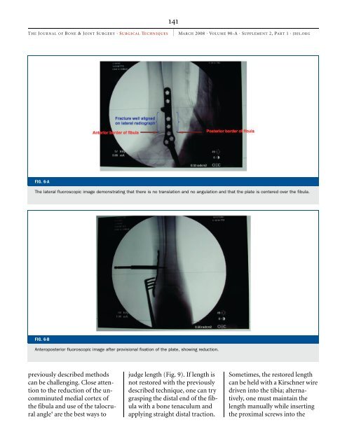
FIG. 6-A<br />
The lateral fluoroscopic image demonstrating that there is no translation and no angulation and that the plate is centered over the fibula.<br />
FIG. 6-B<br />
Anteroposterior fluoroscopic image after provisional fixation <strong>of</strong> the plate, showing reduction.<br />
previously described methods<br />
can be challenging. Close attention<br />
to the reduction <strong>of</strong> the uncomminuted<br />
medial cortex <strong>of</strong><br />
the fibula and use <strong>of</strong> the talocrural<br />
angle 4 are the best ways to<br />
judge length (Fig. 9). If length is<br />
not restored with the previously<br />
described technique, one can try<br />
grasping the distal end <strong>of</strong> the fibula<br />
with a bone tenaculum and<br />
applying straight distal traction.<br />
Sometimes, the restored length<br />
can be held with a Kirschner wire<br />
driven into the tibia; alternatively,<br />
one must maintain the<br />
length manually while inserting<br />
the proximal screws into the