Sex lethal gene initiates female development in germline progenitors
Sex lethal gene initiates female development in germline progenitors
Sex lethal gene initiates female development in germline progenitors

Create successful ePaper yourself
Turn your PDF publications into a flip-book with our unique Google optimized e-Paper software.
Drosophila <strong>Sex</strong> <strong>lethal</strong> Gene Initiates Female Development <strong>in</strong> Germl<strong>in</strong>e<br />
Progenitors<br />
Kazuya Hashiyama et al.<br />
Science 333,<br />
885 (2011);<br />
DOI: 10.1126/science.1208146<br />
This copy is for your personal, non-commercial use only.<br />
If you wish to distribute this article to others,<br />
you can order high-quality copies for your<br />
colleagues, clients, or customers by click<strong>in</strong>g here.<br />
Permission to republish or repurpose articles or portions of articles can be obta<strong>in</strong>ed by<br />
follow<strong>in</strong>g the guidel<strong>in</strong>es here.<br />
The follow<strong>in</strong>g resources related to this article are available onl<strong>in</strong>e at<br />
www.sciencemag.org (this <strong>in</strong>formation is current as of May 16, 2012 ):<br />
Updated <strong>in</strong>formation and services, <strong>in</strong>clud<strong>in</strong>g high-resolution figures, can be found <strong>in</strong> the onl<strong>in</strong>e<br />
version of this article at:<br />
http://www.sciencemag.org/content/333/6044/885.full.html<br />
Support<strong>in</strong>g Onl<strong>in</strong>e Material can be found at:<br />
http://www.sciencemag.org/content/suppl/2011/07/07/science.1208146.DC1.html<br />
A list of selected additional articles on the Science Web sites related to this article can be<br />
found at:<br />
http://www.sciencemag.org/content/333/6044/885.full.html#related<br />
This article cites 30 articles,<br />
8 of which can be accessed free:<br />
http://www.sciencemag.org/content/333/6044/885.full.html#ref-list-1<br />
This article has been cited by 1 articles hosted by HighWire Press; see:<br />
http://www.sciencemag.org/content/333/6044/885.full.html#related-urls<br />
Science (pr<strong>in</strong>t ISSN 0036-8075; onl<strong>in</strong>e ISSN 1095-9203) is published weekly, except the last week <strong>in</strong> December, by the<br />
American Association for the Advancement of Science, 1200 New York Avenue NW, Wash<strong>in</strong>gton, DC 20005. Copyright<br />
2011 by the American Association for the Advancement of Science; all rights reserved. The title Science is a<br />
registered trademark of AAAS.<br />
on May 16, 2012<br />
www.sciencemag.org<br />
Downloaded from
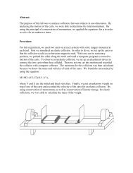
motor doma<strong>in</strong>s at the tail <strong>in</strong>terface should mimic<br />
<strong>in</strong>hibition <strong>in</strong> the absence of tails. Ser 181 residues<br />
<strong>in</strong> the two motor doma<strong>in</strong>s (green <strong>in</strong> Fig. 1A) were<br />
substituted with Cys (<strong>in</strong> a background with the<br />
other reactive cyte<strong>in</strong>es removed). A covalent crossl<strong>in</strong>k<br />
between them could be readily formed by<br />
oxidation to a disulfide and reversed by reduction<br />
with DTT (fig. S6). MT-stimulated adenos<strong>in</strong>e<br />
triphosphatase activity was lost and rega<strong>in</strong>ed <strong>in</strong><br />
parallel with the extent of cross-l<strong>in</strong>k<strong>in</strong>g (fig. S6),<br />
which <strong>in</strong>dicated that cross-l<strong>in</strong>k<strong>in</strong>g strongly <strong>in</strong>hibited<br />
hydrolysis and <strong>in</strong>troduced redox control<br />
of activity. In this preparation, about 10% of<br />
the motor doma<strong>in</strong> monomers were refractory<br />
to cross-l<strong>in</strong>k<strong>in</strong>g.<br />
S<strong>in</strong>gle turnover experiments (Fig. 2A) allow<br />
direct evaluation of the great extent of <strong>in</strong>hibition<br />
of the cross-l<strong>in</strong>ked fraction. In this experiment, a<br />
dimer with S181C substitutions was equilibrated<br />
with the fluorescent ADP analog methylanthraniloyl<br />
ADP (mant-ADP) before oxidation. Mant-<br />
ADP is released from k<strong>in</strong>es<strong>in</strong> with k<strong>in</strong>etics that<br />
are similar to those of unmodified ADP and gives<br />
a fluorescence decrease (13). In the absence of<br />
MTs, ADP release from the un-cross-l<strong>in</strong>ked dimer<br />
is slow (red trace) and further decreased by<br />
cross-l<strong>in</strong>k<strong>in</strong>g (blue trace). MTs greatly accelerate<br />
the rate of mant-ADP release from un-cross-l<strong>in</strong>ked<br />
dimers (black trace), but produces a biphasic<br />
transient with cross-l<strong>in</strong>ked dimers (green trace).<br />
The relative amplitude of the fast phase is small<br />
and similar to the active fraction that is refractory<br />
to cross-l<strong>in</strong>k<strong>in</strong>g <strong>in</strong> this preparation (Fig.<br />
2B). However, the majority of the cross-l<strong>in</strong>ked<br />
preparation exhibits no stimulation of mant-ADP<br />
release by MTs (the slow phase of the green trace<br />
is approximately parallel to the blue trace). Partial<br />
reversal of cross-l<strong>in</strong>k<strong>in</strong>g by DTT (orange trace)<br />
restores the amplitude of the fast phase <strong>in</strong> parallel<br />
to rega<strong>in</strong> of un-cross-l<strong>in</strong>ked prote<strong>in</strong>. Thus, the covalent<br />
cross-l<strong>in</strong>k, which mimics the double lockdown<br />
of the dimer, <strong>in</strong>hibits ADP release from<br />
the motor doma<strong>in</strong>s.<br />
Release of ADP has been hypothesized to be<br />
coupled to neck l<strong>in</strong>ker undock<strong>in</strong>g (11, 12, 14).<br />
Our crystal structure and covalent mimic provide<br />
support for this hypothesis and suggest that<br />
the <strong>in</strong>hibition of ADP release by tails could be<br />
due to block<strong>in</strong>g of neck l<strong>in</strong>ker undock<strong>in</strong>g through<br />
a double lockdown mechanism (Fig. 3). Coupl<strong>in</strong>g<br />
of neck l<strong>in</strong>ker undock<strong>in</strong>g and ADP release<br />
is also supported by the <strong>in</strong>hibition of ADP release<br />
produced by <strong>in</strong>troduc<strong>in</strong>g a cross-l<strong>in</strong>k between<br />
the neck l<strong>in</strong>ker and core motor doma<strong>in</strong><br />
(15). In short, we propose a double lockdown<br />
auto<strong>in</strong>hibition mechanism, whereby cross-l<strong>in</strong>k<strong>in</strong>g<br />
at the coiled-coil and tail <strong>in</strong>terface prevents the<br />
movement of the motor doma<strong>in</strong>s that is needed to<br />
undock the neck l<strong>in</strong>ker and release ADP. This<br />
opens up the possibility that other k<strong>in</strong>es<strong>in</strong>s may be<br />
regulated by a common auto<strong>in</strong>hibitory mechanism.<br />
References and Notes<br />
1. R. D. Vale, T. S. Reese, M. P. Sheetz, Cell 42, 39 (1985).<br />
2. N. Hirokawa, Science 279, 519 (1998).<br />
3. S. A. Endow, F. J. Kull, H. Liu, J. Cell Sci. 123, 3420 (2010).<br />
4. D. D. Hackney, M. F. Stock, Nat. Cell Biol. 2, 257 (2000).<br />
5. M. F. Stock et al., J. Biol. Chem. 274, 14617 (1999).<br />
6. K. J. Verhey, J. W. Hammond, Nat. Rev. Mol. Cell Biol.<br />
10, 765 (2009).<br />
7. K. A. Dietrich et al., Proc. Natl. Acad. Sci. U.S.A. 105,<br />
8938 (2008).<br />
Drosophila <strong>Sex</strong> <strong>lethal</strong> Gene<br />
Initiates Female Development<br />
<strong>in</strong> Germl<strong>in</strong>e Progenitors<br />
Kazuya Hashiyama, 1 Yoshiki Hayashi, 1,2 Satoru Kobayashi 1,2 *<br />
<strong>Sex</strong> determ<strong>in</strong>ation <strong>in</strong> the Drosophila germ l<strong>in</strong>e is regulated by both the sex of the surround<strong>in</strong>g<br />
soma and cell-autonomous cues. How primordial germ cells (PGCs) <strong>in</strong>itiate sexual <strong>development</strong><br />
via cell-autonomous mechanisms is unclear. Here, we demonstrate that, <strong>in</strong> Drosophila, the<br />
<strong>Sex</strong> <strong>lethal</strong> (Sxl) <strong>gene</strong> acts autonomously <strong>in</strong> PGCs to <strong>in</strong>duce <strong>female</strong> <strong>development</strong>. Sxl is transiently<br />
expressed <strong>in</strong> PGCs dur<strong>in</strong>g their migration to the gonads; this expression, which was detected<br />
only <strong>in</strong> XX PGCs, is necessary for PGCs to assume a <strong>female</strong> fate. Ectopic expression of Sxl <strong>in</strong><br />
XY PGCs was sufficient to <strong>in</strong>duce them to enter oo<strong>gene</strong>sis and produce functional eggs when<br />
transplanted <strong>in</strong>to an XX host. Our data provide powerful evidence that Sxl <strong><strong>in</strong>itiates</strong> <strong>female</strong><br />
germl<strong>in</strong>e fate dur<strong>in</strong>g sexual <strong>development</strong>.<br />
Primordial germ cells (PGCs) are able to<br />
differentiate <strong>in</strong>to eggs or sperm. It is thought<br />
that PGCs do not assume a sexual fate until<br />
they reach the gonads, where sexual dimorphism<br />
is imposed by both the sex of the surround<strong>in</strong>g<br />
soma and cell-autonomous cues (1–3). In Drosophila,<br />
pole cells or PGCs differentiate to a male<br />
fate<strong>in</strong>responsetoJAK/STATsignal<strong>in</strong>gfromthe<br />
gonadal soma (4–6). The method by which <strong>female</strong><br />
sexual <strong>development</strong> is <strong>in</strong>itiated <strong>in</strong> pole cells,<br />
however, has not been elucidated. To clarify the<br />
mechanism that <strong><strong>in</strong>itiates</strong> a <strong>female</strong> fate <strong>in</strong> pole<br />
cells, we first identified a <strong>female</strong>-specific marker<br />
for this cell type. Although several sex-specific<br />
markers, <strong>in</strong>clud<strong>in</strong>g mgm-1, disc proliferation abnormal,<br />
and m<strong>in</strong>ichromosome ma<strong>in</strong>tenance 5,<br />
8. D. D. Hackney, N. Baek, A. C. Snyder, Biochemistry 48,<br />
3448 (2009).<br />
9. F. J. Kull, S. A. Endow, J. Cell Sci. 115, 15 (2002).<br />
10. F. Kozielski et al., Cell 91, 985 (1997).<br />
11. W. Hwang, M. J. Lang, M. Karplus, Structure 16, 62<br />
(2008).<br />
12. S. Rice et al., Nature 402, 778 (1999).<br />
13. J. Q. Cheng, W. Jiang, D. D. Hackney, Biochemistry 37,<br />
5288 (1998).<br />
14. C. V. S<strong>in</strong>delar, K. H. Down<strong>in</strong>g, Proc. Natl. Acad. Sci. U.S.A.<br />
107, 4111 (2010).<br />
15. K. Hahlen et al., J. Biol. Chem. 281, 18868 (2006).<br />
Acknowledgments: We thank D. Flot of the European<br />
Synchrotron Radiation Facility and European Molecular<br />
Biology Laboratory–Grenoble, and M. Muller of Swiss<br />
Light Source for assistance and support <strong>in</strong> us<strong>in</strong>g<br />
beaml<strong>in</strong>es ID23-2 and PXI, respectively. We also thank<br />
E. Dodson for valuable crystallography advice, O. Rath for<br />
data collection, V. Ulaganathan for useful discussion,<br />
Y. Yeo and M. Kim for assistance <strong>in</strong> clon<strong>in</strong>g and<br />
preparation of mutant k<strong>in</strong>es<strong>in</strong> motor doma<strong>in</strong>s, and<br />
A. Snyder for assistance <strong>in</strong> preparation of the tail fusion<br />
peptide. H.Y.K.K. holds a National Science Scholarship,<br />
f<strong>in</strong>anced by Agency for Science, Technology, and<br />
Research (A*STAR, S<strong>in</strong>gapore), and this publication<br />
conta<strong>in</strong>s part of her doctoral thesis. We thank Cancer<br />
Research UK, NIH (NS058848) and NSF (MCB-0615549)<br />
for f<strong>in</strong>ancial support. Coord<strong>in</strong>ates and structure factor<br />
files for the dimer and dimer-tail complex have been<br />
deposited <strong>in</strong> the Prote<strong>in</strong> Data Bank under accession<br />
numbers 2Y5W and 2Y65, respectively. The authors<br />
declare no compet<strong>in</strong>g f<strong>in</strong>ancial <strong>in</strong>terests.<br />
Support<strong>in</strong>g Onl<strong>in</strong>e Material<br />
www.sciencemag.org/cgi/content/full/333/6044/883/DC1<br />
Materials and Methods<br />
SOM Text<br />
Figs. S1 to S6<br />
Tables S1 and S2<br />
References (16–38)<br />
25 February 2011; accepted 30 June 2011<br />
10.1126/science.1204824<br />
REPORTS<br />
have been reported, they are all expressed only<br />
<strong>in</strong> male pole cells after gonad formation (stage<br />
15), based on signals from the male gonadal soma<br />
(4, 5, 7). We previously showed that lesswright<br />
(lwr), a <strong>gene</strong> that regulates posttranslational modification<br />
of prote<strong>in</strong>s by small ubiquit<strong>in</strong>-related<br />
modifiers, is expressed <strong>in</strong> pole cells dur<strong>in</strong>g embryo<strong>gene</strong>sis<br />
(8, 9). lwr is not characterized by sexspecific<br />
expression. When a dom<strong>in</strong>ant-negative<br />
form of lwr (lwr DN )(10) was expressed <strong>in</strong> the pole<br />
cells of either sex, however, apoptosis was <strong>in</strong>duced<br />
only <strong>in</strong> <strong>female</strong> (XX) pole cells dur<strong>in</strong>g migration<br />
to the gonads. This effect caused a significant<br />
reduction <strong>in</strong> the number of XX pole cells <strong>in</strong> the<br />
gonads (Fig. 1, fig. S1, and table S1) (11). Introduction<br />
of <strong>female</strong>-specific germl<strong>in</strong>e apoptosis<br />
<strong>in</strong>duced by a dom<strong>in</strong>ant-negative form of lwr<br />
( f-gal ) provides a previously uncharacterized<br />
marker of <strong>female</strong> sexual identity <strong>in</strong> migrat<strong>in</strong>g<br />
pole cells.<br />
1<br />
Okazaki Institute for Integrative Bioscience, National Institute<br />
for Basic Biology, National Institutes of Natural Sciences,<br />
Higashiyama, Myodaiji, Okazaki 444-8787, Japan. 2 Department<br />
of Basic Biology, School of Life Science, Graduate University<br />
for Advanced Studies, Nishigonaka, Myodaiji, Okazaki 444-<br />
8585, Japan.<br />
*To whom correspondence should be addressed. E-mail:<br />
skob@nibb.ac.jp<br />
www.sciencemag.org SCIENCE VOL 333 12 AUGUST 2011 885<br />
on May 16, 2012<br />
www.sciencemag.org<br />
Downloaded from
REPORTS<br />
886<br />
<strong>Sex</strong> determ<strong>in</strong>ation is controlled by the <strong>Sex</strong><br />
<strong>lethal</strong> (Sxl) <strong>gene</strong>, which is first expressed at<br />
the blastodermal stage <strong>in</strong> the embryonic soma<br />
(12–14). Sxl encodes an RNA b<strong>in</strong>d<strong>in</strong>g prote<strong>in</strong><br />
<strong>in</strong>volved <strong>in</strong> alternative splic<strong>in</strong>g and translation.<br />
In the soma of XX embryos, it functions through<br />
transformer (tra)andtransformer-2 (tra-2), which<br />
<strong>in</strong> turn regulate alternative splic<strong>in</strong>g of the doublesex<br />
(dsx) <strong>gene</strong> to produce a <strong>female</strong>-specific form<br />
of Dsx (15, 16). In male (XY) embryos, this<br />
pathway is turned off, and a male-specific form<br />
of Dsx is produced by default (16). These Dsx<br />
prote<strong>in</strong>s determ<strong>in</strong>e the sexual identity of somatic<br />
tissues (15–17). Previous reports, however,<br />
suggested that Sxl does not <strong>in</strong>duce <strong>female</strong><br />
sexual <strong>development</strong> <strong>in</strong> the germ l<strong>in</strong>e, as it does<br />
<strong>in</strong> the soma (18, 19). Although Sxl is autonomously<br />
required for <strong>female</strong> sexual <strong>development</strong><br />
(16, 17, 20), constitutive mutations <strong>in</strong> Sxl (Sxl M )<br />
that cause XY animals to undergo sexual transformation<br />
from male to <strong>female</strong> do not necessarily<br />
<strong>in</strong>terfere with male germl<strong>in</strong>e <strong>development</strong><br />
(18, 19). Moreover, tra, tra-2, anddsx are not required<br />
for <strong>female</strong> germl<strong>in</strong>e <strong>development</strong> (20–22).<br />
F<strong>in</strong>ally, <strong>female</strong>-specific Sxl expression has been<br />
detected later <strong>in</strong> gameto<strong>gene</strong>sis, but not <strong>in</strong> early<br />
germl<strong>in</strong>e <strong>development</strong> (12, 13, 20).<br />
Contrary to previous observations, we found<br />
that Sxl was expressed <strong>in</strong> XX but not XY pole<br />
cells dur<strong>in</strong>g their migration to the gonads (Fig.<br />
2). In the soma, Sxl transcripts are first expressed<br />
from the establishment promoter (Sxl-Pe) <strong>in</strong>a<br />
<strong>female</strong>-specific manner (12, 14). Us<strong>in</strong>g a probe<br />
specific to the early transcript derived from Sxl-<br />
Pe, we detected <strong>in</strong> situ hybridization signals <strong>in</strong><br />
migrat<strong>in</strong>g XX pole cells at around stage 9/10<br />
(Fig. 2, B to E). We used transgenic embryos,<br />
which expressed enhanced green fluorescent<br />
prote<strong>in</strong> (EGFP) under the control of the Sxl-Pe<br />
promoter (Fig. 2, F to I) (23), to further confirm<br />
this <strong>female</strong>-specific Sxl-Pe activation. We used<br />
reverse transcription polymerase cha<strong>in</strong> reaction<br />
and sequenc<strong>in</strong>g analyses <strong>in</strong> pole cells to detect<br />
early Sxl transcripts that had the same sequence<br />
as the transcripts expressed <strong>in</strong> the soma (11).<br />
Next, we determ<strong>in</strong>ed whether Sxl fem<strong>in</strong>ized<br />
early pole cells us<strong>in</strong>g f-gal as a marker for <strong>female</strong><br />
identity. We found that the loss-of-function<br />
mutation Sxl f P7B0 (24) repressed f-gal <strong>in</strong> XX<br />
pole cells (Fig. 1A). This repression is unlikely to<br />
result from sexual transformation of the soma,<br />
because an amorphic tra-2 mutation, which alters<br />
somatic sex (19, 22, 25), did not affect f-gal<br />
(Fig. 1A). Conversely, when we forced the expression<br />
of Sxl together with lwr DN <strong>in</strong> pole cells<br />
from stage 9 onward by us<strong>in</strong>g nanos-Gal4 (26)<br />
and UAS-Sxl (27), f-gal was ectopically observed<br />
<strong>in</strong> XY pole cells (Fig. 1, A, D, and G). We<br />
found that Sxl alone did not <strong>in</strong>duce apoptosis<br />
or <strong>development</strong>al defects <strong>in</strong> pole cells (Fig.<br />
1A) (11). These observations suggest that <strong>female</strong><br />
sexual identity of migrat<strong>in</strong>g pole cells is<br />
regulated cell-autonomously by Sxl.<br />
We then determ<strong>in</strong>ed whether Sxl <strong>in</strong>duced<br />
<strong>female</strong> <strong>development</strong> <strong>in</strong> XY pole cells. Because<br />
XY soma produces signals that direct XX germl<strong>in</strong>e<br />
cells to a male fate (4, 5, 7), we transplanted<br />
XY pole cells express<strong>in</strong>g Sxl <strong>in</strong>to XX <strong>female</strong>s<br />
and exam<strong>in</strong>ed their <strong>development</strong>al fate. Even <strong>in</strong><br />
the presence of a ga<strong>in</strong>-of-function Sxl mutation<br />
(Sxl M1 ) that causes XY soma to transform from<br />
Fig. 1. Female-specific germl<strong>in</strong>e loss <strong>in</strong>duced by a dom<strong>in</strong>ant-negative form of lwr. (A) Averagenumbers<br />
of pole cells <strong>in</strong> the gonads of <strong>female</strong> (magenta) and male (blue) control, lwr DN , Sxl o/e, Sxl o/e lwr DN , Sxl – ,<br />
Sxl – lwr DN , tra-2 – , and tra-2 – lwr DN embryos (stage 15) are shown (29). More than 40 gonads were<br />
exam<strong>in</strong>ed <strong>in</strong> each case. Error bars represent SD. Significance was calculated us<strong>in</strong>g the Student’s t test<br />
(*P 0.05).(B to G) Gonads <strong>in</strong> <strong>female</strong> (B to D) and male (E to G) control (B and E), lwr DN<br />
(C and F), and Sxl o/e lwr DN (D and G) embryos (stage 15) were sta<strong>in</strong>ed for the germl<strong>in</strong>e marker Vasa<br />
(magenta). (H to K) Female (H and I) and male (J and K) control (H and J) and lwr DN (I and K) embryos<br />
(stage 13) were sta<strong>in</strong>ed for Vasa (magenta) and cleaved caspase-3 (green). Arrowheads <strong>in</strong>dicate pole<br />
cells that were positive for cleaved caspase-3. Scale bars, 10 mm.<br />
Fig. 2. Female-specific expression of Sxl <strong>in</strong><br />
migrat<strong>in</strong>g pole cells. (A) Anembryo(stage4)<br />
sta<strong>in</strong>ed for early Sxl transcripts. No specific<br />
signals were observed <strong>in</strong> pole cells (arrowhead).<br />
(Right) Zoomed-<strong>in</strong> view. (B and C) Female<br />
(B) and male (C) embryos (stage 9)<br />
sta<strong>in</strong>ed for early Sxl transcripts. Early transcripts<br />
were detected <strong>in</strong> pole cells only <strong>in</strong> <strong>female</strong>s.<br />
Arrowheads <strong>in</strong>dicate pole cells. (D and E) Confocal images of a <strong>female</strong> embryo (stage 10) double sta<strong>in</strong>ed<br />
for Vasa (D) and early Sxl transcripts (E). (F to I) Female (F and G) and male (H and I) embryos (stage 9)<br />
carry<strong>in</strong>g Sxl-Pe-EGFP were sta<strong>in</strong>ed for Vasa (magenta) (F and H) and GFP (green) (G and I). GFP was<br />
detected <strong>in</strong> pole cells of <strong>female</strong> embryos (arrowheads). Scale bars, 10 mm.<br />
12 AUGUST 2011 VOL 333 SCIENCE www.sciencemag.org<br />
on May 16, 2012<br />
www.sciencemag.org<br />
Downloaded from
male to <strong>female</strong>, XY (or XO) pole cells enter the<br />
spermatogenic pathway when transplanted <strong>in</strong>to<br />
XX <strong>female</strong>s (18, 19). These results suggest that<br />
Sxl is not sufficient to activate <strong>female</strong> germl<strong>in</strong>e<br />
<strong>development</strong>. Sxl M1 mutations, however, do not<br />
affect transcription from the Sxl-Pe promoter,<br />
but <strong>in</strong>stead structurally alter the late transcript<br />
from the Sxl ma<strong>in</strong>tenance promoter (Sxl-Pm),<br />
which allows Sxl prote<strong>in</strong> production <strong>in</strong> both<br />
males and <strong>female</strong>s (28). Consistent with this observation,<br />
we detected Sxl transcripts derived<br />
from Sxl-Pe <strong>in</strong> the pole cells of only <strong>female</strong><br />
Sxl M1 embryos (fig. S7, D and E). Thus, the<br />
Sxl M1 mutation does not result <strong>in</strong> Sxl expression<br />
<strong>in</strong> XY pole cells as early as <strong>in</strong> XX pole cells.<br />
Instead, we used nanos-Gal4 and UAS-Sxl<br />
to <strong>in</strong>duce Sxl expression <strong>in</strong> XY pole cells. We<br />
transplanted three types of XY pole cells, each<br />
characterized by a different duration of Sxl expression:<br />
(i) XY pole cells <strong>in</strong> which Sxl was<br />
expressed from stage 9 until stage 16/17 us<strong>in</strong>g<br />
maternal nanos-Gal4 (XY-mSxl), (ii) XY pole<br />
cells <strong>in</strong> which Sxl was expressed from stage 15/16<br />
onward us<strong>in</strong>g zygotic nanos-Gal4 (XY-zSxl),<br />
and (iii) XY pole cells <strong>in</strong> which Sxl was expressed<br />
from stage 9 onward us<strong>in</strong>g both maternal and<br />
zygotic nanos-Gal4 (XY-mzSxl) (29). We found<br />
that XY-mzSxl and XY-mSxl pole cells entered<br />
the oogenic pathway and produced mature oocytes<br />
<strong>in</strong> XX <strong>female</strong>s (Fig. 3, C and D, and Table 1).<br />
These oocytes contributed to progeny production<br />
(Fig. 3, E to H, and Table 1). Thus, the<br />
XY pole cells produced functional eggs, even<br />
though oo<strong>gene</strong>sis and egg production were reduced<br />
compared with XX pole cells (Table 1).<br />
In contrast, XY-zSxl pole cells did not enter the<br />
oogenic pathway <strong>in</strong> almost all (92.3%) of the<br />
XX <strong>female</strong> hosts (Table 1) and <strong>in</strong>stead were<br />
characterized by a tumorous phenotype, an <strong>in</strong>dication<br />
of XY germl<strong>in</strong>e cells that have ma<strong>in</strong>ta<strong>in</strong>ed<br />
male characteristics (Fig. 3, A and B)<br />
(18). Control XY pole cells from the embryos<br />
express<strong>in</strong>g Sxl only <strong>in</strong> the soma (XY-nullo-Sxl)<br />
showed a similar phenotype to that of XY-zSxl<br />
pole cells (Table 1). These observations demonstrate<br />
that Sxl expression <strong>in</strong> XY pole cells dur<strong>in</strong>g<br />
embryo<strong>gene</strong>sis <strong>in</strong>duces functional egg differentiation<br />
<strong>in</strong> the <strong>female</strong> soma.<br />
We then used Sxl-specific double-stranded<br />
RNA (UAS-Sxl RNAi ) under the control of maternal<br />
nanos-Gal4 to reduce Sxl activity <strong>in</strong> XX<br />
pole cells dur<strong>in</strong>g embryo<strong>gene</strong>sis. Introduc<strong>in</strong>g<br />
UAS-Sxl RNAi resulted <strong>in</strong> tumorous and agametic<br />
phenotypes <strong>in</strong> <strong>female</strong> adults, <strong>in</strong>dicat<strong>in</strong>g that the<br />
XX germ l<strong>in</strong>e lost <strong>female</strong> characteristics (fig. S8)<br />
(18, 19). Taken together, our results show that Sxl<br />
acts as a master <strong>gene</strong> necessary and sufficient to<br />
<strong>in</strong>duce <strong>female</strong> <strong>development</strong> <strong>in</strong> pole cells.<br />
We found that XY-mzSxl pole cells adopted<br />
a male fate and executed spermato<strong>gene</strong>sis when<br />
they developed <strong>in</strong> an XY male soma (Fig. 3, I<br />
and J, and fig. S9). This observation suggests that<br />
themalesomaplaysadom<strong>in</strong>antrole<strong>in</strong>determ<strong>in</strong><strong>in</strong>g<br />
the male germl<strong>in</strong>e fate, overrid<strong>in</strong>g the fem<strong>in</strong>iz<strong>in</strong>g<br />
effect of Sxl. Another possibility is that<br />
the XX <strong>female</strong> soma plays a critical role <strong>in</strong> ma<strong>in</strong>ta<strong>in</strong><strong>in</strong>g<br />
the Sxl-<strong>in</strong>itiated <strong>female</strong> germl<strong>in</strong>e fate.<br />
Indeed, an XX germ l<strong>in</strong>e <strong>in</strong> the male soma shows<br />
a male <strong>gene</strong>-expression profile, whereas an XY<br />
germ l<strong>in</strong>e <strong>in</strong> the <strong>female</strong> soma exhibits a <strong>female</strong><br />
expression profile, although these germ l<strong>in</strong>es<br />
does not execute gameto<strong>gene</strong>sis (4, 30). Thus,<br />
<strong>female</strong> germl<strong>in</strong>e <strong>development</strong> requires <strong>in</strong>teractions<br />
between the germl<strong>in</strong>e and somatic cells, <strong>in</strong><br />
addition to germl<strong>in</strong>e-autonomous mechanisms<br />
<strong>in</strong>volv<strong>in</strong>g Sxl.<br />
In mice, germl<strong>in</strong>e sexual identity is also regulated<br />
by both germl<strong>in</strong>e-autonomous and somatic<br />
signals (6, 7). In the coelenterate Hydra,<br />
the germl<strong>in</strong>e sex is not <strong>in</strong>fluenced by the surround<strong>in</strong>g<br />
soma, and the germ l<strong>in</strong>e determ<strong>in</strong>es<br />
the phenotypic sex of the polyp (31, 32). Thus,<br />
germl<strong>in</strong>e-autonomous regulation of sex has probably<br />
been present throughout the evolution of<br />
animals, and somatic control may have evolved<br />
with the emergence of mesodermal tissues, <strong>in</strong>clud<strong>in</strong>g<br />
gonadal soma. Sxl does not appear to play a<br />
key role <strong>in</strong> sex determ<strong>in</strong>ation <strong>in</strong> non-drosophilid<br />
Table 1. Sxl <strong>in</strong>duced <strong>female</strong> fate <strong>in</strong> XY pole cells. Pole cell transplantation experiments are<br />
described <strong>in</strong> the support<strong>in</strong>g onl<strong>in</strong>e material (29).<br />
Donors<br />
No. of<br />
transplanted<br />
embryos<br />
No. of <strong>female</strong><br />
adults with<br />
transplanted<br />
germl<strong>in</strong>e cells<br />
No. of <strong>female</strong><br />
adults with<br />
transplanted<br />
germl<strong>in</strong>e cells<br />
execut<strong>in</strong>g oo<strong>gene</strong>sis*<br />
REPORTS<br />
No. of <strong>female</strong><br />
adults with<br />
transplanted<br />
germl<strong>in</strong>e cells<br />
produc<strong>in</strong>g progeny<br />
XY 203 13 0 0<br />
XY-zSxl 141 13 1† 1<br />
XY-mzSxl 250 18 13‡ 8<br />
XY-mSxl 181 21 8‡ 5<br />
XY-nullo-Sxl 152 15 0 0<br />
XX 242 25 25 25<br />
*Females with egg chambers (stages 3 to 10) derived from donor pole cells were counted. The number of egg chambers derived from<br />
transplanted XX pole cells <strong>in</strong> each ovary was 1 or 2, which was identical to that observed <strong>in</strong> <strong>female</strong>s transplanted with XY-mzSxl or<br />
XY-mSxl pole cells. †P ≥ 0.5; significance was calculated versus XY donors us<strong>in</strong>g Fisher’s exact probability test. ‡P UAS-Sxl) (J)males.Innos-Gal4>UAS-Sxl testes, spermato<strong>gene</strong>sis proceeded<br />
normally,andspermwasproduced.Scalebars,40mm.<br />
www.sciencemag.org SCIENCE VOL 333 12 AUGUST 2011 887<br />
on May 16, 2012<br />
www.sciencemag.org<br />
Downloaded from
REPORTS<br />
888<br />
animals (15, 17). Nevertheless, future studies<br />
should determ<strong>in</strong>e whether Sxl homologs are expressed<br />
<strong>in</strong> the germ l<strong>in</strong>e of non-drosophilids.<br />
Moreover, it would be of particular <strong>in</strong>terest to<br />
identify downstream targets of Sxl <strong>in</strong> the Drosophila<br />
germ l<strong>in</strong>e and to test whether these <strong>gene</strong>s have a<br />
widespread role <strong>in</strong> germl<strong>in</strong>e sex determ<strong>in</strong>ation.<br />
References and Notes<br />
1. T. DeFalco, B. Capel, Annu. Rev. Cell Dev. Biol. 25,<br />
457 (2009).<br />
2. G. Durcova-Hills, B. Capel, Curr. Top. Dev. Biol. 83,<br />
185 (2008).<br />
3. S. M. Murray, S. Y. Yang, M. Van Doren, Curr. Op<strong>in</strong>.<br />
Cell Biol. 22, 722 (2010).<br />
4. M. Wawersik et al., Nature 436, 563 (2005).<br />
5. A. L. Casper, M. Van Doren, Development 136,<br />
3821 (2009).<br />
6. M. Wawersik, Cell Cycle 5, 1385 (2006).<br />
7. A. Casper, M. Van Doren, Development 133,<br />
2783 (2006).<br />
8. S. Shigenobu, Y. Kitadate, C. Noda, S. Kobayashi,<br />
Proc. Natl. Acad. Sci. U.S.A. 103, 13728 (2006).<br />
9. K. Hashiyama, S. Shigenobu, S. Kobayashi, Gene Expr.<br />
Patterns 9, 50 (2009).<br />
10. L. Huang, S. Ohsako, S. Tanda, Dev. Biol. 280,<br />
407 (2005).<br />
11. Support<strong>in</strong>g onl<strong>in</strong>e text is available on Science Onl<strong>in</strong>e.<br />
12. D. Bopp, L. R. Bell, T. W. Cl<strong>in</strong>e, P. Schedl, Genes Dev. 5,<br />
403 (1991).<br />
13. L. N. Keyes, T. W. Cl<strong>in</strong>e, P. Schedl, Cell 68,<br />
933 (1992).<br />
14. H. K. Salz, J. W. Erickson, Fly (Aust<strong>in</strong>) 4, 60<br />
(2010).<br />
15. L. O. Penalva, L. Sánchez, Microbiol. Mol. Biol. Rev.<br />
67, 343 (2003).<br />
16. N. Camara, C. Whitworth, M. Van Doren, Curr. Top.<br />
Dev. Biol. 83, 65 (2008).<br />
17. L. Sánchez, Int. J. Dev. Biol. 52, 837 (2008).<br />
18. M. Ste<strong>in</strong>mann-Zwicky, H. Schmid, R. Nöthiger, Cell 57,<br />
157 (1989).<br />
19. M. Ste<strong>in</strong>mann-Zwicky, Dev. Genet. 15, 265 (1994).<br />
20. B. Oliver, Int. Rev. Cytol. 219, 1 (2002).<br />
21. J. L. Marsh, E. Wieschaus, Nature 272, 249 (1978).<br />
22. T. Schüpbach, Dev. Biol. 89, 117 (1982).<br />
23. L. U. Hempel, B. Oliver, BMC Dev. Biol. 7, 113<br />
(2007).<br />
24. G. Deshpande, G. Calhoun, J. L. Yanowitz, P. D. Schedl,<br />
Cell 99, 271 (1999).<br />
25. W. Mattox, M. E. McGuff<strong>in</strong>, B. S. Baker, Genetics 143,<br />
303 (1996).<br />
26. M. Van Doren, A. L. Williamson, R. Lehmann, Curr. Biol.<br />
8, 243 (1998).<br />
27. J. I. Horab<strong>in</strong>, Development 132, 4801 (2005).<br />
28. M. Bernste<strong>in</strong>, R. A. Lersch, L. Subrahmanyan, T. W. Cl<strong>in</strong>e,<br />
Genetics 139, 631 (1995).<br />
Nicot<strong>in</strong>ic Acetylchol<strong>in</strong>e Receptor b2<br />
Subunits <strong>in</strong> the Medial Prefrontal<br />
Cortex Control Attention<br />
Kar<strong>in</strong>e Guillem, 1 Bernard Bloem, 1 * Rogier B. Poorthuis, 1 * Maarten Loos, 2 August B. Smit, 2<br />
Uwe Maskos, 3,4 Sab<strong>in</strong>e Spijker, 2 † Huibert D. Mansvelder 1 †‡<br />
More than one-third of all people are estimated to experience mild to severe cognitive<br />
impairment as they age. Acetylchol<strong>in</strong>e (ACh) levels <strong>in</strong> the bra<strong>in</strong> dim<strong>in</strong>ish with ag<strong>in</strong>g, and<br />
nicot<strong>in</strong>ic ACh receptor (nAChR) stimulation is known to enhance cognitive performance. The<br />
prefrontal cortex (PFC) is <strong>in</strong>volved <strong>in</strong> a range of cognitive functions and is thought to mediate<br />
attentional focus. We found that mice carry<strong>in</strong>g nAChR b2-subunit deletions have impaired<br />
attention performance. Efficient lentiviral vector–mediated reexpression of functional<br />
b2-subunit–conta<strong>in</strong><strong>in</strong>g nAChRs <strong>in</strong> PFC neurons of the prelimbic area (PrL) completely restored<br />
the attentional deficit but did not affect impulsive and motivational behavior. Our f<strong>in</strong>d<strong>in</strong>gs<br />
show that b2-subunit expression <strong>in</strong> the PrL PFC is sufficient for endogenous nAChR-mediated<br />
chol<strong>in</strong>ergic regulation of attentional performance.<br />
Cortical acetylchol<strong>in</strong>e (ACh) release from<br />
the basal forebra<strong>in</strong> is essential for proper<br />
sensory process<strong>in</strong>g and cognition (1–3)<br />
and tunes neuronal and synaptic activity <strong>in</strong> the<br />
1<br />
Department of Integrative Neurophysiology, Center for Neurogenomics<br />
and Cognitive Research (CNCR), Neuroscience Campus<br />
Amsterdam, VU University, 1081 HV Amsterdam, Netherlands.<br />
2<br />
Department of Molecular and Cellular Neurobiology, CNCR,<br />
Neuroscience Campus Amsterdam, VU University, 1081 HV<br />
Amsterdam, Netherlands. 3 Unité Neurobiologie Intégrative<br />
des Systèmes Chol<strong>in</strong>ergiques, Département de Neuroscience,<br />
Institut Pasteur, F-75724 Paris cedex 15, France. 4 CNRS,<br />
URA2182, F-75724 Paris cedex 15, France.<br />
*These authors contributed equally to this work.<br />
†These authors contributed equally to this work.<br />
‡To whom correspondence should be addressed. E-mail:<br />
huibert.mansvelder@cncr.vu.nl<br />
underly<strong>in</strong>g cortical networks (4, 5). Loss of<br />
chol<strong>in</strong>ergic function dur<strong>in</strong>g ag<strong>in</strong>g and Alzheimer’s<br />
disease results <strong>in</strong> cognitive decl<strong>in</strong>e,<br />
notably a loss of memory and the ability to susta<strong>in</strong><br />
attention (6, 7). Interfer<strong>in</strong>g with the chol<strong>in</strong>ergic<br />
system strongly affects cognition (3, 8–13).<br />
Rapid changes <strong>in</strong> prefrontal cortical ACh levels<br />
at the scale of seconds are correlated with<br />
attend<strong>in</strong>g and detect<strong>in</strong>g cues (14, 15). Various<br />
types of nicot<strong>in</strong>ic ACh receptor (nAChR) subunits<br />
are expressed <strong>in</strong> the prefrontal cortex (PFC)<br />
(16–18), and <strong>in</strong> particular nAChRs conta<strong>in</strong><strong>in</strong>g<br />
b2 subunits are thought to enhance attention (13).<br />
However, the causal relation between nAChR b2<br />
subunits (henceforth b2*-nAChRs) expressed <strong>in</strong><br />
the medial PFC (mPFC) and attention performance<br />
has not yet been demonstrated.<br />
12 AUGUST 2011 VOL 333 SCIENCE www.sciencemag.org<br />
29. Materials and methods are available as support<strong>in</strong>g<br />
material on Science Onl<strong>in</strong>e.<br />
30. L. U. Hempel, R. Kalamegham, J. E. Smith III, B. Oliver,<br />
Curr. Top. Dev. Biol. 83, 109 (2008).<br />
31. C. L. Littlefield, Dev. Biol. 102, 426 (1984).<br />
32. R. D. Campbell, J. Exp. Zool. 234, 451 (1985).<br />
Acknowledgments: We thank the researchers who provided<br />
us with flies and antibodies and members of our<br />
laboratory for their valuable comments. We also thank<br />
the Drosophila Genetic Resource Center (Kyoto), the<br />
Bloom<strong>in</strong>gton and Vienna Drosophila RNAi Stock Centers,<br />
and the Developmental Studies Hybridoma Bank for fly<br />
stocks and antibodies. This work was supported <strong>in</strong> part by<br />
a Grant-<strong>in</strong>-Aid for Scientific Research from the M<strong>in</strong>istry of<br />
Education, Culture, Sports, Science and Technology<br />
(Japan) to S.K. and a Research Fellowship for Young<br />
Scientists from the Japan Society for the Promotion of<br />
Science to K.H.<br />
Support<strong>in</strong>g Onl<strong>in</strong>e Material<br />
www.sciencemag.org/cgi/content/full/science.1208146/DC1<br />
Materials and Methods<br />
SOM Text<br />
Figs. S1 to S9<br />
Tables S1 to S5<br />
References<br />
10 May 2011; accepted 20 June 2011<br />
Published onl<strong>in</strong>e 7 July 2011;<br />
10.1126/science.1208146<br />
We first determ<strong>in</strong>ed whether absence of nicot<strong>in</strong>ic<br />
b2 subunits affects attentional behavior<br />
<strong>in</strong> the five-choice serial reaction time task (5-<br />
CSRTT), a well-established test setup that taxes<br />
various aspects of attentional control over performance<br />
(19). Mice lack<strong>in</strong>g b2 subunits of<br />
nAChRs (b2 −/− ) and their wild-type littermates<br />
(WT) were tra<strong>in</strong>ed to detect and respond to a<br />
brief light stimulus randomly presented <strong>in</strong> one<br />
of five nose poke holes to receive a food pellet<br />
(20). b2 −/− mice showed normal locomotor activity<br />
<strong>in</strong> an open field test (fig. S1), normal sensorimotor<br />
gat<strong>in</strong>g <strong>in</strong> a prepulse <strong>in</strong>hibition test<br />
(fig. S2), and normal acquisition <strong>in</strong> the 5-CSRTT<br />
(fig. S3). After complete acquisition of the 5-<br />
CSRTT, animals were tra<strong>in</strong>ed at the stimulus<br />
duration of 1 s (SD1) for 10 more days until they<br />
reached stable performance (fig. S4). Basel<strong>in</strong>e<br />
5-CSRTT performance was then calculated from<br />
the 6th until the 10th session at SD1 (Fig. 1, A<br />
and B). b2 −/− mice exhibited significantly more<br />
omissions than their WT littermates [F(1,27) =<br />
12.45; P < 0.01] (Fig. 1A), whereas the level of<br />
accuracy was not significantly different [F(1,27) =<br />
2.56; not significant (NS)] (Fig. 1B). We found<br />
no effect of genotype on any other measures,<br />
such as number of <strong>in</strong>itiated trials [F(1,27) = 1.99;<br />
NS], number of premature responses [F(1,27) =<br />
0.003; NS], correct response latency [F(1,27) =<br />
2.03; NS], or latency to collect earned food pellets<br />
[F(1,27) = 0.12; NS] (table S1), suggest<strong>in</strong>g<br />
that <strong>in</strong>creased omissions reflected impairments<br />
<strong>in</strong> stimulus detection processes <strong>in</strong> b2 −/− mice<br />
rather than motor or motivational deficits. b2 −/−<br />
mice and their WT littermates did not differ <strong>in</strong><br />
the number of food pellets earned by respond<strong>in</strong>g<br />
to a s<strong>in</strong>gle cue light nor <strong>in</strong> the maximal number<br />
of responses <strong>in</strong> a progressive ratio for earn<strong>in</strong>g<br />
food pellets (fig. S5). In contrast to b2 −/− mice,<br />
on May 16, 2012<br />
www.sciencemag.org<br />
Downloaded from