Lacrimal Canaliculitis, A Case Report - KSOS
Lacrimal Canaliculitis, A Case Report - KSOS
Lacrimal Canaliculitis, A Case Report - KSOS

You also want an ePaper? Increase the reach of your titles
YUMPU automatically turns print PDFs into web optimized ePapers that Google loves.
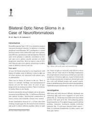
422 Kerala Journal of Ophthalmology Vol. XX, No. 4<br />
Fig. 4. showing positive<br />
beaded, coccoid, thin<br />
branching filaments<br />
on gram staining<br />
positive beaded, coccoid, thin branching filaments.<br />
(Fig.4) The patient recovered rapidly and was left with<br />
a slit punctum and adjoining canaliculus. The remaining<br />
portion of the lower canaliculus was intact and was<br />
patent. Post-operatively, the swelling, conjunctivitis and<br />
discharge disappeared (Fig.5).<br />
Discussion<br />
Fig. 5. P ost-operative<br />
appearance of the eye<br />
Actinomyces israelii species is a gram-positive, castforming,<br />
non–acid-fast, non–spore-forming anaerobic<br />
bacillus. Its filamentous growth and mycelia like<br />
colonies have a striking resemblance to fungi. They are<br />
soil organisms, often found in decaying organic matter<br />
(eg, wet hay, straw). It is primarily a commensal<br />
microbe found in normal oral cavities, in tonsillar<br />
crypts, in dental plaques, and in caries teeth and enters<br />
the lacrimal system through the nasal passage or<br />
indirectly by means of saliva into the conjunctiva. The<br />
anaerobic environment also helps in the growth of the<br />
Actinomyces in the canaliculus.<br />
Other ocular manifestations include keratitis,<br />
conjunctivitis, blepharitis, dacryocystitis, postsurgical<br />
endophthalmitis, and infected porous orbital implant.<br />
Cervico-facial actinomycosis has also been reported.<br />
<strong>Canaliculitis</strong> usually presents as chronic watering,<br />
redness and discharge from eye. A pouted punctum is<br />
clinically diagnostic, although it occurs in less than<br />
50 % of all patients who are affected. Typically, the<br />
discharge is particulate and contains concretions.The<br />
plica may be swollen and congested, and canalicular<br />
swelling and overlying lid erythema are often<br />
present.The lower lid is more commonly affected, and<br />
the lacrimal sac and the duct are usually not<br />
involved.The disease is most commonly unilateral.<br />
Among the reported cases almost all were unilateral,<br />
involving the lower lid. Bilateral and upperlid<br />
involvement as in this case is a rare presentation.<br />
Concretions on the lacrimal canaliculus can also be due<br />
to Candida albicans , Aspergillus niger , Fusobacterium<br />
species, and Nocardia asteroids.<br />
Lab Studies<br />
Canalicular discharge and canaliculiths may be sent for<br />
the following studies:<br />
Gram stain/Giemsa stain<br />
Culture and sensitivity (ie, blood agar, Sabouraud,<br />
anaerobic media)<br />
Special stains (ie, calcofluor white)<br />
Treatment<br />
Actinomycetes are usually susceptible to penicillins and<br />
cephalosporins.<br />
Surgical Care<br />
Failure of resolution of canaliculitis by topical treatment<br />
necessitates surgical exploration of the canalicular<br />
system and removal of any casts. Extensive surgery is<br />
not always required. A 2-snip punctoplasty, cast<br />
removal, curettage, and probing is usually done.<br />
Subsequent lacrimal irrigation with 1 MU of penicillin<br />
in 10 mL of sterile water may be helpful.<br />
References<br />
1. Jordan DR. Dacryoadenitis, Dacryocystitis, and<br />
<strong>Canaliculitis</strong>, chapter 57. In: Cornea- Cornea and<br />
External Disease: Clinical Diagnosis and Management.<br />
Krachmer JH, Mannis MJ, Holland EJ, Eds. (St. Louis,<br />
Mosby). 1997;687-693.<br />
2. Richards WW. Actinomycotic lacrimal canaliculitis.<br />
American J Ophthalmol 1975;75:155-157.<br />
3. Pine L, Hardin H, Turner L, Roberts SS. Actinomycotic<br />
lacrimal canaliculitis - A report of two cases with a<br />
review of the characteristics which identify the causal<br />
organism. American J Ophthalmol 1960;49:1278-1298.<br />
4. Sridhar MS, Gopinathan U, Garg P, Sharma S, Rao GN.<br />
Ocular Nocardia infections with special emphasis on<br />
the cornea. Surv Ophthalmol 2001;45:361-378.<br />
5. Sharma S. Ocular Microbiology. 1st ed. (Aravind Eye<br />
Hospitals Publication, Madurai) 1988:79-84.