PhRC NEWSLETTER PHOTONICS'La - Nanyang Technological ...
PhRC NEWSLETTER PHOTONICS'La - Nanyang Technological ...
PhRC NEWSLETTER PHOTONICS'La - Nanyang Technological ...

Create successful ePaper yourself
Turn your PDF publications into a flip-book with our unique Google optimized e-Paper software.
FEATURE<br />
ARTICLE<br />
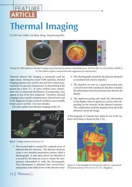
Thermal Imaging<br />
Fu Chit Yaw, Eddie Tan Khay Ming, Yang Kwang Wei<br />
During the SARS epidemic, thermal imaging camera has become almost a household name. Just how does it work and how reliable is<br />
it? The authors explain, and provide some suggestions for improvement.<br />
Thermal infrared (IR) imaging is commonly used for<br />
night vision. During the recent SARS epidemic, thermal<br />
imaging cameras were used for fast and non-contact measurement<br />
of a person’s temperature to determining if the<br />
person has a fever [1]. In many medical cases, temperature<br />
rise or abnormal distribution of temperature may<br />
appear as one of the first symptoms. Therefore, thermal<br />
imaging is also a useful complementary clinical tool to aid<br />
in the diagnosis of other medical condition, most notably<br />
breast cancer (see Ref. 2 for more details).<br />
Let’s first explore how thermal imaging works [3]:<br />
Figure 1: Image courtesy of Infrared, Inc.<br />
1. The focused light is scanned by a phased array of<br />
infrared-detector elements. The detector elements<br />
create a very detailed temperature pattern called a<br />
thermograph. It only takes about one-thirtieth of<br />
a second for the detector array to obtain the temperature<br />
information to make the thermograph.<br />
This information is obtained from several thousand<br />
points in the field of view of the detector array.<br />
22 hotonics'a<br />
2. The thermograph created by the detector elements<br />
is translated into electric impulses.<br />
3. The impulses are sent to a signal-processing unit,<br />
a circuit board with a dedicated chip that translates<br />
the information from the elements into data for the<br />
display.<br />
4. The signal-processing unit sends the information<br />
to the display, where it appears as various colors depending<br />
on the intensity of the infrared emission.<br />
The combination of all the impulses from all of the<br />
elements creates the image.<br />
A thermograph of a human face, taken by one of the authors<br />
(Joel Yang) is shown in Fig. 2 [4].<br />
Figure 2: A thermograph of a human face take by a commercial<br />
IR camera [courtesy Photonitech Pte Ltd., Singapore]