The Relationship Between the Curve of Spee ... - ResearchGate
The Relationship Between the Curve of Spee ... - ResearchGate
The Relationship Between the Curve of Spee ... - ResearchGate
You also want an ePaper? Increase the reach of your titles
YUMPU automatically turns print PDFs into web optimized ePapers that Google loves.
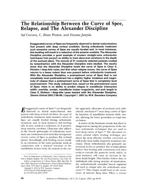
<strong>The</strong> <strong>Relationship</strong> <strong>Between</strong> <strong>the</strong> <strong>Curve</strong> <strong>of</strong> <strong>Spee</strong>,<br />
Relapse, and <strong>The</strong> Alexander Discipline<br />
Sal Carcara, C. Brian Preston, and Ossama Jureyda<br />
E<br />
Exaggerated curves <strong>of</strong> <strong>Spee</strong> are frequently observed in dental malocclusions<br />
that present with deep vertical overbites. During orthodontic treatment<br />
such excessive curves <strong>of</strong> <strong>Spee</strong> are usually leveled and, in most instances,<br />
this leveling will result in a reduction <strong>of</strong> <strong>the</strong> anterior overbite. <strong>The</strong> Alexander<br />
Discipline provides a good example <strong>of</strong> modern straight-wire orthodontic<br />
techniques that purport an ability to treat abnormal variations in <strong>the</strong> depth<br />
<strong>of</strong> <strong>the</strong> occlusal plane. <strong>The</strong> records <strong>of</strong> 31 randomly selected patients treated<br />
by nonextraction with <strong>the</strong> Alexander Discipline were studied. <strong>The</strong> results<br />
show that <strong>the</strong> Alexander Discipline levels <strong>the</strong> curve <strong>of</strong> <strong>Spee</strong> in Class II,<br />
Division I deep-bite cases and that when relapse occurs, <strong>the</strong> curve <strong>of</strong> <strong>Spee</strong><br />
returns to a lesser extent than was present before orthodontic treatment.<br />
With <strong>the</strong> Alexander Discipline, a pretreatment curve <strong>of</strong> <strong>Spee</strong> that is not<br />
completely level posttreatment has a slightly higher incidence and magni-<br />
tude <strong>of</strong> relapse than a pretreatment curve <strong>of</strong> <strong>Spee</strong> that is completely level<br />
posttreatment. This study indicated that, based on <strong>the</strong> pretreatment curve<br />
<strong>of</strong> <strong>Spee</strong>, <strong>the</strong>re is no ability to predict relapse in mandibular intercanine<br />
width, overbite, overjet, mandibular incisor irregularity, and arch length in<br />
Class II, Division I deep-bite cases treated with <strong>the</strong> Alexander Discipline.<br />
(Semin Orthod 2001;7:90-99.) Copyright © 2001 by W.B. Saunders Company<br />
xaggerated curves <strong>of</strong> <strong>Spee</strong> 1,2 are frequently<br />
observed in dental malocclusions that<br />
present with deep vertical overbites. As a part <strong>of</strong><br />
orthodontic treatment, such excessive curves <strong>of</strong><br />
<strong>Spee</strong> are usually leveled during orthodontic<br />
treatment, and, in most instances, this leveling<br />
will, in turn, result in a reduction <strong>of</strong>, if present,<br />
deep anterior overbites. Clinicians who adhere<br />
to <strong>the</strong> Tweed I philosophy <strong>of</strong> orthodontic treatment<br />
use continuous arch wires that incorporate<br />
reverse curves <strong>of</strong> <strong>Spee</strong> to produce flat occlusal<br />
planes. Accordingly, arch leveling occurs mostly<br />
by an extrusion <strong>of</strong> <strong>the</strong> lower premolar teeth in<br />
conjunction with a minimal intrusion <strong>of</strong> <strong>the</strong><br />
mandibular incisor teeth. In contrast to <strong>the</strong> ear-<br />
From <strong>the</strong> Department <strong>of</strong> Orthodontics, State University <strong>of</strong> New<br />
York at Buffalo, Buffizlo, NY," and Private practice, New York, NY.<br />
Address correspondence to C Brian Preston, BDS, Dip Orth, M<br />
Dent, PhD, Department <strong>of</strong> Orthodontics, School <strong>of</strong> Dental Medicine,<br />
Squire Hall, 3435 Main St, Buffalo, NY 14214-3008.<br />
Copyright Q 2001 by 14(B. Saunders Company<br />
1073-8746/01/0702-0006535. 00/0<br />
doi: 10.1053/sodo. 2001.23550<br />
lier approach, advocates <strong>of</strong> sectional arch orth-<br />
odontic mechanics z,~ treat deep curves <strong>of</strong> <strong>Spee</strong><br />
by intrusion <strong>of</strong> mandibular incisors while, usu-<br />
ally, allowing <strong>the</strong> lower premolars to erupt into<br />
occlusion.<br />
A review <strong>of</strong> <strong>the</strong> literature reveals that <strong>the</strong>re is<br />
disagreement among <strong>the</strong> proponents <strong>of</strong> <strong>the</strong> var-<br />
ious orthodontic techniques that are used to<br />
level deep curves <strong>of</strong> <strong>Spee</strong>. 4-s <strong>The</strong> discussion re-<br />
volves around which leveling technique pro-<br />
duces <strong>the</strong> most effective overbite correction as<br />
well as <strong>the</strong> most stable long-term treatment out-<br />
comes. Those orthodontists who primarily use<br />
sectional arches to produce fiat occlusal planes<br />
believe that leveling with continuous arch wires<br />
tends to extrude <strong>the</strong> posterior teeth, which, in<br />
most instances, will cause an increase in <strong>the</strong><br />
lower facial height. <strong>The</strong>y fur<strong>the</strong>r believe that, in<br />
individuals with strong muscles <strong>of</strong> mastication,<br />
<strong>the</strong> orthodontically extruded buccal segments<br />
will tend to relapse after <strong>the</strong> orthodontic treat-<br />
ment. 4,5,9 This relapse would lead to <strong>the</strong> recur-<br />
rence <strong>of</strong> anterior deep bites. When a reverse<br />
90 Seminars in Orthodontics, Vol 7, No 2 ([une), 2001: pp 90-99
curve <strong>of</strong> <strong>Spee</strong> is placed in a continuous arch wire<br />
for <strong>the</strong> purpose <strong>of</strong> arch leveling, this results in<br />
an almost automatic tendency for <strong>the</strong> mandibu-<br />
lar incisors to flare labially. 1°,11 Contrary to this<br />
viewpoint, Ferguson a2 states that a reverse curve<br />
<strong>of</strong> <strong>Spee</strong> in an arch wire does not in itself cause<br />
<strong>the</strong> lower incisor teeth to flare unless <strong>the</strong> arch is<br />
allowed to act beyond <strong>the</strong> stage at which <strong>the</strong><br />
occlusal plane is flat. Orthodontists who use<br />
Tweed's leveling technique 6,13-15 argue that <strong>the</strong><br />
extrusion <strong>of</strong> premolars and molars provides a<br />
stable change while, on <strong>the</strong> o<strong>the</strong>r hand, <strong>the</strong><br />
intrusion <strong>of</strong> <strong>the</strong> lower incisors <strong>of</strong>ten relapses to<br />
produce an increased overbite.<br />
Radiographic cephalometric studies showed<br />
that both <strong>the</strong> Ricketts and modified Tweed tech-<br />
niques can successfully correct deep dental over-<br />
bites. 16,a7 <strong>The</strong>se studies concentrated on over-<br />
bite correction only and nei<strong>the</strong>r analyzed study<br />
models to evaluate how effectively <strong>the</strong> curves <strong>of</strong><br />
<strong>Spee</strong> were leveled, nor did <strong>the</strong>y evaluate <strong>the</strong><br />
long-term stability <strong>of</strong> <strong>the</strong> results that were pro-<br />
duced.<br />
<strong>The</strong> present article reflects <strong>the</strong> findings <strong>of</strong> a<br />
study that was designed to evaluate <strong>the</strong> long-<br />
term outcomes <strong>of</strong> a representative sample <strong>of</strong><br />
orthodontic patients who were treated accord-<br />
ing to <strong>the</strong> Alexander Discipline. Irrespective <strong>of</strong><br />
<strong>the</strong> philosophy and mechanical principles <strong>of</strong> <strong>the</strong><br />
orthodontic technique used, one <strong>of</strong> <strong>the</strong> primary<br />
objectives <strong>of</strong> orthodontic treatment is to obtain a<br />
level occlusal plane, is In this article, leveling will<br />
be defined as <strong>the</strong> process <strong>of</strong> bringing <strong>the</strong> incisal<br />
edges <strong>of</strong> <strong>the</strong> anterior teeth and <strong>the</strong> buccal cusp<br />
tips <strong>of</strong> <strong>the</strong> posterior teeth into <strong>the</strong> same horizon-<br />
tal plane. 19<br />
<strong>The</strong> anatomic definition <strong>of</strong> <strong>the</strong> anteroposte-<br />
rior curve <strong>of</strong> occlusion is generally accepted by<br />
orthodontists as describing <strong>the</strong> curve <strong>of</strong><br />
<strong>Spee</strong>. 2°-94 Some studies in <strong>the</strong> orthodontic liter-<br />
ature propose o<strong>the</strong>r ways to define and measure<br />
<strong>the</strong> curve <strong>of</strong> <strong>Spee</strong> on orthodontic study mod-<br />
els. 25-27 Three-dimensional digitizers 2-~,26 have<br />
been used to calculate <strong>the</strong> depth <strong>of</strong> <strong>the</strong> mandib-<br />
ular curve <strong>of</strong> <strong>Spee</strong> ma<strong>the</strong>matically. Koyama, 27 in<br />
a more practical approach to <strong>the</strong> problem, used<br />
a caliper to measure <strong>the</strong> curvature <strong>of</strong> <strong>the</strong> occlu-<br />
sal plane in both jaws and found <strong>the</strong> greatest<br />
pretreatment depth <strong>of</strong> <strong>the</strong> curvature to be lo-<br />
cated in <strong>the</strong> bicuspid region.<br />
In a mechanical sense, <strong>the</strong> presence <strong>of</strong> a<br />
curve <strong>of</strong> <strong>Spee</strong> may make it possible for a denti-<br />
<strong>The</strong> <strong>Curve</strong> <strong>of</strong> <strong>Spee</strong>, Relapse, and <strong>the</strong> Alexander Discipline 91<br />
tion to resist <strong>the</strong> forces <strong>of</strong> occlusion during mas-<br />
tication. 2s-~-~ Although several <strong>the</strong>ories have<br />
been proposed to explain <strong>the</strong> presence <strong>of</strong> a<br />
curve <strong>of</strong> <strong>Spee</strong> in natural dentitions, its role dur-<br />
ing normal mandibular function has been ques-<br />
tionedY 9,~4,35 It has been proposed that an im-<br />
balance between <strong>the</strong> anterior and <strong>the</strong> posterior<br />
components <strong>of</strong> occlusal force can cause <strong>the</strong><br />
lower incisors to overerupt, <strong>the</strong> premolars to<br />
infraerupt, and <strong>the</strong> lower molars to be mesially<br />
inclined. ~6,37 According to Root 2s and Fidler et<br />
al, 3s when a skeletal open bite is not present, <strong>the</strong><br />
curve <strong>of</strong> <strong>Spee</strong> in Class II malocclusions is deeper<br />
than in o<strong>the</strong>r malocclusions. Although an exag-<br />
gerated curve <strong>of</strong> <strong>Spee</strong> is <strong>of</strong>ten obsmwed in Class<br />
II, Division I relationships, it is not unique to this<br />
type <strong>of</strong> malocclusion. -~9<br />
Andrews as noted that <strong>the</strong> occlusal planes in<br />
120 nonorthodontically treated and ostensibly<br />
normal occlusions varied from being generally<br />
flat to having a slight curve <strong>of</strong> <strong>Spee</strong>. This finding<br />
led him to believe that <strong>the</strong> presence <strong>of</strong> a curve <strong>of</strong><br />
<strong>Spee</strong> could be associated with postorthodontic<br />
treatment relapse. Andrews concluded, "even<br />
though not all <strong>of</strong> <strong>the</strong> orthodontic normals had<br />
flat planes <strong>of</strong> occlusion, I believe that a flat plane<br />
should be a treatment goal as a form <strong>of</strong> over-<br />
treatment. ''is A deep curve <strong>of</strong> <strong>Spee</strong> may make it<br />
almost impossible to achieve a Class I canine<br />
relationship ls,28 though it may also result in oc-<br />
clusal interferences that will manifest during<br />
mandibular function. -~2,$4<br />
To date, <strong>the</strong>re are little or no data that quan-<br />
tify <strong>the</strong> amount <strong>of</strong> arch leveling that occurs with<br />
orthodontic treatment, or <strong>the</strong> long-term, pos-<br />
torthodontic treatment relapse <strong>of</strong> <strong>the</strong> curve <strong>of</strong><br />
<strong>Spee</strong>. It is perhaps worthwhile noting that very<br />
little research has been undertaken to deter-<br />
mine <strong>the</strong> most effective, and stable, method <strong>of</strong><br />
leveling a deep curve <strong>of</strong> <strong>Spee</strong>.<br />
Numerous studies have been performed to<br />
quantify <strong>the</strong> amount and type <strong>of</strong> postretention<br />
relapse that occurs after orthodontic treat-<br />
ment. 6A5-17,38,42-52 In general, <strong>the</strong>se studies have<br />
noted posttreatment increases in overjet, over-<br />
bite, mandibular incisor crowding, along with<br />
decreases in arch length and arch width. Inves-<br />
tigations have also been undertaken to deter-<br />
mine whe<strong>the</strong>r untreated normal occlusions un-<br />
dergo <strong>the</strong> same changes that are observed in<br />
treated cases. 5°,51 At <strong>the</strong> same time, very little<br />
research has been performed to evaluate <strong>the</strong>
92 Carcara, Preston, and Jureyda<br />
long-term stability <strong>of</strong> leveling <strong>the</strong> curve <strong>of</strong> <strong>Spee</strong>,<br />
and few, if any, studies have attempted to corre-<br />
late <strong>the</strong> pretreatment curve <strong>of</strong> <strong>Spee</strong> with postre-<br />
tention changes in o<strong>the</strong>r aspects <strong>of</strong> <strong>the</strong> occlu-<br />
sion.<br />
<strong>The</strong> primary purpose <strong>of</strong> <strong>the</strong> present investi-<br />
gation was to determine <strong>the</strong> effectiveness <strong>of</strong> <strong>the</strong><br />
Alexander continuous arch wire technique in<br />
leveling <strong>the</strong> curve <strong>of</strong> <strong>Spee</strong> in Class II, Division I<br />
deep bite cases. A second purpose <strong>of</strong> <strong>the</strong> study<br />
was to determine <strong>the</strong> long-term stability <strong>of</strong> <strong>the</strong><br />
leveling <strong>of</strong> <strong>the</strong> curve <strong>of</strong> <strong>Spee</strong> achieved with <strong>the</strong><br />
Alexander Discipline. A third objective <strong>of</strong> <strong>the</strong><br />
research was to determine whe<strong>the</strong>r a relation-<br />
ship exists between <strong>the</strong> presence <strong>of</strong> a deep curve<br />
<strong>of</strong> <strong>Spee</strong> before orthodontic treatment and <strong>the</strong><br />
relapse that takes place in a number <strong>of</strong> occlusal<br />
traits. <strong>The</strong> traits studied included <strong>the</strong> mandibu-<br />
lar intercanine width, overbite, overjet, mandib-<br />
ular incisor irregularity, and arch length.<br />
<strong>The</strong> sample for this retrospective study con-<br />
sisted <strong>of</strong> 31 patients, 22 female and 9 male,<br />
randomly selected from <strong>the</strong> records <strong>of</strong> orth-<br />
odontic patients treated in <strong>the</strong> private practice<br />
<strong>of</strong> Dr. R.G. "Wick" Alexander, in Arlington,<br />
Texas. <strong>The</strong> average age <strong>of</strong> <strong>the</strong> patients at <strong>the</strong><br />
start <strong>of</strong> treatment was 12 years and 6 months.<br />
<strong>The</strong> average treatment time for <strong>the</strong> sample was 2<br />
years and 1 month whereas <strong>the</strong> average time<br />
from T1 to T2 record taking was 2 years and 5<br />
months. Each case was treated by nonextraction<br />
and met specific criteria for inclusion in <strong>the</strong><br />
study. <strong>The</strong>se selection criteria included <strong>the</strong> pres-<br />
ence <strong>of</strong> a Class II skeletal (ANB > 4 °) and at<br />
least a half-cusp Class II molar relationship, an<br />
overbite <strong>of</strong> 50% or greater as measured from <strong>the</strong><br />
initial (T1) study models, and a curve <strong>of</strong> <strong>Spee</strong><br />
measuring 2 mm or more. 37 Only cases with<br />
complete records were selected for this study.<br />
<strong>The</strong>se records consisted <strong>of</strong> dental casts taken<br />
pretreatment (T1), post-treatment (T2), and<br />
postretention (T3). <strong>The</strong> posttreatment (T2)<br />
records were taken 2 months after debonding at<br />
a mean age <strong>of</strong> 14 years and 11 months. <strong>The</strong> final<br />
(T3) records were taken at an average <strong>of</strong> 7 years<br />
and 5 months after <strong>the</strong> removal <strong>of</strong> <strong>the</strong> fixed<br />
retainer, which was at an average <strong>of</strong> 11 years and<br />
5 months after <strong>the</strong> debonding <strong>of</strong> <strong>the</strong> patient. All<br />
31 patients were treated by a single operator, Dr.<br />
R.G. "Wick" Alexander, who used a fully pread-<br />
justed fixed orthodontic appliance with a 0.018"<br />
slot size according to <strong>the</strong> Alexander Discipline.<br />
Dr. Alexander's patients were selected for this<br />
study because he is <strong>the</strong> recognized authority for<br />
this technique, a goal <strong>of</strong> his treatment is to level<br />
any curve <strong>of</strong> <strong>Spee</strong> that is present in <strong>the</strong> mandib-<br />
ular arch, and complete long-term records were<br />
available for <strong>the</strong> present study.<br />
<strong>The</strong> Alexander technique was also selected<br />
for this study, over o<strong>the</strong>r preadjusted appliance<br />
techniques, because <strong>of</strong> its unique prescription,<br />
and its biomechanical principles that assist with<br />
mandibular incisor control during arch leveling.<br />
<strong>The</strong> unique features <strong>of</strong> <strong>the</strong> prescription include<br />
a -5 ° torque built into <strong>the</strong> mandibular incisor<br />
bracket base to maintain <strong>the</strong> lower incisors up-<br />
right over <strong>the</strong> basal bone. In addition, a -6 °<br />
distal tip is incorporated into <strong>the</strong> mandibular<br />
first molar buccal tube to facilitate molar up-<br />
righting, 1 and to create arch length to help re-<br />
duce lower incisor flaring. <strong>The</strong> early use <strong>of</strong> rect-<br />
angular wire, as is required in this system, makes<br />
it easier, than is <strong>the</strong> case with some o<strong>the</strong>r orth-<br />
odontic techniques, to control <strong>the</strong> position <strong>of</strong><br />
<strong>the</strong> lower incisors from <strong>the</strong> outset <strong>of</strong> treatment.<br />
After <strong>the</strong> initial leveling phase <strong>of</strong> treatment<br />
<strong>the</strong> upper and lower first arch wires are replaced<br />
with "working arch wires" constructed from<br />
0.016 × 0.022 inches or 0.017 X 0.025 inches<br />
stainless steel. <strong>The</strong> maxillary arch wire has an<br />
accentuated curve <strong>of</strong> <strong>Spee</strong>, and <strong>the</strong> mandibular<br />
arch wire has a reverse curve <strong>of</strong> <strong>Spee</strong>, placed into<br />
it to facilitate arch leveling. O<strong>the</strong>r than <strong>the</strong> ini-<br />
tial arch wires, all remaining arch wires include<br />
omega loops placed 1 to 2 mm anterior to <strong>the</strong><br />
first or second molar tubes. <strong>The</strong>se omega loops<br />
allow all <strong>of</strong> <strong>the</strong> arch wires, after <strong>the</strong> initial arch<br />
wires, to be actively tied back with 0.014-inch<br />
stainless steel ligatures. <strong>The</strong> finishing arch wires<br />
in both arches are constructed from 0.017 X<br />
0.025 inch stainless steel wires. <strong>The</strong> upper and<br />
lower arch wires are bent to incorporate an ac-<br />
centuated or a reverse curve <strong>of</strong> <strong>Spee</strong> respec-<br />
tively. A goal <strong>of</strong> <strong>the</strong> Alexander technique is to<br />
have <strong>the</strong> 0.017 × 0.025 inch stainless steel fin-<br />
ishing arch wire placed in both arches as early as<br />
possible during treatment. <strong>The</strong> early placement<br />
<strong>of</strong> this relatively heavy lower arch wire makes it<br />
possible for <strong>the</strong> curve <strong>of</strong> <strong>Spee</strong> to be flat during<br />
most <strong>of</strong> <strong>the</strong> active treatment. Each stainless steel<br />
arch wire is heat-treated before insertion to in-<br />
crease <strong>the</strong> stiffness <strong>of</strong> <strong>the</strong> wire. 5S At <strong>the</strong> end <strong>of</strong><br />
treatment <strong>the</strong> bands are removed and retention<br />
appliances are inserted. In all <strong>of</strong> <strong>the</strong> 31 patients
selected for this study <strong>the</strong> mandibular canine-to-<br />
canine fixed retainer was removed after <strong>the</strong><br />
third molars were ei<strong>the</strong>r extracted or had<br />
erupted normally into occlusion. This occurred<br />
at a mean time <strong>of</strong> 3 years and 4 months after<br />
appliance removal. At <strong>the</strong> time <strong>of</strong> <strong>the</strong> removal <strong>of</strong><br />
<strong>the</strong> fixed retainer, selective interproximal strip-<br />
ping was performed on each patient to decrease<br />
<strong>the</strong> tendency for relapse <strong>of</strong> lower incisor crow&<br />
ing. 42<br />
Three sets <strong>of</strong> study casts (T1, T2, and T3)<br />
were collected for each <strong>of</strong> <strong>the</strong> 31 randomly se-<br />
lected patients. <strong>The</strong> 93 sets <strong>of</strong> study models were<br />
each assigned a random number that made it<br />
possible for a single investigator to measure each<br />
set in a random blind fashion. <strong>The</strong> curve <strong>of</strong> <strong>Spee</strong><br />
in this study was measured in <strong>the</strong> mandibular<br />
buccal occlusion between <strong>the</strong> center <strong>of</strong> <strong>the</strong> in-<br />
cisal edge <strong>of</strong> <strong>the</strong> central incisor anteriorly and<br />
<strong>the</strong> distobuccal cusp tip <strong>of</strong> <strong>the</strong> first molar poste-<br />
riorly. 27 By using a standard palatometer (GPM,<br />
Switzerland), <strong>the</strong> depth <strong>of</strong> <strong>the</strong> curve <strong>of</strong> <strong>Spee</strong> was<br />
measured on each side <strong>of</strong> <strong>the</strong> mandibular arch<br />
as being <strong>the</strong> vertical distance from <strong>the</strong> buccal<br />
cusp tip <strong>of</strong> <strong>the</strong> most infraoccluded premolar, to<br />
<strong>the</strong> occlusal plane previously described. 97<br />
<strong>The</strong> curves <strong>of</strong> <strong>Spee</strong> were measured on both<br />
<strong>the</strong> left, and <strong>the</strong> right, sides <strong>of</strong> each <strong>of</strong> <strong>the</strong> 93<br />
mandibular models included in this study. <strong>The</strong><br />
resulting sets <strong>of</strong> 93 left and 93 right measure-<br />
ments were compared statistically by means <strong>of</strong> a<br />
paired t test. <strong>The</strong> results indicated that <strong>the</strong>re<br />
were no significant statistical differences (P ><br />
.05) between <strong>the</strong>se pairs <strong>of</strong> measurements, curve<br />
<strong>of</strong> <strong>Spee</strong> on <strong>the</strong> right side versus curve <strong>of</strong> <strong>Spee</strong> on<br />
<strong>the</strong> left side, for each <strong>of</strong> <strong>the</strong> 31 patients at T1,<br />
T2, and T3. <strong>The</strong> average <strong>of</strong> <strong>the</strong> right and left<br />
cmwes <strong>of</strong> <strong>Spee</strong> for each patient at <strong>the</strong> three<br />
different time intervals was <strong>the</strong>refore used for<br />
fur<strong>the</strong>r definitive statistical analysis and compar-<br />
ison.<br />
<strong>The</strong> following measurements were made by a<br />
single operator in a random blind fashion and<br />
directly on study casts for each patient at three<br />
time intervals (T1, T2, T3): mandibular interca-<br />
nine width, 46 overbite, 46 overjet, 46 mandibular<br />
incisor irregularity index, 4:~ and mandibular<br />
arch length. 44<br />
To test whe<strong>the</strong>r <strong>the</strong> curve <strong>of</strong> <strong>Spee</strong> remained<br />
unchanged from T1 to T2, and from T2 to T3,<br />
paired t tests were calculated. To compare <strong>the</strong><br />
incidence <strong>of</strong> relapse (T2-T3) <strong>of</strong> <strong>the</strong> curve <strong>of</strong><br />
<strong>The</strong> Crave <strong>of</strong> <strong>Spee</strong>, Relapse, and <strong>the</strong> Alexander Discipline 93<br />
<strong>Spee</strong> in <strong>the</strong> patients that were completely level at<br />
T2 with those that were not completely level at<br />
T2, a two-sample t test was calculated to compare<br />
<strong>the</strong> proportion <strong>of</strong> relapse occurrence. To com-<br />
pare <strong>the</strong> magnitude <strong>of</strong> relapse (T2-T3) <strong>of</strong> <strong>the</strong><br />
curve <strong>of</strong> <strong>Spee</strong> in <strong>the</strong> patients that were com-<br />
pletely level at T2 with those that were not com-<br />
pletely level at T2, two independent samples' t<br />
test was calculated.<br />
<strong>The</strong> treatment effects (T1 vs. T2) and relapse<br />
(T2 vs. T3) <strong>of</strong> five variables (mandibular inter-<br />
canine width, overbite, overjet, mandibular inci-<br />
sor irregularity, and arch length), were calcu-<br />
lated with paired t tests. A Pearson correlation<br />
coefficient and regression analysis was <strong>the</strong>n per-<br />
formed to determine <strong>the</strong> predictive power <strong>of</strong> <strong>the</strong><br />
pretreatment curve <strong>of</strong> <strong>Spee</strong> (T1) on <strong>the</strong> relapse<br />
<strong>of</strong> <strong>the</strong> five variables studied (T2-T3).<br />
<strong>The</strong> mean pretreatment (T1) curve <strong>of</strong> <strong>Spee</strong><br />
for <strong>the</strong> 31 patients included in this stud}, was<br />
2.41 mm with a standard deviation <strong>of</strong> -+ 0.48 mm<br />
and a range <strong>of</strong> 2.00 to 3.75 mm. <strong>The</strong> mean<br />
posttreatment (T2) curve <strong>of</strong> <strong>Spee</strong> for this sample<br />
was 0.11 mm with a standard deviation <strong>of</strong> _+ 0.19<br />
mm and a range <strong>of</strong> 0.00 to 0.50 mm. <strong>The</strong> differ-<br />
ences between <strong>the</strong> pretreatment (T1) and post-<br />
treatment (T2) curves <strong>of</strong> <strong>Spee</strong> were highly<br />
statistically significant (P < .0001). It was con-<br />
eluded that in this sample <strong>of</strong> patients a mean-<br />
ingful degree <strong>of</strong> arch leveling was achieved with<br />
<strong>the</strong> Alexander Discipline.<br />
<strong>The</strong> mean treatment-induced reduction in<br />
<strong>the</strong> curve <strong>of</strong> <strong>Spee</strong> was 2.30 mm with a standard<br />
deviation <strong>of</strong> _+ 0.47 mm. <strong>The</strong> range <strong>of</strong> reduction<br />
<strong>of</strong> <strong>the</strong> depth <strong>of</strong> <strong>the</strong> curve <strong>of</strong> <strong>Spee</strong> from T1 to T2<br />
was 1.50 to 3.75 mm. This corresponds to a<br />
95.43% average reduction in <strong>the</strong> curve <strong>of</strong> <strong>Spee</strong><br />
during treatment. Twenty-two <strong>of</strong> <strong>the</strong> 31 patients<br />
studied (approximately 71%) were completely<br />
(100%) level after treatment (T2), whereas 9<br />
patients (approximately 29%) had a residual<br />
curve <strong>of</strong> <strong>Spee</strong> at <strong>the</strong> end <strong>of</strong> <strong>the</strong> orthodontic<br />
treatment.<br />
<strong>The</strong> mean posttreatment (T2) curve <strong>of</strong> <strong>Spee</strong><br />
for <strong>the</strong> 31 patients treated with <strong>the</strong> Alexander<br />
Discipline was 0.11 mm with a standard devia-<br />
tion <strong>of</strong> -+ 0.19 mm and a range <strong>of</strong> 0.00 to 0.50<br />
mm. <strong>The</strong> mean postretention (T3) curve <strong>of</strong><br />
<strong>Spee</strong> for this sample was 0.48 mm with a stan-<br />
dard deviation <strong>of</strong> + 0.50 mm and a range <strong>of</strong> 0.00<br />
to 1.75 mm. <strong>The</strong> mean increase in <strong>the</strong> curve <strong>of</strong><br />
<strong>Spee</strong> from T2 to T3 was 0.37 mm with a standard
94 Carcara, Preston, and Jureyda<br />
deviation <strong>of</strong> + 0.40 mm and a range <strong>of</strong> 0.13 to<br />
1.25 mm. <strong>The</strong> differences between <strong>the</strong> posttreat-<br />
ment (T2) and postretention (T3) curves <strong>of</strong><br />
<strong>Spee</strong>, though small, were statistically significant<br />
(P < .0001).<br />
<strong>The</strong> posttreatment (T2) curve <strong>of</strong> <strong>Spee</strong> data<br />
for <strong>the</strong> sample (N = 31) revealed two subpopu-<br />
lations. Twenty-two patients at T2 had curves <strong>of</strong><br />
<strong>Spee</strong> that were completely leveled whereas nine<br />
patients had residual curves <strong>of</strong> <strong>Spee</strong> at this time.<br />
A comparison <strong>of</strong> <strong>the</strong> occurrence <strong>of</strong> relapse in<br />
<strong>the</strong> curves <strong>of</strong> <strong>Spee</strong> in <strong>the</strong>se two subpopulations<br />
was calculated by using a two-sample t test. <strong>The</strong><br />
results <strong>of</strong> this test revealed that <strong>the</strong>re was a sta-<br />
tistically significant difference (P < .05) in <strong>the</strong><br />
occurrence <strong>of</strong> relapse <strong>of</strong> <strong>the</strong> curve <strong>of</strong> <strong>Spee</strong> in<br />
<strong>the</strong>se two subpopulations. A statistically greater<br />
occurrence <strong>of</strong> relapse (88.9% vs. 50%, P < .05)<br />
was seen between those patients that were com-<br />
pletely leveled at T2 and those that were not<br />
A comparison <strong>of</strong> <strong>the</strong> magnitude <strong>of</strong> relapse in<br />
<strong>the</strong> curve <strong>of</strong> <strong>Spee</strong> that takes place in <strong>the</strong>se two<br />
groups between posttreatment and postreten-<br />
tion was calculated by using two independent<br />
samples' t test. <strong>The</strong> results <strong>of</strong> this test revealed a<br />
statistically significant difference (P < .0001) in<br />
<strong>the</strong> amount <strong>of</strong> relapse <strong>of</strong> <strong>the</strong> curve <strong>of</strong> <strong>Spee</strong> in<br />
<strong>the</strong>se two subpopulations (P < .0001). Eleven <strong>of</strong><br />
22 patients that were completely level at T2 sub-<br />
sequently relapsed an average <strong>of</strong> 0.28 mm at T3,<br />
which is equal to a relapse <strong>of</strong> 11.68% <strong>of</strong> <strong>the</strong> T1<br />
curve <strong>of</strong> <strong>Spee</strong>. By comparison, eight <strong>of</strong> <strong>the</strong> nine<br />
cases that were not completely leveled at T2<br />
relapsed an average <strong>of</strong> 0.39 mm at T3, which is<br />
equal to 22.46% <strong>of</strong> <strong>the</strong> T1 curve <strong>of</strong> <strong>Spee</strong>.<br />
<strong>The</strong> overall mean period <strong>of</strong> <strong>the</strong> time that<br />
elapsed from taking <strong>the</strong> initial records (T1) to<br />
taking <strong>the</strong> final records (T3) was 14 years and 4<br />
months with a range <strong>of</strong> 7 to 28 years, 8 months.<br />
Over this period (T1-T3) <strong>the</strong> overall effect on<br />
<strong>the</strong> curve <strong>of</strong> <strong>Spee</strong> was an average reduction <strong>of</strong><br />
-1.93 mm, which represents a mean 80.62%<br />
reduction in <strong>the</strong> original depth <strong>of</strong> this curve.<br />
<strong>The</strong> means and standard deviations for each<br />
<strong>of</strong> <strong>the</strong> five variables measured on <strong>the</strong> study casts<br />
(mandibular intercanine width, overbite, over-<br />
jet, mandibular incisor irregularity, and arch<br />
length) at T1, T2, and T3 are reported in Table<br />
1. <strong>The</strong> means and standard deviations for treat-<br />
ment changes (T2-T1), posttreatment changes<br />
(T3-T2), and overall changes (TI-T3) are<br />
shown in Table 2.<br />
Mandibular Intercanine Width<br />
A total <strong>of</strong> 77.5% <strong>of</strong> <strong>the</strong> cases showed statistically<br />
significant increases in <strong>the</strong> mandibular interca-<br />
nine width during treatment (~ = +1.37 mm,<br />
P = .0002). <strong>The</strong> same 24 cases (77.5%) in which<br />
intercanine widths were increased during treat-<br />
merit showed a marginally significant postreten-<br />
tion reduction (9` = -0.62 ram, P = .0505) in<br />
<strong>the</strong>ir intercanine widths. It should be noted that<br />
when <strong>the</strong> mandibular fixed cuspid-to-cuspid re-<br />
tainer was removed, interproximal enamel re-<br />
duction was performed.<br />
Overbite<br />
In all 31 patients, <strong>the</strong> overbite was reduced sig-<br />
nificantly during treatment (9` = - 2.67 mm, P <<br />
.0001). In 74% <strong>of</strong> <strong>the</strong> cases <strong>the</strong> overbite in-<br />
creased significantly postretention (9, = +0.75<br />
mm, P < .0001). <strong>The</strong> posttreatment mean over-<br />
bite was 2.09 mm, and <strong>the</strong> postretention mean<br />
overbite was 2.84 mm.<br />
Overjet<br />
In all 31 cases <strong>the</strong> overjet was reduced signifi-<br />
cantly during treatment (9` = -4.09 mm, P <<br />
.0001). In 87.1% <strong>of</strong> <strong>the</strong> cases <strong>the</strong> overjet in-<br />
creased significantly postretention (9` = + 1.09<br />
Table 1. Pretreatment (T1), Posttreatment (T2), and Postretention (T3) Model Measurements<br />
Pretreatment (T1) Posttreatment (T2) Postretention (T3)<br />
Measure (mm) Mean SD Mean SD Mean SD<br />
Mandibular width intercanine 25.75 2.1 26.11 1.4 25.5 2.36<br />
Overbite 4.76 0.95 2.09 0.65 2.84 0.85<br />
Overjet 6.27 2.97 2.18 0.56 3.27 0.93<br />
Mandibular incisor irregularity 3.97 3.35 0.31 0.46 1.28 1.35<br />
Arch length 62.22 4.64 64.01 3.17 61.85 3.41<br />
Abbreviation: SD, standard deviation.
<strong>The</strong> <strong>Curve</strong> <strong>of</strong> <strong>Spee</strong>, Relapse, and <strong>the</strong> Alexander Discipline 95<br />
Table 2. Treatment, Posttreatment, and Total Changes in Model Measurements<br />
Treatment Changes Postt~atment Total Changes<br />
(T2- T1) Changes (T3- T2) (T3- T1)<br />
Measure (mm) Mean SD Mean SD Mean 5~<br />
Mandibular intercanine width 1.37 1.85 -0.62 1.69 0.75 2.38<br />
Overbite - 2.67 1.05 0.75 0.89 - 1.92 1.06<br />
Overjet -4.09 2.96 1.09 0.84 -3.00 2.93<br />
Mandibular incisor irregularity - 3.66 3.25 0.98 1.19 - 2.69 2.94<br />
Arch length 1.79 4.57 -2.16 2.11 -0.37 4.42<br />
Abbreviation: SD, standard deviation.<br />
mm, P < .0001). <strong>The</strong> posttreatment mean over-<br />
jet was 2.18 mm and <strong>the</strong> postretention mean<br />
overjet was 3.27 mm.<br />
Irregularity Index<br />
<strong>The</strong> mean pretreatment incisor irregularity was<br />
3.97 mm; 54.5% <strong>of</strong> cases had minimal irregular-<br />
ity 52 before treatment (6.5 mm).<br />
Treatment produced a significant decrease in<br />
<strong>the</strong> incisor irregularity (z~ = -3.66 mm, P <<br />
.0001). Incisor irregularity increased signifi-<br />
cantly posttreatment (:~ = +0.98 ram, P <<br />
.0001). However, 90% <strong>of</strong> cases at T3 had mini-<br />
mal incisor irregularity, and 10% had moderate<br />
irregularity. All 31 cases showed a net improve-<br />
ment in incisor irregularity from T1 to T3.<br />
Arch Length<br />
A total <strong>of</strong> 64.5% <strong>of</strong> <strong>the</strong> cases showed a slightly<br />
significant increase in arch length because <strong>of</strong><br />
treatment (:~ = +1.79 mm, P = .04) whereas<br />
87.1% <strong>of</strong> <strong>the</strong> cases showed a significant decrease<br />
in arch length postretention (g: = -2.16, P <<br />
.0001).<br />
<strong>The</strong> Pearson correlation coefficient was cal-<br />
culated by comparing <strong>the</strong> T1 curve <strong>of</strong> <strong>Spee</strong> with<br />
<strong>the</strong> posttreatment changes (T3-T2) observed<br />
for each <strong>of</strong> five variables (mandibular interca-<br />
nine distance, overbite, overjet, mandibular in-<br />
cisor irregularity, and arch length) and revealed<br />
no statistical correlation (P > .05). Follow-up<br />
regression analyses revealed no ability to predict<br />
relapse in any <strong>of</strong> <strong>the</strong> five factors mentioned ear-<br />
lier based on <strong>the</strong> T1 curve <strong>of</strong> <strong>Spee</strong> (P > .05).<br />
Discussion<br />
<strong>The</strong> important contribution that leveling <strong>the</strong><br />
curve <strong>of</strong> <strong>Spee</strong> makes toward <strong>the</strong> success <strong>of</strong> orth-<br />
odontic treatment has been well documented in<br />
<strong>the</strong> literature. 18,19,25-2s,~9,34,4° <strong>The</strong>re is, however,<br />
no general agreement as to <strong>the</strong> most appropri-<br />
ate biomechanical principles that should be<br />
used to accomplish stable long-term arch level-<br />
ing. Two important studies have been per-<br />
formed to compare <strong>the</strong> use <strong>of</strong> sectional versus<br />
continuous arch leveling mechanics in <strong>the</strong> treat-<br />
ment <strong>of</strong> deep-bite cases. 16,17 Dake and Sinclair 1~<br />
in a comparison <strong>of</strong> Ricketts' and Schudy's treat-<br />
ments <strong>of</strong> adolescent Class II deep-bite low-angle<br />
nonextraction cases, concluded that both oper-<br />
ators' techniques were effective in overbite cor-<br />
rection, and that <strong>the</strong>se changes remained stable<br />
after an average posttreatment period <strong>of</strong> 4 years<br />
and 6 months.<br />
Weiland et al, 17 in a study <strong>of</strong> 50 adult low-<br />
angle deep-bite cases, concluded that in adult<br />
patients <strong>the</strong> Burstone 4-~' segmental arch tech-<br />
nique is superior to conventional continuous<br />
arch wire techniques when arch leveling by inci-<br />
sor intrusion is indicated. <strong>The</strong> earlier-men-<br />
tioned studies 16,~7 compared <strong>the</strong> effectiveness <strong>of</strong><br />
overbite correction as measured on cephalomet-<br />
ric radiographs. Nei<strong>the</strong>r study used study models<br />
to measure <strong>the</strong> curve <strong>of</strong> <strong>Spee</strong> nor to measure <strong>the</strong><br />
effectiveness or long-term stability <strong>of</strong> leveling <strong>the</strong><br />
curve <strong>of</strong> <strong>Spee</strong>. <strong>The</strong> present study was prompted<br />
by <strong>the</strong> recognition <strong>of</strong> a need for a long-term<br />
study-model analysis <strong>of</strong> <strong>the</strong> effectiveness and sta-<br />
bility <strong>of</strong> leveling <strong>the</strong> curve <strong>of</strong> <strong>Spee</strong>.<br />
Findings reported in <strong>the</strong> literature dealing<br />
with <strong>the</strong> stability <strong>of</strong> orthodontic treatment are<br />
<strong>of</strong>ten contradictory, in large part, because <strong>of</strong> <strong>the</strong><br />
fact that investigators group malocclusions that<br />
require different treatment strategies toge<strong>the</strong>r.<br />
Fur<strong>the</strong>r, <strong>the</strong> orthodontic records that are used<br />
in outcomes studies <strong>of</strong>ten belong to patients<br />
who were treated by both experienced and inex-<br />
perienced operators. It is also an unfortunate<br />
fact that detailed outcomes goals are not regu-
96 Carcara, Preston, and Jureyda<br />
lady established for orthodontic patients before<br />
<strong>the</strong> start <strong>of</strong> <strong>the</strong>ir treatment. Lastly, assessments<br />
<strong>of</strong> relapse are <strong>of</strong>ten qualitative and do not allow<br />
for quantitative comparison. By defining strict<br />
guidelines for <strong>the</strong> selection <strong>of</strong> cases treated by a<br />
single experienced operator with clearly defined<br />
goals, <strong>the</strong> present study attempted to overcome<br />
at least some <strong>of</strong> <strong>the</strong> earlier shortcomings.<br />
<strong>The</strong> effectiveness <strong>of</strong> arch leveling achieved<br />
with <strong>the</strong> Alexander Discipline was determined<br />
by comparing T1 and T2 curve <strong>of</strong> <strong>Spee</strong> data by<br />
using a paired t test. Results <strong>of</strong> <strong>the</strong> paired t test<br />
indicated a statistically significant change (P <<br />
.0001) in <strong>the</strong> curve <strong>of</strong> <strong>Spee</strong> during treatment. It<br />
was concluded that <strong>the</strong> Alexander Discipline is<br />
an effective preadjusted continuous arch wire<br />
technique for leveling a curve <strong>of</strong> <strong>Spee</strong> in Class II,<br />
Division I nonextraction deep-bite cases in<br />
which <strong>the</strong> initial curve <strong>of</strong> <strong>Spee</strong> was in <strong>the</strong> range<br />
<strong>of</strong> 2 to 4 mm. Seventy-one percent <strong>of</strong> <strong>the</strong> cases<br />
studied were leveled completely, whereas 29%<br />
had a slight residual curve <strong>of</strong> <strong>Spee</strong> at T2. For <strong>the</strong><br />
latter cases <strong>the</strong> mean curve <strong>of</strong> <strong>Spee</strong> remaining at<br />
<strong>the</strong> end <strong>of</strong> treatment was 0.11 mm. A residual<br />
curve <strong>of</strong> <strong>Spee</strong> <strong>of</strong> 0.11 mm is probably clinically<br />
insignificant based on <strong>the</strong> qualitative observa-<br />
tions <strong>of</strong> <strong>the</strong> posttreatment study models. <strong>The</strong> T2<br />
models all exhibited Class I molar and canine<br />
relationships with properly finished buccal oc-<br />
clusions, and normal overjets and overbites. 18<br />
A question that cannot be answered by this<br />
study is how <strong>the</strong> curve <strong>of</strong> <strong>Spee</strong> was leveled. Sev-<br />
eral investigators 4,5,9,1°,u,16,~7 have reported on<br />
<strong>the</strong> negative effects <strong>of</strong> continuous arch wire me-<br />
chanics. <strong>The</strong>se effects include a flaring <strong>of</strong> <strong>the</strong><br />
lower incisors, an extrusion <strong>of</strong> <strong>the</strong> mandibular<br />
molars, and an opening <strong>of</strong> <strong>the</strong> occlusal mandib-<br />
ular plane. Some features <strong>of</strong> <strong>the</strong> Alexander Dis-<br />
cipline, including <strong>the</strong> -5 ° <strong>of</strong> torque in <strong>the</strong> lower<br />
incisors and <strong>the</strong> -6 ° <strong>of</strong> distal tip in <strong>the</strong> mandib-<br />
ular molars, are specific and probably unique<br />
among <strong>the</strong> preadjusted appliance prescriptions.<br />
<strong>The</strong>se unique features, along with biomechani-<br />
cal principles such as <strong>the</strong> use <strong>of</strong> heat-treated<br />
arch wires with omega stops tied back to <strong>the</strong><br />
molar tubes, could play a role in preventing <strong>the</strong><br />
untoward side effects seen with some o<strong>the</strong>r<br />
straight-wire techniques.<br />
<strong>The</strong> long-term stability <strong>of</strong> arch leveling<br />
achieved with <strong>the</strong> Alexander Discipline was de-<br />
termined by comparing <strong>the</strong> T2 and T3 curve <strong>of</strong><br />
<strong>Spee</strong> data by using a paired t test. A statistically<br />
significant change (P < .0001) was seen in <strong>the</strong><br />
curves <strong>of</strong> <strong>Spee</strong> after <strong>the</strong> removal <strong>of</strong> <strong>the</strong> mandib-<br />
ular retention appliances. <strong>The</strong> curves <strong>of</strong> <strong>Spee</strong><br />
increased fi'om a mean <strong>of</strong> 0.11 mm posttreat-<br />
ment to a mean <strong>of</strong> 0.48 mm postretention. In<br />
o<strong>the</strong>r words, <strong>the</strong> curve <strong>of</strong> <strong>Spee</strong> relapsed on av-<br />
erage 0.37 mm over a period <strong>of</strong> 7 years and 5<br />
months after <strong>the</strong> fixed lingual canine-to-canine<br />
mandibular retainer was removed. Although <strong>the</strong><br />
relapse in <strong>the</strong> curve <strong>of</strong> <strong>Spee</strong> may be statistically<br />
significant, it has been explained, in a clinical<br />
sense, by several investigators as being a normal<br />
physiologic process. 18,27,~x,~2 It was concluded<br />
that <strong>the</strong> Alexander Discipline efficiently "over<br />
treats" Class II, Division I deep-bite malocclu-<br />
sions so that when <strong>the</strong> relapse occurs <strong>the</strong> curve<br />
<strong>of</strong> <strong>Spee</strong> returns to a lesser extent than was ini-<br />
tially present. <strong>The</strong> overall long-term (T1-T3)<br />
effect <strong>of</strong> orthodontic treatment with <strong>the</strong> Alex-<br />
ander Discipline is an average <strong>of</strong> 80.62% reduc-<br />
tion in <strong>the</strong> pretreatment curve <strong>of</strong> <strong>Spee</strong>. Twelve<br />
<strong>of</strong> <strong>the</strong> 31 cases studied remained 100% level over<br />
a time span <strong>of</strong> 5 to 25 years after <strong>the</strong> conclusion<br />
<strong>of</strong> orthodontic treatment. This study shows that<br />
in this sample <strong>the</strong> observed relapse <strong>of</strong> <strong>the</strong> curve<br />
<strong>of</strong> <strong>Spee</strong> (:~ = 0.48 mm) was minimal and that it<br />
occurred slowly over an extended period <strong>of</strong><br />
time. <strong>The</strong> effects <strong>of</strong> this degree <strong>of</strong> relapse <strong>of</strong> <strong>the</strong><br />
curve <strong>of</strong> <strong>Spee</strong> are probably clinically insignifi-<br />
cant with regard to proper mandibular function,<br />
es<strong>the</strong>tics, and occlusion.<br />
<strong>The</strong> results <strong>of</strong> a two-sample t test used to<br />
compare <strong>the</strong> proportion <strong>of</strong> relapse that took<br />
place revealed a significant difference in <strong>the</strong><br />
incidence <strong>of</strong> relapse that occurred in <strong>the</strong> 22<br />
cases that were completely leveled at <strong>the</strong> end <strong>of</strong><br />
treatment (T2) and <strong>the</strong> 9 cases that were not<br />
(P < .05). In addition, <strong>the</strong> results <strong>of</strong> <strong>the</strong> two<br />
independent samples' t tests also revealed a sig-<br />
nificant difference in <strong>the</strong> magnitude <strong>of</strong> relapse<br />
that occurred in <strong>the</strong> 22 cases that were com-<br />
pletely leveled and <strong>the</strong> 9 cases that were not (P <<br />
.0001). Half <strong>of</strong> <strong>the</strong> 22 cases that were completely<br />
leveled at <strong>the</strong> end <strong>of</strong> treatment showed some<br />
relapse at T3. <strong>The</strong> amount <strong>of</strong> this relapse was<br />
11.68% <strong>of</strong> <strong>the</strong> original curve <strong>of</strong> <strong>Spee</strong> (T1) or<br />
0.28 mm. In contrast, eight <strong>of</strong> nine (88.9%) <strong>of</strong><br />
<strong>the</strong> cases that were not completely leveled at T2<br />
relapsed, and <strong>the</strong> amount <strong>of</strong> relapse was 22.46%<br />
(0.39 mm) <strong>of</strong> <strong>the</strong> original curve <strong>of</strong> <strong>Spee</strong>. It was<br />
concluded that in those cases treated with <strong>the</strong><br />
Alexander Discipline that were not completely
leveled posttreatment, <strong>the</strong>re is a slightly higher<br />
incidence and magnitude <strong>of</strong> relapse than in<br />
those cases that were completely leveled.<br />
To establish whe<strong>the</strong>r significant treatment<br />
and posttreatment changes had taken place in<br />
mandibular intercanine width, overbite, overjet,<br />
mandibular incisor irregularity, and arch length,<br />
preliminary statistical analyses were performed.<br />
Paired t tests were calculated to compare <strong>the</strong><br />
pretreatment and posttreatment data and <strong>the</strong><br />
posttreatment and postretention data.<br />
For each <strong>of</strong> <strong>the</strong> five variables measured from<br />
<strong>the</strong> study casts, statistically significant changes<br />
occurred during treatment with <strong>the</strong> Alexander<br />
Discipline (P < .05). An evaluation <strong>of</strong> <strong>the</strong> effects<br />
<strong>of</strong> treatment on <strong>the</strong>se five variables was not <strong>the</strong><br />
primary goal <strong>of</strong> this research project. <strong>The</strong> find-<br />
ings did, however, detect that in association with<br />
<strong>the</strong> treatment <strong>the</strong>re was a general decrease in<br />
overbite, overjet, and incisor irregularity, and an<br />
increase in mandibular intercanine width and<br />
arch length. With one exception, arch length,<br />
<strong>the</strong>se results are similar to those reported by<br />
Elms et al. 46 <strong>The</strong> increase in <strong>the</strong> arch length<br />
during orthodontic treatment that was observed<br />
in <strong>the</strong> present study was not statistically signifi-<br />
cant in <strong>the</strong> Elms et a146 study. In <strong>the</strong> present<br />
study, four <strong>of</strong> <strong>the</strong> five variables (overbite, overjet,<br />
incisor irregularity, and arch length) showed sta-<br />
tistically significant (P < .05) posttreatment<br />
changes. In <strong>the</strong> present study, <strong>the</strong> mandibular<br />
intercanine width showed marginally significant<br />
(P = .0505) posttreatment changes. Although it<br />
was not a major goal <strong>of</strong> this study to investigate<br />
<strong>the</strong> relapse <strong>of</strong> mandibular occlusal traits, signif-<br />
icant posttreatment changes were detected for<br />
all five variables studied. Although <strong>the</strong>se results<br />
are similar to those found by Elms et al, 46 <strong>the</strong><br />
posttreatment changes noted for overbite, over-<br />
jet, and <strong>the</strong> irregularity index were marginally<br />
greater in <strong>the</strong> present study than are those re-<br />
ported by Elms et al. 46<br />
Most <strong>of</strong> <strong>the</strong> posttreatment changes noted in<br />
<strong>the</strong> mandibular intercanine width and arch<br />
length were small and probably reflect normal<br />
physiologic changes that occur with increasing<br />
age, as reported in <strong>the</strong> literature. 44,5°,51 It must,<br />
however, not be overlooked that overexpansion<br />
<strong>of</strong> <strong>the</strong> intercanine arch width in <strong>the</strong> mandible is<br />
a potential source <strong>of</strong> relapse after orthodontic<br />
treatment. Although a statistically significant<br />
posttreatment increase in incisor irregularity was<br />
<strong>The</strong> <strong>Curve</strong> <strong>of</strong> <strong>Spee</strong>, Relapse, and <strong>the</strong> Alexander Discipline 97<br />
observed, it is a fact that 90% <strong>of</strong> <strong>the</strong> cases at T3<br />
had minimal incisor irregularity ( .05).<br />
Follow-up regression analyses revealed no ability<br />
to predict relapse in mandibular intercanine<br />
width, overbite, overjet, mandibular incisor ir-<br />
regularity, and arch length based on <strong>the</strong> depth<br />
<strong>of</strong> <strong>the</strong> pretreatment curve <strong>of</strong> <strong>Spee</strong>. It should be<br />
noted that in each <strong>of</strong> <strong>the</strong>se cases, interproximal<br />
enamel reduction was performed on <strong>the</strong> man-<br />
dibular anterior teeth. <strong>The</strong> variable with <strong>the</strong><br />
highest correlation was overjet r = -0.268), yet<br />
only 7.2% <strong>of</strong> <strong>the</strong> variability seen in <strong>the</strong> overjet<br />
change can be accounted for by <strong>the</strong> pretreat-<br />
ment curve <strong>of</strong> <strong>Spee</strong> (r 2 = 0.072). It is possible<br />
that if a sample with larger pretreatment curves<br />
<strong>of</strong> <strong>Spee</strong> were studied, a positive correlation<br />
could be seen between <strong>the</strong> larger pretreatment<br />
curves <strong>of</strong> <strong>Spee</strong>, and <strong>the</strong> relapse in o<strong>the</strong>r aspects<br />
<strong>of</strong> <strong>the</strong> occlusion posttreatment.
98 Carcara, Preston, and Jureyda<br />
Although this study has shown <strong>the</strong> clinical<br />
effectiveness <strong>of</strong> using continuous arch wire me-<br />
chanics to level <strong>the</strong> curve <strong>of</strong> <strong>Spee</strong>, it must be<br />
kept in mind that not every straight-wire appli-<br />
ance has <strong>the</strong> unique prescription that is part <strong>of</strong><br />
<strong>the</strong> Mexander Discipline, namely <strong>the</strong> -5 °<br />
torque in <strong>the</strong> mandibular incisor and <strong>the</strong> -6 °<br />
distal tip built into <strong>the</strong> molar tubes. <strong>The</strong>se<br />
unique appliance features may play a large role<br />
in allowing for an effective, and controlled, man-<br />
dibular arch leveling as shown in this study. In<br />
addition, <strong>the</strong> mechanical principles <strong>of</strong> actively<br />
tying back a heat-treated curved arch wire may<br />
contribute to <strong>the</strong> success <strong>of</strong> <strong>the</strong> arch leveling<br />
achieved with <strong>the</strong> Alexander Discipline. It is un-<br />
wise to assume that every straight-wire appliance,<br />
using continuous arch wire mechanics to level<br />
<strong>the</strong> curve <strong>of</strong> <strong>Spee</strong>, will be as successful as <strong>the</strong> one<br />
studied here. Fur<strong>the</strong>rmore, this study investi-<br />
gated <strong>the</strong> cases <strong>of</strong> not only an experienced cli-<br />
nician but also <strong>the</strong> authority on <strong>the</strong> Alexander<br />
Discipline.<br />
Because only study models were evaluated,<br />
this investigation was unable to ascertain <strong>the</strong><br />
exact process by which <strong>the</strong> curve <strong>of</strong> <strong>Spee</strong> is lev-<br />
eled with <strong>the</strong> continuous arch wire mechanics <strong>of</strong><br />
<strong>the</strong> Alexander Discipline. Also, <strong>the</strong> exact process<br />
by which <strong>the</strong> slight relapse <strong>of</strong> <strong>the</strong> curve <strong>of</strong> <strong>Spee</strong>,<br />
noted in this study, occurred was not ascer-<br />
tained. A comprehensive cephalometric ap-<br />
praisal <strong>of</strong> <strong>the</strong> mechanism <strong>of</strong> arch leveling and<br />
relapse <strong>of</strong> <strong>the</strong> curve <strong>of</strong> <strong>Spee</strong> in this sample has<br />
been undertaken by Bernstein. 54 Additionally,<br />
study model and cephalometric investigations <strong>of</strong><br />
<strong>the</strong> curve <strong>of</strong> <strong>Spee</strong> leveling process by using inci-<br />
sor intrusion mechanics should also be per-<br />
formed. If <strong>the</strong> sample <strong>of</strong> such a study is carefully<br />
matched to <strong>the</strong> present one, valid comparisons<br />
could be made to ultimately determine <strong>the</strong> most<br />
effective biomechanics necessary to level <strong>the</strong><br />
curve <strong>of</strong> <strong>Spee</strong> and to maintain it level in <strong>the</strong> long<br />
term.<br />
Conclusions<br />
1. <strong>The</strong> Alexander Discipline is an effective con-<br />
tinuous arch wire technique for leveling <strong>the</strong><br />
curve <strong>of</strong> <strong>Spee</strong> in Class II Division I deep-bite<br />
cases treated by nonextraction in which <strong>the</strong><br />
initial curve <strong>of</strong> <strong>Spee</strong> is 2 to 4 mm.<br />
2. <strong>The</strong> Alexander Discipline efficiently over-<br />
treats a pretreatment curve <strong>of</strong> <strong>Spee</strong> <strong>of</strong> 2 to 4<br />
mm in Class II Division I deep-bite cases<br />
such that when relapse occurs, <strong>the</strong> curve <strong>of</strong><br />
<strong>Spee</strong> returns to a lesser extent than was<br />
present before orthodontic treatment.<br />
3. Postretention changes in overbite, overjet,<br />
and irregularity index were small and<br />
showed net improvement.<br />
4. With <strong>the</strong> Alexander Discipline, a pretreat-<br />
ment curve <strong>of</strong> <strong>Spee</strong> <strong>of</strong> 2 to 4 mm that is not<br />
completely level posttreatment has a slightly<br />
higher incidence and magnitude <strong>of</strong> relapse<br />
than a pretreatment curve <strong>of</strong> <strong>Spee</strong> that is<br />
completely level posttreatment.<br />
5. <strong>The</strong>re is no ability to predict relapse in man-<br />
dibular intercanine width, overbite, overjet,<br />
mandibular incisor irregularity, and arch<br />
length in Class II Division I deep-bite cases<br />
treated with <strong>the</strong> Alexander Discipline based<br />
on <strong>the</strong> pretreatment curve <strong>of</strong> <strong>Spee</strong>.<br />
References<br />
1. Tweed CH. Clinical orthodontics. St. Louis: CV Mosby,<br />
1966, pp 84-180.<br />
2. Ricketts RM. Bioprogressive <strong>the</strong>rapy as an answer to<br />
orthodontic needs. Part I. AmJ Orthod 1969;70:241-268.<br />
3. Ricketts RM. Facial and denture changes during orth-<br />
odontic treatment as analyzed from <strong>the</strong> temperoman-<br />
dibular joint. Am J Orthod 1955;41:163-167.<br />
4. Wylie WL. Overbite and vertical facial dimensions in<br />
terms <strong>of</strong> muscle balance. Angle Orthod 1944;14:13-17.<br />
5. Bench RW, Gugino CF, Hilgers ~. Bioprogressive <strong>the</strong>r-<br />
apy. Part 2. J Clin Orthod 1977;11:661-682.<br />
6. Merritt J. A cephalometric study <strong>of</strong> <strong>the</strong> treatment and<br />
retention <strong>of</strong> deep overbite cases [master's <strong>the</strong>sis]. Hous-<br />
ton, TX: University <strong>of</strong> Texas, 1964.<br />
7. Schudy FF. <strong>The</strong> association <strong>of</strong> anatomical entities as<br />
applied to clinical orthodontics. Angle Orthod 1966;36:<br />
190-203.<br />
8. Graber TM. Orthodontics: Principles and practice. Phil-<br />
adelphia: W.B. Saunders, 1969.<br />
9. Otto RE, AnholmJM, Engel GA. A comparative analysis<br />
<strong>of</strong> intrusion <strong>of</strong> incisor teeth achieved in adults and chil-<br />
dren according to facial types. AmJ Orthod 1980;77:437-<br />
446.<br />
10. Berman MS. Straight wire myths-BJO Interview. Br J<br />
Orthod 1988;151:57-61.<br />
11. Woods M. A reassessment <strong>of</strong> space requirements for<br />
lower arch leveling. J Clin Orthod 1986;20:770-778.<br />
12. FergusonJW. Lower incisor torque-<strong>the</strong> effects <strong>of</strong> rectan-<br />
gular archwires with a reverse curve <strong>of</strong> <strong>Spee</strong>. BrJ Orthod<br />
1990;17:311-315.<br />
13. Schudy FF. Cant <strong>of</strong> <strong>the</strong> occlusal plane and axial inclina-<br />
tion <strong>of</strong> teeth. Angle Orthod 1963;23:69-82.<br />
14. Schudy FF. Vertical growth versus antero-posterior<br />
growth as related to function. Angle Orthod 1964;34:<br />
756-793.<br />
15. Lett RL. Overbite correction and relapse as analyzed by
some cephalometric and treatment related variables<br />
[master's <strong>the</strong>sis]. Minneapolis, MN: University <strong>of</strong> Min-<br />
nesota, 1969.<br />
16. Dake ML, Sinclair PM. A comparison <strong>of</strong> <strong>the</strong> Ricketts and<br />
Tweed-type arch leveling techniques. AmJ Orthod 1989;<br />
95:72-78.<br />
17. Weiland FJ, Bartleon HP, Droschl H. Evaluation <strong>of</strong> con-<br />
tinuous arch and segmented arch leveling techniques in<br />
adult patients: A clinical study. Am J Orthod 1996;110:<br />
647-652.<br />
18. Andrews LF. <strong>The</strong> six keys to normal occlusion. Am J<br />
Orthod 1972;9:296-309.<br />
19. Baldridge DW. Leveling <strong>the</strong> curve <strong>of</strong> <strong>Spee</strong>: Its effect on<br />
mandibular archlength. J Clin Orthod 1969;64:26-41.<br />
20. <strong>Spee</strong> FG. <strong>The</strong> gliding path <strong>of</strong> <strong>the</strong> mandible along <strong>the</strong><br />
skull. J Am Dent Assoc 1980;100:670-675.<br />
21. Gysi A. <strong>The</strong> problem <strong>of</strong> articulation. Dental Cosmos<br />
1910;52:1-19, 148-169.<br />
22. Christensen C. <strong>The</strong> problem <strong>of</strong> <strong>the</strong> bite. Dental Cosmos<br />
1905;47:1184-1195.<br />
23. Orthlieb J. <strong>The</strong> curve <strong>of</strong> <strong>Spee</strong>: Understanding <strong>the</strong> sagit-<br />
tal organization <strong>of</strong> mandibular teeth. J Craniomandib<br />
Pract 1997;15:333-340.<br />
24. <strong>The</strong> Academy <strong>of</strong> Prosthodontics. Glossary <strong>of</strong> prosth-<br />
odontic terms. J Pros<strong>the</strong>t Dent 1994;71:50-112.<br />
25. Germane N, Staggers JA, Rubenstein L, et al. Arch<br />
length consideration due to <strong>the</strong> curve <strong>of</strong> <strong>Spee</strong>: A math-<br />
ematical model. AmJ Orthod 1992;102:251-255.<br />
26. Braun S, Hnat WP, Johnson BE. <strong>The</strong> curve <strong>of</strong> <strong>Spee</strong><br />
revisited. Am J Orthod 1996;110:206-210.<br />
27. Koyama T. A comparative analysis <strong>of</strong> <strong>the</strong> curve <strong>of</strong> <strong>Spee</strong><br />
(lateral aspect) before and after orthodontic treatment-<br />
with particular reference to overbite patients. J Nihon<br />
Univ Sch Dent 1979;21:25-34.<br />
28. Root T. Level anchorage. Monrovia, CA: Unitek Corp,<br />
1988.<br />
29. Sicher H. Oral anatomy. St. Louis: CV Mosby, 1949.<br />
30. Hemley S. Orthodontic <strong>the</strong>ory and practice (ed. 2). New<br />
York: Grune and Stratton, 1953.<br />
31. Wheeler RC. A textbook <strong>of</strong> dental anatomy and physiol-<br />
ogy (ed. 2). Philadelphia: W.B. Saunders, 1950.<br />
32. Ash MM. Wheeler's dental anatomy, physiology and oc-<br />
clusion (ed. 6). Philadelphia: W.B. Saunders, 1984.<br />
33. OsbornJW. Orientation <strong>of</strong> <strong>the</strong> masseter muscle and <strong>the</strong><br />
curve <strong>of</strong> <strong>Spee</strong> in relation to crushing forces on <strong>the</strong> molar<br />
teeth <strong>of</strong> Primates. Am J Phys Anthropol 1993;92:99-106.<br />
34. Dawson P. Evaluation, diagnosis and treatment <strong>of</strong> occlu-<br />
sal problems. St. Louis: CV Mosby, 1974.<br />
35. Diamond M. Dental anatomy (ed. 3). NewYork: McMil-<br />
lan, 1952.<br />
36. Strang RH. A textbook <strong>of</strong> orthodontia (ed. 3). Philadel-<br />
phia: Lea and Feibiger, 1950.<br />
37. Gresham H. A manual <strong>of</strong> orthodontics. Christ Church,<br />
New Zealand: N.M. Peryer, 1957.<br />
<strong>The</strong> <strong>Curve</strong> <strong>of</strong> <strong>Spee</strong>, Relapse, and <strong>the</strong> Alexander Discipline 99<br />
38. Fidler BC, Artun J, Joondeph DR, et al. Long-term sta-<br />
bility <strong>of</strong> angle class II, Division I malocclusions with<br />
successful occlusal results at <strong>the</strong> end <strong>of</strong> active treatment.<br />
AmJ Orthod 1995;107:276-285.<br />
39. Braun ML, Schmidt WG. A cephalometric appraisal <strong>of</strong><br />
<strong>the</strong> curve <strong>of</strong> <strong>Spee</strong> in class 1 and class II division I occlu-<br />
sion for males and females. Am J Orthod 1956;42:255-<br />
278.<br />
40. Hellsing E. Increased overbite and craniomandibular<br />
disorders-a clinical approach. AmJ Orthod 1990;98:516-<br />
522.<br />
41. McNamara JA, Seligman DA, Okeson JP. Occlusion,<br />
orthodontic treatment, and temporomandibular disor-<br />
ders: A review. J Or<strong>of</strong>ac Pain 1995;9:73-90.<br />
42. Williams R. Eliminating lower retention. J Clin Orthod<br />
1985;22:342-349.<br />
43. Little RM. <strong>The</strong> irregularity index: A quantitative score <strong>of</strong><br />
mandibular anterior teeth. Am J Orthod 1975;68:554-<br />
563.<br />
44. Bishara SE,JakobsenJR, TrederJE, et al. Changes in <strong>the</strong><br />
maxillary and mandibular tooth-size-archlength rela-<br />
tionship from early adolescence to early adulthood.<br />
Am J Orthod 1989; 1:46-59.<br />
45. Burstone CJ. <strong>The</strong> mechanics <strong>of</strong> <strong>the</strong> segmental arch tech-<br />
nique. Angle Orthod 1966;36:99-120.<br />
46. Elms TN, Buschang PH, Alexander RG. Long-term sta-<br />
bility <strong>of</strong> class II division I nonextraction cervical face-bow<br />
<strong>the</strong>rapy: I. Model analysis. Am J Orthod 1996;109:271-<br />
276.<br />
47. Glenn, G, Sinclair PM, Alexander RG. Nonextraction<br />
orthodontic <strong>the</strong>rapy: Postreatment dental and skeletal<br />
stability. Am J Orthod 1987;92:321-328.<br />
48. Little RM, Reidel RA, Artun J. An evaluation <strong>of</strong> changes<br />
in mandibular anterior alignment from 10 to 20 years<br />
post-retention. Am J Orthod 1988;5:423-428.<br />
49. Puneky PJ, Sadowsky C, BeGole EA. Tooth morphology<br />
and lower incisor alignment many years after orthodon-<br />
tic <strong>the</strong>rapy. AnlJ Orthod 1984;10:299-305.<br />
50. Sinclair PM. Little RM. Maturation <strong>of</strong> untreated normal<br />
occlusion. AmJ Orthod 1983;2:114-123.<br />
51. DeKock WH. Dental arch depth and width studied lon-<br />
gitudinally from 12 years <strong>of</strong> age to adulthood. Am J<br />
Orthod 1972;62:56-66.<br />
52. Little RM, Wallen TR, Reidel RA. Stability and relapse <strong>of</strong><br />
mandibular anterior alignment-first premolar cases<br />
treated by traditional edgewise orthodontics. Am J<br />
Orthod 1981;80:349-364.<br />
53. Alexander RG. <strong>The</strong> Alexander Discipline, contemporary<br />
concepts and philosophies. Glendora, CA: Ormco, 1986.<br />
54. Bernstein: Leveling <strong>the</strong> curve <strong>of</strong> <strong>Spee</strong> with a continuous<br />
archwire technique-A long-term cephalometric analysis<br />
[master's <strong>the</strong>sis]. Buffalo, NY: University <strong>of</strong> Buffalo,<br />
1999.


