PhD Thesis Demeter Zoltan
PhD Thesis Demeter Zoltan
PhD Thesis Demeter Zoltan

You also want an ePaper? Increase the reach of your titles
YUMPU automatically turns print PDFs into web optimized ePapers that Google loves.
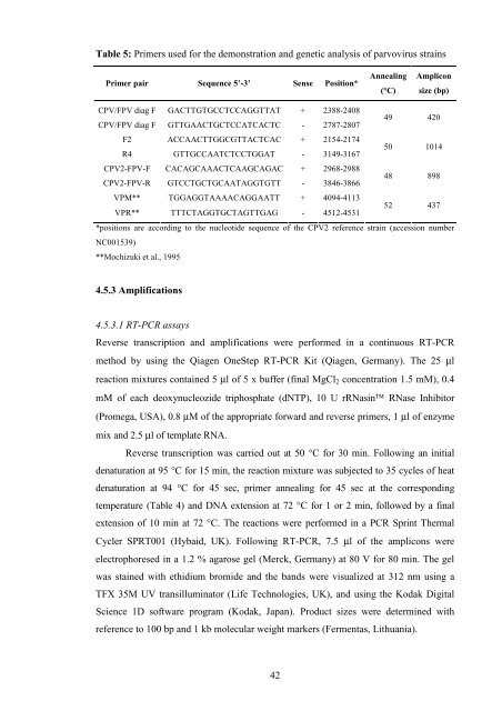
Table 5: Primers used for the demonstration and genetic analysis of parvovirus strains<br />
Primer pair Sequence 5’-3’ Sense Position* Annealing<br />
CPV/FPV diag F GACTTGTGCCTCCAGGTTAT + 2388-2408<br />
CPV/FPV diag F GTTGAACTGCTCCATCACTC - 2787-2807<br />
F2 ACCAACTTGGCGTTACTCAC + 2154-2174<br />
R4 GTTGCCAATCTCCTGGAT - 3149-3167<br />
CPV2-FPV-F CACAGCAAACTCAAGCAGAC + 2968-2988<br />
CPV2-FPV-R GTCCTGCTGCAATAGGTGTT - 3846-3866<br />
VPM** TGGAGGTAAAACAGGAATT + 4094-4113<br />
VPR** TTTCTAGGTGCTAGTTGAG - 4512-4531<br />
42<br />
(°C)<br />
Amplicon<br />
size (bp)<br />
49 420<br />
50 1014<br />
48 898<br />
52 437<br />
*positions are according to the nucleotide sequence of the CPV2 reference strain (accession number<br />
NC001539)<br />
**Mochizuki et al., 1995<br />
4.5.3 Amplifications<br />
4.5.3.1 RT-PCR assays<br />
Reverse transcription and amplifications were performed in a continuous RT-PCR<br />
method by using the Qiagen OneStep RT-PCR Kit (Qiagen, Germany). The 25 μl<br />
reaction mixtures contained 5 μl of 5 x buffer (final MgCl2 concentration 1.5 mM), 0.4<br />
mM of each deoxynucleozide triphosphate (dNTP), 10 U rRNasin RNase Inhibitor<br />
(Promega, USA), 0.8 μM of the appropriate forward and reverse primers, 1 μl of enzyme<br />
mix and 2.5 μl of template RNA.<br />
Reverse transcription was carried out at 50 °C for 30 min. Following an initial<br />
denaturation at 95 °C for 15 min, the reaction mixture was subjected to 35 cycles of heat<br />
denaturation at 94 °C for 45 sec, primer annealing for 45 sec at the corresponding<br />
temperature (Table 4) and DNA extension at 72 °C for 1 or 2 min, followed by a final<br />
extension of 10 min at 72 °C. The reactions were performed in a PCR Sprint Thermal<br />
Cycler SPRT001 (Hybaid, UK). Following RT-PCR, 7.5 μl of the amplicons were<br />
electrophoresed in a 1.2 % agarose gel (Merck, Germany) at 80 V for 80 min. The gel<br />
was stained with ethidium bromide and the bands were visualized at 312 nm using a<br />
TFX 35M UV transilluminator (Life Technologies, UK), and using the Kodak Digital<br />
Science 1D software program (Kodak, Japan). Product sizes were determined with<br />
reference to 100 bp and 1 kb molecular weight markers (Fermentas, Lithuania).