Molecular Devices Image Express Brochure_rev_B_LR.pdf
Molecular Devices Image Express Brochure_rev_B_LR.pdf
Molecular Devices Image Express Brochure_rev_B_LR.pdf

Create successful ePaper yourself
Turn your PDF publications into a flip-book with our unique Google optimized e-Paper software.
Corporate<br />
8222<br />
ephys<br />
8181<br />
analyst<br />
8243<br />
Flex<br />
8303<br />
FlIpR<br />
8303<br />
lifeSci<br />
8142<br />
Imaging<br />
8263<br />
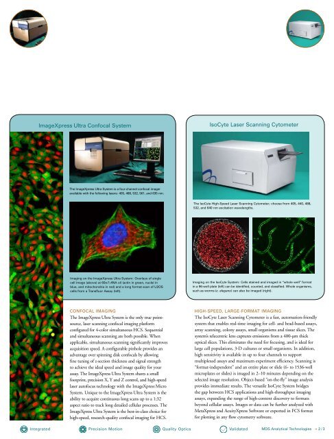
<strong>Image</strong>Xpress Ultra Confocal System<br />
Integrated<br />
the <strong>Image</strong>Xpress Ultra System is a four-channel confocal imager<br />
available with the following lasers: 405, 488, 532, 561, and 635 nm.<br />
Imaging on the <strong>Image</strong>Xpress Ultra System: overlays of single<br />
cell image (above) at 60x/1.4na oil (actin in green, nuclei in<br />
blue, and mitochondria in red) and a long format scan of U2oS<br />
cells from a transfluor assay (left).<br />
ConFoCal ImagIng<br />
The <strong>Image</strong>Xpress Ultra System is the only true pointsource,<br />
laser scanning confocal imaging platform<br />
configured for 4-color simultaneous HCS. Sequential<br />
and simultaneous scanning are both possible. When<br />
applicable, simultaneous scanning significantly improves<br />
Corporate<br />
acquisition 8222 speed. A configurable pinhole provides an<br />
advantage over spinning disk confocals by allowing<br />
fine ephys tuning of z-section thickness and signal strength<br />
8181<br />
to achieve the ideal speed and image quality for your<br />
assay. analyst The <strong>Image</strong>Xpress Ultra System shares a small<br />
8243<br />
footprint, precision X, Y and Z control, and high-speed<br />
laser autofocus technology with the <strong>Image</strong>Xpress Micro<br />
Flex<br />
8303 System. Unique to the <strong>Image</strong>Xpress Ultra System is the<br />
ability to acquire continuous long scans up to a 1:32<br />
FlIpR<br />
aspect 8303 ratio to track long detailed cellular processes. The<br />
<strong>Image</strong>Xpress Ultra System is the best-in-class choice for<br />
high-speed, research-quality confocal imaging for HCS.<br />
lifeSci<br />
8142<br />
Imaging<br />
8263<br />
precision motion<br />
Quality optics<br />
IsoCyte laser Scanning Cytometer<br />
the IsoCyte high-Speed laser Scanning Cytometer; choose from 405, 440, 488,<br />
532, and 640 nm excitation wavelengths.<br />
Imaging on the IsoCyte System: Cells stained and imaged in “whole well” format<br />
in a 96-well plate (left) can be identified, counted, and classified. whole organisms,<br />
such as worms (c. elegans) can also be imaged (right).<br />
hIgh-SpeeD, laRge-FoRmat ImagIng<br />
The IsoCyte Laser Scanning Cytometer is a fast, automation-friendly<br />
system that enables real-time imaging for cell- and bead-based assays,<br />
array scanning, colony assays, small organisms and tissue slices. The<br />
system’s telecentric lens captures emissions from a 400-µm thick<br />
optical slices. This eliminates the need for focusing, and is ideal for<br />
Corporate<br />
large 8222cell<br />
populations, 3-D cultures or small organisms. In addition,<br />
high sensitivity is available in up to four channels to support<br />
multiplexed ephys assays and maximum experiment efficiency. Scanning is<br />
8181<br />
“format-independent” and an entire plate or slide (6- to 1536-well<br />
microplates analyst or slides) is imaged in 2–10 minutes depending on the<br />
8243<br />
selected image resolution. Object-based “on-the-fly” image analysis<br />
provides immediate results. The versatile IsoCyte System bridges<br />
Flex<br />
the 8303 gap between HCS applications and high-throughput imaging<br />
assays, expanding the range of high-content discovery to formats<br />
FlIpR<br />
beyond 8303 cellular assays. <strong>Image</strong>s or data can be further analyzed with<br />
MetaXpress and AcuityXpress Software or exported in FCS format<br />
for plotting in any flow cytometry software.<br />
lifeSci<br />
8142<br />
Imaging<br />
8263<br />
validated<br />
mDS analytical technologies > 2 / 3