Frog axis cheat sheet
Frog axis cheat sheet
Frog axis cheat sheet

You also want an ePaper? Increase the reach of your titles
YUMPU automatically turns print PDFs into web optimized ePapers that Google loves.
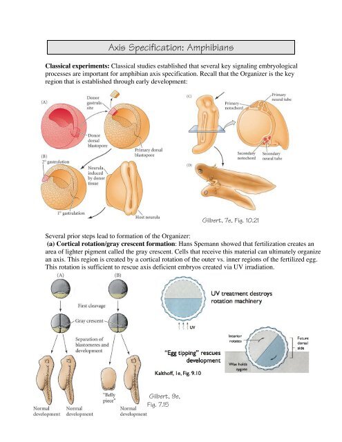
Axis Specification: Amphibians<br />
Classical experiments: Classical studies established that several key signaling embryological<br />
processes are important for amphibian <strong>axis</strong> specification. Recall that the Organizer is the key<br />
region that is established through early development:<br />
Several prior steps lead to formation of the Organizer:<br />
(a) Cortical rotation/gray crescent formation: Hans Spemann showed that fertilization creates an<br />
area of lighter pigment called the gray crescent. Cells that receive this material can ultimately organize<br />
an <strong>axis</strong>. This region is created by a cortical rotation of the outer vs. inner regions of the fertilized egg.<br />
This rotation is sufficient to rescue <strong>axis</strong> deficient embryos created via UV irradiation.<br />
Gilbert, 9e,<br />
Fig. 7.15<br />
Gilbert, 7e, Fig. 10.21
<strong>Frog</strong> <strong>axis</strong> <strong>cheat</strong> <strong>sheet</strong> – Zoo 470 2013 – Page 2<br />
(b) Vegetal cells signal to induce mesoderm: Dorsal vegetal cells are capable of inducing<br />
an <strong>axis</strong> and rescuing UV-treated embryos:<br />
(c) This leads to the “Nieuwkoop Center”, a region on the dorsal side that induces<br />
Organizer, whereas non-dorsal cells induce non-dorsal mesoderm:<br />
Note: Based on cell marking experiments,<br />
dorsal endoderm generates dorsal-type<br />
mesoderm in the experiment shown in (B), whereas<br />
non-dorsal types of endoderm induce non-dorsal<br />
types of mesoderm.<br />
Gilbert, 8e,<br />
Fig. 10.20<br />
Gilbert, 8e,<br />
Fig. 10.21
<strong>Frog</strong> <strong>axis</strong> <strong>cheat</strong> <strong>sheet</strong> – Zoo 470 2013 – Page 3<br />
Modern Experiments<br />
The challenge of modern developmental biology is to correlate classical experiments with molecular<br />
mechanisms. While much progress has been made in this area, there are still areas that are poorly<br />
understood.<br />
The establishment of the D-V <strong>axis</strong> involves several steps:<br />
Step 1: Fertilization induces<br />
cortical rotation. Cortical<br />
rotation is driven by<br />
microtubules, which in part are<br />
formed via nucleation from the<br />
sperm aster. The sperm enters<br />
180° away from the future dorsal<br />
side of the embryo. In some<br />
species, this creates a lighter area<br />
on the surface known as the gray<br />
crescent.<br />
Step 2: Cortical rotation activates dorsal development, possibly through Dishevelled, GBP, and<br />
XWnt11. This leads to more β-catenin on the dorsal side of the zygote. Excess β-catenin introduced<br />
ventrally by overexpression can result in <strong>axis</strong> duplication (see figure below). Conversely, loss of βcatenin<br />
via morpholinos in dorsal vegetal cells leads to a ventralized embryo (not shown).<br />
Wolpert et al, 2e, Fig. 3.8<br />
Gilbert, 9e, Fig. 7.1<br />
Gilbert, 9e, Fig. 7.21
<strong>Frog</strong> <strong>axis</strong> <strong>cheat</strong> <strong>sheet</strong> – Zoo 470 2013 – Page 4<br />
Step 3: β-catenin accumulates in the nuclei of dorsal cells. This leads to the expression of genes by<br />
Nieuwkoop Center cells in the next step.<br />
Step 4: The Nieuwkoop Center<br />
forms. Ventral vegetal cells<br />
differentiate as endoderm, due in part<br />
to VegT (and possibly Vg1). Dorsal<br />
vegetal cells differentiate differently,<br />
due to the added effect of β-catenin.<br />
Tcf/Lef proteins, in combination with<br />
β-catenin, lead to expression of<br />
siamois and twin (transcription<br />
factors) and other genes in these<br />
cells.<br />
Step 6: Mesoderm is induced in overlying<br />
marginal zone tissue by vegetal cells. The<br />
character of the induced mesoderm reflects the<br />
character of the underlying vegetal cells.<br />
Gilbert, 9e, Fig. 7.21<br />
Gilbert, 8e, Fig. 10.25<br />
Step 5: Graded expression of Nodal-related proteins arises in vegetal<br />
cells. This creates a full spectrum of D-V differences.<br />
Wolpert, 2e, Fig. 3.35
<strong>Frog</strong> <strong>axis</strong> <strong>cheat</strong> <strong>sheet</strong> – Zoo 470 2013 – Page 5<br />
Step 7: The Organizer expresses proteins<br />
that counteract the effects of BMPs and<br />
Wnts, leading to dorsal mesoderm<br />
formation. Complete loss of BMP<br />
signaling leads to excess dorsal structures,<br />
suggesting that the non-signaled default is<br />
dorsal mesoderm. However, ventral cells<br />
produce BMPs and XWnt8, among other<br />
secreted proteins. These normally cause<br />
cells that would be dorsal by default to be<br />
ventral. Near the Organizer, however, these<br />
signals are counteracted by BMP (noggin,<br />
chordin) and Wnt (Frbz1, Cerberus, and<br />
Dickkopf) inhibitors (see Gilbert 9e, Table<br />
7.2), leading to dorsal structures.<br />
Step 8: Gastrulation moves mesoderm and endoderm into position for further inductive events<br />
[Note: We will cover gastrulation in detail later!]<br />
Gilbert, 9e, Fig. 7.6
<strong>Frog</strong> <strong>axis</strong> <strong>cheat</strong> <strong>sheet</strong> – Zoo 470 2013 – Page 6<br />
Step 9: The same signals lead to neural induction: In the ectoderm, BMPs and Wnt promote<br />
epidermis, but the organizer counteracts these signals in ectoderm near the Organizer, leading to neural<br />
induction<br />
Gilbert, 9e, Fig. 7.27 & 7.32<br />
Step 9: Refinement of neural induction occurs: Early in gastrulation, the organizer appears to send<br />
signals to nearby ectoderm prior to involution of the dorsal mesoderm (“planar induction”). Later, after<br />
involution, regional induction results in anterior and posterior neural tissues (“vertical induction”).
<strong>Frog</strong> <strong>axis</strong> <strong>cheat</strong> <strong>sheet</strong> – Zoo 470 2013 – Page 7<br />
Experiments indicate that both sorts of signals are important. Embryological experiments show that both<br />
of these kinds of signals probably act. Planar signals can be shown to occur via Keller explants, in which<br />
the ectoderm is adjacent to mesoderm., and cannot involute to lie underneath the ectoderm.<br />
Vertical signals can be shown to<br />
operate through “Einsteck”<br />
implants, in which anterior or<br />
posterior mesoderm is inserted into<br />
a host gastrula. The induced tissue<br />
is appropriate to the nature of the<br />
inserted mesoderm. This suggests<br />
that after involution, anteriorposterior<br />
refinement of neural<br />
pattern occurs via vertical<br />
inductive signaling.<br />
Late in gastrulation a posteriorizing signal,<br />
mediated by retinoic acid (RA), Wnts, and<br />
fibroblast growth factors (FGFs) reinforces<br />
posterior neural fates. Secreted Wnt<br />
antagonists such as Cerberus, produced by the<br />
anterior mesendoderm reinforce head-specific<br />
neural fates, as opposed to trunk spinal cord,<br />
where mainly BMPs are blocked.<br />
“Planar” induction: Keller explant<br />
(Wolpert 2e, Fig. 4.23)<br />
“Vertical” induction: Otto Mangold’s “Einsteck” experiment<br />
(Woilpert et al 2e, Fig. 4.20)<br />
Gilbert, 9e, Fig. 7.35
<strong>Frog</strong> <strong>axis</strong> <strong>cheat</strong> <strong>sheet</strong> – Zoo 470 2013 – Page 8<br />
That regional differences like this occur can be demonstrated using explants. Ectoderm exposed to Wnt<br />
and BMP signals, to neither, or to both have differing effects:<br />
Gilbert, 9e, Fig. 7.30