EPIGENETIC SIGNALS AT GASTRULATION IN TIIE SEA URCHIN ...
EPIGENETIC SIGNALS AT GASTRULATION IN TIIE SEA URCHIN ...
EPIGENETIC SIGNALS AT GASTRULATION IN TIIE SEA URCHIN ...

You also want an ePaper? Increase the reach of your titles
YUMPU automatically turns print PDFs into web optimized ePapers that Google loves.
Developmental Biology, pages 251-255<br />
© 1990 Wiley'-Liss, Inc_<br />
<strong>EPIGENETIC</strong> <strong>SIGNALS</strong> <strong>AT</strong> GASTRUL<strong>AT</strong>ION <strong>IN</strong> <strong>TIIE</strong> <strong>SEA</strong> URCH<strong>IN</strong><br />
David R. McClay, James A. Coffman, and Jeffrey D. Hardin<br />
Department of Zoology, Duke University, Durham, NC 27706<br />
ABSTRACT Archenteron fonnation occurs in two phases in the<br />
sea urchin embryo. In the first phase, cell shape changes plus<br />
localized cell rearrangements begin the invagination and lead to<br />
elongation of the archenteron to more than half its final length. In<br />
the second phase secondary mesenchyme cells extend filopodia<br />
which attach at the animal pole and contract to provide the force<br />
necessary for the fmal elongation of the archenteron. Experiments<br />
show the filopodial behavior to continue until a specific target region<br />
at the animal pole is reached. This mechanism assures proper<br />
placement of the archenteron relative to the stomadaem. During the<br />
two phases of archenteron invagination the sequence of gene<br />
expression appears to involve at least two critical periods of<br />
epigenetic interactions as shown experimentally.<br />
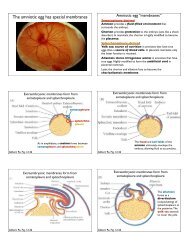
An inward bending of the vegetal plate is the first visible sign of<br />
archenteron formation in the sea urchin embryo. The presumptive<br />
endoderm cells in the vegetal region then begin to rearrange so that the initial<br />
inward bulge becomes a tube (Figure I). Many experiments have shown<br />
that these morphogenetic events are autonomous to the vegetal plate. For<br />
example, the movements occur in isolated vegetal plates (Moore and Burt,<br />
1939; Ettensohn, 1984). Also, cell marking experiments show that only<br />
cells at the vegetal plate participate in invagination movements, Le., there<br />
are no morphogenetic movements pushing new cells into the vegetal region<br />
from elsewhere (Ettensohn, 1984; Hardin, 1989). Thus, to explain how<br />
invagination occurs, one must learn how the cells rearrange themselves and<br />
how invagination is properly directed.<br />
The shape of the archenteron emerges first as a result of endodermal cell<br />
rearrangements, and later as a result of secondary mesenchyme cell pulling.<br />
Two sorts of experiments demonstrate that cell rearrangements occur. First,<br />
as the archenteron increases in length, it decreases in diameter, and there are<br />
progressively fewer cells in each cross section (Ettensohn, 1985; Hardin<br />
and Cheng, 1986). The only way to explain this observation is to infer a<br />
cell rearrangement. Second, in cell marking experiments, a round patch of
252 McClay et aI.<br />
cells in the vegetal plate prior to invagination becomes resolved into a line of<br />
cells along the length of the archenteron (Figure 1) (Wray, 1987; Hardin,<br />
1989). These data show that archenteron invagination begins with a series<br />
of directed cell rearrangements. The mechanism(s) responsible for the cell<br />
rearrangement and for the directed cell movements are unknown. It is<br />
known, however, that isolated endoderm cells become motile at the<br />
beginning of archenteron formation (McClay, 1986; J. Hardin, unpublished<br />
observations). Whether this motility is important for the cell rearrangements<br />
and/or for the invagination remains to be shown.<br />
-<br />
early mid late quiescent<br />
FIGURE 1. Stages in archenteron elongation. As the archenteron<br />
elongates, the number of cells around the circumference steadily decreases,<br />
and labeled patches of cells narrow and lengthen. Late in gastrulation,<br />
filopodia extend upward, pulling on the archenteron to complete its<br />
elongation. Upon reaching the "target" region the filopodia cease their<br />
activity.<br />
The latter part of archenteron formation is the responsibility of the<br />
secondary mesenchyme cells (SMCs), which appear to pull the archenteron<br />
through the fmall/3 to 1/2 of its elongation phase. The SMCs extend<br />
filopodia that reach to a final length of about 30-35 11m. By attaching to the<br />
wall of the blastocoel and contracting, it is presumed that the fllopodia pull<br />
the archenteron to its final length. In support of this hypothesis, if filopodia<br />
are ablated the archenteron does not extend beyond about 2/3 its fmallength<br />
(Hardin, 1988). Also, in exogastrulae the archenteron extends to about 2(3<br />
its final length, but then can extend no further (Hardin and Cheng, 1986).<br />
Thus, the final sequence of archenteron elongation requires SMCs which<br />
use filopodial contraction to stretch the archenteron to its final length.<br />
Observation of filopodial behavior showed that SMCs extended<br />
filopodia during much of gastrulation. At the end of gastrulation filopodia!<br />
extension ceased rather quickly when a putative target region was reached at
Sea Urchin Gastrulation 253<br />
the animal pole. It was as if contact with a certain region of the blastocoel<br />
provided a signal to tenninate the.filopodial extension behavior (Hardin.et<br />
a/., 1989). Experiments were deSigned to ask whether there was a specific<br />
target, and to ask whether SMCs were programmed to extend filopodia until<br />
the target was reached. The archenteron was forced into contact with many<br />
regions of the blastocoel. In each case the archenteron made temporary<br />
contact but then moved further toward the animal pole until the putative<br />
target region was reached. Only contact with the "correct" target had the<br />
affect of stopping the filopodial extension behavior. Thus there appears to<br />
be a specific region that serves as a target for ftlopodial extension.<br />
We then asked whether the filopodial extension was somehow<br />
programmed to continue until a target was reached. If that were true then it<br />
might be predicted that precocious contact with the target would bring about<br />
a precocious end to the ftlopodial extensions. Accordingly when the animal<br />
pole was pushed into contact with the archenteron several hours before<br />
contact normally would have been made, the filopodial extension stopped.<br />
Another prediction tested was that of delayed contact with the target. By<br />
experimentally elongating the embryos the archenterons were prevented or<br />
delayed from coming into contact with the target region. When contact was<br />
delayed for several hours SMCs continued to extend ftlopodia. Release of<br />
the embryos from the elongated shape allowed the filopodia to touch the<br />
target, and filopodial extension stopped shortly thereafter. Thus it would<br />
appear that the SMCs were programmed to extend filopodia until the cells<br />
made contact with a target region.<br />
If SMCs were prevented from reaching the target for long periods of<br />
time (several hours), the SMCs eventually left the tip of the archenteron,<br />
migrated to the blastocoel wall and then on to the target region on their own.<br />
They then appear to differentiate normally. These data suggested that<br />
contact with the target might be a critical period for differentiation of SMCs<br />
and led us to examine events that might provide epigenetic information for<br />
differentiation of the cells of the archenteron.<br />
Ifepigenesis were involved in archenteron formation, we reasoned that<br />
there might be certain time periods that were more critical than others for the<br />
events that led to the completion of gastrulation. Our observations have<br />
suggested that much of archenteron formation occurs autonomously, and<br />
there are two critical periods that require interaction with parts of the embryo<br />
external to the archenteron itself. At the beginning of invagination, and at<br />
the end of the process, contact with the extracellular matrix appears to be<br />
important for differentiation of the endoderm and of the secondary<br />
mesenchyme. Support for this hypothesis is provided by the following<br />
experiments.<br />
The embryo is surrounded on the outside by the hyaline layer. A basal<br />
lamina lines the blastocoel. Ifeither of these layers is disrupted, or<br />
prevented from being in contact with the embryo at the beginning of<br />
gastrulation, invagination of the archenteron does not occur (Wessell and<br />
McClay, 1987; Butler et a/., 1987; Adelson and Humphreys, 1988). In the<br />
normal embryo, Endo 1 is expressed in the mid- and hindgut regions at the<br />
early to mid gastrula stage. Ecto V becomes localized to the ventral ectoderm
254 McClay et aI.<br />
and foregut at the end of gastrulation. In the inhibited embryos, these<br />
markers fail to be expressed. However, if the blocks to matrix contact are<br />
removed, invagination begins, goes to completion, and Endo 1 and Ecto V<br />
are expressed in the correct sequence. Thus presumptive endoderm requires<br />
some kind of interaction with the extracellular matrix for archenteron<br />
formation to begin. Ifthe inhibitors are added after invagination begins, the<br />
ftrst phase of elongation proceeds to completion, but the ftnal elongation<br />
and attachment of SMCs to the target often fail to occur (Butler et aI., 1987;<br />
Hardin and McClay, in preparation). In these embryos Endo 1 and Ecto V<br />
are expressed in sequence even though the tip of the archenteron has failed<br />
to reach the target region (Hardin and McClay, in preparation).<br />
Many SMCs remain undifferentiated during invagination and appear to<br />
remain undifferentiated until after their behavior changes as a result of<br />
coming into contact with the animal pole target Experimentally, this<br />
conclusion was reached based on the ability of SMCs to convert to become<br />
primary mesenchyme cells throughout much of the invagination process<br />
(Ettensohn and McClay, 1988). The lineage conversion capability is a<br />
property of a subset of SMCs since pigment cells differentiate from SMCs<br />
during invagination and were not observed to convert to the PMC lineage<br />
(Gibson and Burke, 1985; Ettensohn and McClay, 1988). The precise time<br />
at which SMCs lose their competence to become PMCs is unknown. It can<br />
be observed, however, that differentiation of these cells into muscle or<br />
coelomic pouch does not occur until after the target event at the end of<br />
gastrulation. Thus, at some time following target recognition, which itself<br />
may involve contact with the extracellular matrix, SMCs become restricted<br />
with respect to lineage and begin to differentiate.<br />
We have shown that once initiated, differentiation within the archenteron<br />
is largely autonomous. During the process of invagination cells rearrange, a<br />
sequence of genes are expressed, and the primitive gut elongates.<br />
Secondary mesenchyme cells then help to complete the process of<br />
invagination, and their contact with a site that is anatomically related to the<br />
region of the future mouth somehow signals the completion of invagination.<br />
Once this target is reached the SMCs then begin to differentiate. Thus<br />
morphogenesis of the sea urchin embryo involves a combination of both<br />
genetically programmed and epigenetically regulated cell differentiation and<br />
gene expression.<br />
REFERENCES<br />
1. Adelson, D.L. and Humphreys, T. (1988). Sea urchin<br />
morphogenesis and cell-hyalin adhesion are perturbed by a monoclonal<br />
antibody speciftc for hyalin. Development 104, 391-402.<br />
2. Alliegro, M.C., Ettensohn, c.A., Burdsal, C.A., Erickson, H.P., and<br />
McClay, D.R. (1988) Echinonectin: a new embryonic substrate adhesion<br />
protein. 1. Cell Bioi. 107, 2319-2327
Sea Urcbin Gastrulation 255<br />
3. Butler, E., Hardin, J. and Benson, S. (1987). The role of lysyl<br />
oxidase and collagen crosslinking during sea urchin development. Exp.<br />
Cell Res. 173, 174-182.<br />
4. Coffman, J., Nelson, S., and McClay, D.R. (1985). A cell surface<br />
protein that identifies the ventral surface of the ectoderm of sea urchin<br />
gastrulae. J. Cell Bioi. 101, 469a.<br />
5. Ettensohn, C.A. (1984). Primary invagination of the vegetal plate<br />
during sea urchin gastrulation. Amer. Zool. 24,571-588.<br />
6. Ettensohn, C.A. (1985). Gastrulation in the sea urchin is accompanied<br />
by the rearrangement of invaginating epithelial cells. Dev. Bioi. 112,<br />
383-390.<br />
7. Ettensohn, C.A. and McClay, D.R. (1988) Cell lineage conversion in<br />
the sea urchin embryo. Dev. Bioi. 125, 396-409.<br />
8. Gibson, A.W. and Burke, R.D. (1985). The origin of pigment cells in<br />
embryos of the sea urchin Strongylocentrotus purpuratus. Dev. Bioi. 107,<br />
414-419.<br />
9. Hardin, J. (1988). The role of secondary mesenchyme cells during sea<br />
urchin gastrulation studied by laser ablation. Develop. 103, 317-324.<br />
10. Hardin, J. (1989) Local shifts in position and polarized motility drive<br />
cell rearrangement during sea urchin gastrulation. Submitted for publication.<br />
11. Hardin, J.D. and Cheng, L.Y. (1986). The mechanisms and<br />
mechanics of archenteron elongation during sea urchin gastrulation. Dev.<br />
Bioi. 115, 490-501.<br />
12. Hardin, J., Morrill, J., and McClay, D. (1989). Filopodia use local<br />
guidance cues during sea urchin gastrulation. In preparation.<br />
13. Moore, A.R. and Burt, A.S. (1939). On the locus and nature of the<br />
forces causing gastrulation in the embryos of Dendraster excentricus. J.<br />
Exp. Zool. 82, 159-171.<br />
14. Wessell, G.M. and McClay, D.R. (1987). Gastrulation in the sea<br />
urchin embryo requires the deposition of crosslinked collagen within the<br />
extracellular matrix. Dev. Bioi. 121, 149-165.<br />
15. Wray, G.A. (1987). "Heterochrony and Homology in the Evolution of<br />
Echinoid Development". Ph.D. dissertation, Duke University, Durham,<br />
N.C.