ZEI Template Brochure 071030 - Mitegen
ZEI Template Brochure 071030 - Mitegen
ZEI Template Brochure 071030 - Mitegen

Create successful ePaper yourself
Turn your PDF publications into a flip-book with our unique Google optimized e-Paper software.
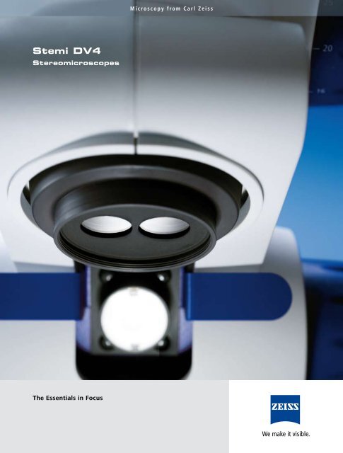
Stemi DV4<br />
Stereomicroscopes<br />
The Essentials in Focus<br />
Microscopy from Carl Zeiss
The fruit of the cocklebur “uses” hooks<br />
to attach itself to animals‘ furs, which<br />
then disperse it‘s seeds.<br />
For Education and Routine<br />
in Science and Industry<br />
On a circuit board, residues of fluxing<br />
agents can be clearly identified between<br />
the contacts.<br />
Chicken embryos in<br />
transmitted-light darkfield<br />
Applying protective lacquer to<br />
a plaster model in a dental laboratory<br />
Brilliant images and easy to use<br />
microscope functions make<br />
microscopy lessons a genuine<br />
pleasure for students and<br />
teachers alike.<br />
Stemi DV4 in the classroom<br />
High-quality optics and a variable<br />
LED illumination in a compact unit<br />
are important criteria when it comes<br />
to tasks in industrial quality control.<br />
Stemi DV4 with tilting mechanism<br />
being used in quality control<br />
Thanks to true-color images,<br />
combined with the possibility<br />
of switching between and mixing<br />
reflected and transmitted light,<br />
rapid and precise diagnoses can be<br />
achieved in biological laboratories.<br />
Stemi DV4 in a developmental<br />
biology laboratory<br />
A fiberoptic reflected light spot<br />
illuminator integrated into the body<br />
of the Stemi DV4 SPOT ensures that<br />
specimens are always well illuminated<br />
– whatever the position and<br />
direction of observation.<br />
Stemi DV4 SPOT on stand U<br />
in a dental laboratory
The Essentials<br />
in Focus<br />
The innovative nature of this stereomicroscope can be seen<br />
not only in the color and unique, compact design of<br />
the instrument, but also in its attractive price – a result<br />
of state-of-the-art production and assembly technologies.<br />
Sturdy, hard-wearing technology, very easy operation of all<br />
microscope funktions and no compromises whatsoever<br />
in the definition and brilliance of the microscopic images – that<br />
is the best way to describe the Stemi DV4 stereomicroscopes<br />
from Carl Zeiss.<br />
The pancratic system, as the heart of the stereomicroscope,<br />
has a decisive influence on the optical<br />
performance of the overall system.<br />
The Stemi DV4 is the first instrument in its class with<br />
a mechanically corrected zoom system, a feature that<br />
was previously only incorporated into stereomicroscopes<br />
in higher price categories. The result: needle-sharp<br />
images across the entire zoom range.
Stemi DV4 stereomicroscope<br />
with sample carousel 16<br />
A range of well-chosen accessories make the Stemi DV4<br />
an even more attractive option for a whole range<br />
of applications. Thanks to the sample carousel 16,<br />
up to sixteen different specimens can be positioned under<br />
the microscope safely, quickly and extremely easily.
Stemi DV4 stereomicroscope<br />
The patented zoom system of the Stemi DV4<br />
(Double Lens Vario, zoom factor 4) delivers images<br />
that are needle-sharp and, thanks to rigorous stray-light<br />
minimization, brilliant and rich in contrast. Across the<br />
entire magnification range, from 8x to 32x and across the<br />
whole field of view.<br />
Stemi DV4 SPOT stereomicroscope<br />
The fiberoptic cold-light illuminator additionally<br />
integrated into the microscope body of the Stemi DV4<br />
ensures that the object field is fully illuminated at<br />
all times. The SPOT reflected light irradiating almost<br />
vertically on the specimen, provides a shadow-free,<br />
high contrast illumination.
9<br />
Stand C LED:<br />
Combine Illumination Modes<br />
for Outstanding Results<br />
As good results in microscopy depend on<br />
efficient illumination, we have equipped the<br />
stand with a modern LED illuminator.<br />
You have a choice between the following<br />
standard features:<br />
- reflected-light brightfield<br />
- transmitted-light brightfield<br />
- mixed light<br />
In addition, there is an option for:<br />
- transmitted-light darkfield<br />
- polarization contrast in reflected and transmitted light<br />
This means that you have the common illumination<br />
modes in stereomicroscopy at your fingertips.<br />
You can decide which ones to use and how to combine<br />
them practically during observation – it can all be<br />
done rapidly at the touch of a button.<br />
Stand C LED features an integrated LED illuminator<br />
for applications in reflected and transmitted light.<br />
This powerful daylight-quality illuminator shows specimens<br />
with high contrast in their natural colors. In transmitted<br />
light, six LEDs ensure bright, homogeneous illumination<br />
at any magnification, from overview to detail.<br />
At the touch of a button:<br />
rapid switching between illumination modes.
Using the example of a lacewing (Chrysopa<br />
carnea, dead specimen), the different illumination<br />
options that stand C LED offers can be<br />
clearly illustrated.<br />
Reflected-light brightfield<br />
A standard illumination mode on a<br />
stereomicroscope, used to illuminate<br />
the surface of opaque specimens.<br />
Transmitted-light brightfield<br />
Also a standard mode.<br />
For observing transparent<br />
specimens and contours.<br />
If a certain amount of<br />
transmitted light is added,<br />
the disruptive shadows<br />
beneath the specimen fade.<br />
If a certain amount of<br />
reflected light is added,<br />
structures on the insect’s<br />
body also become visible.<br />
Transmitted-light dark field (optional)<br />
Extremely fine structures are illumi-<br />
nated in their natural colors against<br />
a dark background.<br />
If a certain amount of<br />
reflected light is added,<br />
structures on the insect’s<br />
body also become visible.<br />
The ready-for-use design<br />
of the Stemi DV4 stereomicroscope<br />
with full range of illumination<br />
options as standard<br />
7
The Basis of Your<br />
Modular System<br />
Exact positioning<br />
Quick, simple and with no risk.<br />
Sample carousel 16, which can be retrofitted<br />
to stand C LED, allows you to<br />
position up to 16 different specimens with<br />
precision under the stereomicroscope.<br />
A click-stop mechanism<br />
ensures precise positioning<br />
under the microscope, whilst<br />
a low-reflection glass plate<br />
protects the specimens.<br />
Measurement at a glance<br />
Using the eyepiece micrometer you can<br />
read measurement values directly from the<br />
scale, either at the 8x overview or 32x<br />
detail magnification.<br />
The 8x/32x/18 eyepiece<br />
micrometer is retrofitted in<br />
the 10x standard eyepiece.<br />
Digital documentation<br />
For the documentation of microscopic<br />
images you can attach the camera to one<br />
of the two eyepiece tubes in place of the<br />
eyepiece.<br />
Digital compact cameras,<br />
SLR- or video cameras can be<br />
connected to the microscope<br />
quickly and easily using the<br />
appropriate eyepiece adapters.<br />
10
Specifications<br />
Front lens system<br />
Factor FWD*<br />
(mm)<br />
0,3x 287<br />
0,4x 211<br />
0,3x ... 0,5x 233 ... 90<br />
0,63x 130<br />
w/o 92<br />
1,25x 60<br />
1,6x 48<br />
2x<br />
31<br />
Front lens system<br />
Factor FWD*<br />
(mm)<br />
0,3x 287<br />
0,4x 211<br />
0,3x ... 0,5x 233 ... 90<br />
0,63x 130<br />
w/o 92<br />
1,25x 60<br />
1,6x 48<br />
2x<br />
31<br />
Eyepiece 10x/20 Br. foc.<br />
Magnification Object field<br />
(mm)<br />
2,4x ... 9,6x<br />
83,3 ... 20,8<br />
3,2x ... 12,8x 62,5 ... 15,6<br />
2,4x ... 16,0x 39,7 ... 12,5<br />
5,0x ... 20,2x 40,0 ... 9,9<br />
8,0x ... 32,0x 25,0 ... 6,3<br />
10,0x ... 40,0x 20,0 ... 5,0<br />
12,8x ... 51,2x 15,6 ... 3,9<br />
16,0x ... 64,0x 12,5 ... 3,1<br />
Eyepiece 15x/13 Br. foc.<br />
Magnification Object field**<br />
(mm)<br />
3,6x ... 14,4x 55,4 ... 13,9<br />
7,6x ... 30,2x 41,6 ... 10,4<br />
3,6x ... 24,0x 55,4 ... 8,3<br />
7,5x ... 30,3x 26,4 ... 6,6<br />
12,0x ... 48,0x 16,6 ... 4,2<br />
15,0x ... 60,0x 13,3 ... 3,3<br />
19,2x ... 76,8x 10,4 ... 2,6<br />
24,0x ... 96,0x 8,3 ... 2,1<br />
*FWD = Free Working Distance<br />
* * values based on real field number 13,3<br />
110 110<br />
230 230 300 300<br />
92<br />
92<br />
350<br />
350<br />
Mass 5 kg
“A fundamental key to success was therefore the fact that<br />
Carl Zeiß, right from the outset, made every effort to introduce<br />
extremely precise technology in his small workshop to ensure<br />
that the uncertain dexterity of the human hand was subjected<br />
to rigorous methods of testing at every point in the process.”<br />
Carl Zeiss MicroImaging GmbH<br />
07740 Jena, Germany<br />
BioSciences, Industrial | Göttingen Location<br />
Phone : +49 551 5060 660<br />
Telefax : +49 551 5060 464<br />
E-Mail : micro@zeiss.de<br />
www.zeiss.de/stereo<br />
Ernst Abbe<br />
(from his commemorative speech at the<br />
Optical Workshop’s 50th anniversary celebration,<br />
given on December 12, 1896 in Jena)<br />
Subject to change.<br />
Printed on environmentally friendly paper,<br />
bleached without the use of chlorine.<br />
60-2-0034/e – printed 04.09