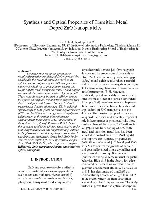
Synthesis and Optical Properties of Transition Metal Doped ZnO ...
Synthesis and Optical Properties of Transition Metal Doped ZnO ...
Synthesis and Optical Properties of Transition Metal Doped ZnO ...
You also want an ePaper? Increase the reach of your titles
YUMPU automatically turns print PDFs into web optimized ePapers that Google loves.
<strong>Synthesis</strong> <strong>and</strong> <strong>Optical</strong> <strong>Properties</strong> <strong>of</strong> <strong>Transition</strong> <strong>Metal</strong><br />
<strong>Doped</strong> <strong>ZnO</strong> Nanoparticles<br />
Ruh Ullah1, Joydeep Dutta2<br />
1Department <strong>of</strong> Electronic Engineering NUST Institute <strong>of</strong> Information Technology Chaklala Scheme III,<br />
2Center o f Excellence in Nanotechnology, Industrial Systems Engineering School <strong>of</strong> Engineering &<br />
2Technologies Asian Institute <strong>of</strong> Technolo<br />
1email: ruhullah@niit.edu.pk, ruhullahg@gmail.com<br />
2email: joy@ait.ac.th<br />
1. Abstract<br />
Enhancement in the optical absorption <strong>of</strong><br />
metal <strong>and</strong> transition metal doped <strong>ZnO</strong> nanoparticles<br />
could make this material capable to work as an<br />
efficient photocatalysts. <strong>Doped</strong> <strong>ZnO</strong> nanoparticles<br />
were synthesized via co-precipitation techniques.<br />
Doping <strong>of</strong> <strong>ZnO</strong> with manganese (Mn2 +) <strong>and</strong> cupper<br />
was intended to enhance the surface defects <strong>of</strong> <strong>ZnO</strong>.<br />
These can subsequently be used as efficient centers<br />
for optical ab sorption. Nanoparticles prepared with<br />
these techniques, which were characterized with<br />
transmission electron microscopy (TEM), infrared<br />
spectroscopy (FTIR), photo-co-relation spectroscopy<br />
(PCS) <strong>and</strong> UV/VIS-spectroscopy showed significant<br />
enhancement in the optical absorption when<br />
compared with the undoped <strong>ZnO</strong>. Enhancement in<br />
the optical absorption <strong>of</strong> Mn-doped <strong>ZnO</strong> indicates<br />
that it can be used as an efficient photocatalyst under<br />
visible light irradiation <strong>and</strong> might have applications<br />
in the photoelectrochemical hydrogen production. It<br />
was found that manganese-doped <strong>ZnO</strong> (<strong>ZnO</strong>:Mn2+ )<br />
absorbs more visible light in comparison to cupper-<br />
doped <strong>ZnO</strong> (<strong>ZnO</strong>:Cu2+ ) when exposed to tungsten<br />
bulb. Key words: <strong>ZnO</strong>, manganese doping, photocatalysts,<br />
optical absorption<br />
2. INTRODUCTION<br />
optoelectronic devices [2], ferromagnetic<br />
devices <strong>and</strong> heterogeneous photocatalysts<br />
[3-4]. <strong>ZnO</strong> is an interesting wide b<strong>and</strong> gap<br />
(3.3ev) metal oxide semiconductor martial<br />
<strong>and</strong> is currently under investigation owing to<br />
its tremendous applications in response to its<br />
tunable properties [5-6]. Magnetic,<br />
electrical, optical <strong>and</strong> catalytic properties <strong>of</strong><br />
<strong>ZnO</strong> are mostly size <strong>and</strong> surface dependent.<br />
Attempts [6-9] have been made to improve<br />
these properties <strong>and</strong> enhance the industrial<br />
applications <strong>of</strong> <strong>ZnO</strong> nanoparticles\nano-<br />
devices. Since surface properties such as<br />
oxygen deficiencies <strong>and</strong> area play important<br />
role in heterogeneous photocatalysis, these<br />
can be enhanced by doping <strong>ZnO</strong> with metal<br />
ion [9]. In addition, doping <strong>of</strong> <strong>ZnO</strong> with<br />
metal <strong>and</strong> transition metal ions has been<br />
reported to control the size <strong>of</strong> <strong>ZnO</strong> crystal<br />
<strong>and</strong> improve the magnetic properties <strong>of</strong><br />
<strong>ZnO</strong>. Viswanatha et al, [10] has doped <strong>ZnO</strong><br />
with Mn to control the growth <strong>of</strong> particle<br />
<strong>and</strong> get smaller sized single crystallites <strong>and</strong><br />
was deemed to have applications in<br />
spintronics owing to some unusual magnetic<br />
behavior. Blue shift in the absorption edge<br />
compared to the bulk was attributed to the<br />
quantum confinement effect. S. Sakthivel et<br />
al. [11] has demonstrated that <strong>ZnO</strong> can<br />
comparatively absorb more light than TiO2<br />
in the region where the light absorption<br />
occurs due to b<strong>and</strong> gap excitation. The study<br />
<strong>ZnO</strong> has been extensively studied as<br />
a potential material for various applications<br />
such as sensors, varistors, piezoelectric [1]<br />
transducers, surface acoustic wave devices,<br />
phosphors, transparent conducting oxides,<br />
further suggests that, the optical absorption<br />
1-4244-1494-6/07/$25.00 © 2007 IEEE 306
<strong>of</strong> <strong>ZnO</strong> can be enhanced by creating more<br />
defects on its surface. R. Wang et al. [9] has<br />
demonstrated that silver ion doped <strong>ZnO</strong> has<br />
improved photocatalytic activities owing to<br />
the increased surface defects caused by the<br />
enhanced oxygen vacancies. Doping <strong>of</strong> <strong>ZnO</strong><br />
with metal <strong>and</strong> transitional metals might<br />
shift the optical absorption <strong>of</strong> <strong>ZnO</strong> to the<br />
visible region i.e. to the longer wavelength.<br />
This shifting <strong>of</strong> optical absorption <strong>of</strong> doped<br />
<strong>ZnO</strong> will make this material capable to<br />
operate at lower excitation energy <strong>and</strong> can<br />
generate electron hole pair upon visible light<br />
irradiation from solar spectrum. In a<br />
technical study by K. Vanheusden et al. [13]<br />
it has been observed that lead (Pb) doping in<br />
<strong>ZnO</strong> narrows the effective b<strong>and</strong> gap <strong>of</strong> <strong>ZnO</strong><br />
nanopowders <strong>and</strong> decreases both the<br />
photoluminescence <strong>and</strong> the free-carrier<br />
concentration. Doping <strong>of</strong> <strong>ZnO</strong> films with<br />
Cobalt (Co) [12] has been reported to<br />
significantly decrease the bang gap <strong>of</strong> <strong>ZnO</strong><br />
up to 2.75 ev. This decrease in the b<strong>and</strong> gap<br />
<strong>of</strong> cobalt-doped <strong>ZnO</strong> films resultantly<br />
causes hyperchromic shift in its optical<br />
absorption. Incorporating cupper <strong>and</strong><br />
manganese ions in <strong>ZnO</strong> thin films<br />
separately, bring opposite effect on the grain<br />
size <strong>of</strong> <strong>ZnO</strong>, cupper doping increases while<br />
the manganese doping decreases the grain<br />
size [14]. It has been reported that both the<br />
dopant Cu <strong>and</strong> Mn resulted a slight decrease<br />
in the optical b<strong>and</strong> gap <strong>of</strong> <strong>ZnO</strong> films.<br />
Similarly [5] b<strong>and</strong> gap tailing effect has<br />
been observed for aluminum doped <strong>ZnO</strong>,<br />
which caused reduction in the b<strong>and</strong> gap <strong>of</strong><br />
<strong>ZnO</strong>, likely due to the doping <strong>of</strong> donors. The<br />
doping <strong>of</strong> <strong>ZnO</strong> nanocrystals with various<br />
ions was accomplished by Y. S. Wang et al.<br />
[15] using a family <strong>of</strong> dopants such as Cd,<br />
Mg, Mn, <strong>and</strong> Fe ions. Shift in the b<strong>and</strong> gap<br />
was attributed to the effect <strong>of</strong> dopant, which<br />
causes decrease in the b<strong>and</strong> gap with Cd<br />
doping while increase in the b<strong>and</strong> gap for<br />
other dopants. An enhancement in the<br />
optical absorption has been found for<br />
various dopants level in case <strong>of</strong> Fe <strong>and</strong> Mn<br />
doped <strong>ZnO</strong> nanocrystals. It is well known<br />
from the various studies that doping <strong>of</strong> <strong>ZnO</strong><br />
with metal <strong>and</strong> transition metals could<br />
decrease the effective b<strong>and</strong> gap <strong>and</strong> can<br />
subsequently increase optical absorption <strong>of</strong><br />
this b<strong>and</strong> gap tunable material. We<br />
therefore, doped <strong>ZnO</strong> with manganese using<br />
the co-precipitation techniques. This<br />
material can be used as a better<br />
photocatalyst <strong>and</strong> might have applications in<br />
photoelectrochemical hydrogen production.<br />
This study focused on the optical<br />
characteristic <strong>of</strong> manganese doped <strong>ZnO</strong><br />
nanoparticles, synthesized by coprecipitation<br />
techniques.<br />
3. EXPERIMENT<br />
The synthesis method described<br />
earlier in our previous work [16] was<br />
pursued with a little modification for<br />
preparation <strong>of</strong> doped <strong>ZnO</strong> nanoparticles.<br />
Two types <strong>of</strong> dopants, manganese <strong>and</strong><br />
cupper were investigated at the earlier<br />
stages. In a typical process, 4 mili moles <strong>of</strong><br />
Zinc acetate dehydrate were dissolved in 40<br />
ml <strong>of</strong> ethanol <strong>and</strong> heated at 50C along with<br />
vigorous stirring for half an hour, thus<br />
making precursor solution A. Since then 4<br />
mili moles <strong>of</strong> Sodium hydroxide were<br />
dissolved in 40 ml <strong>of</strong> ethanol <strong>and</strong> heated at<br />
50 C along with vigorous stirring for one<br />
hour, making precursor solution B. The<br />
dopant solutions were also prepared by<br />
dissolving 0.02 mili moles <strong>of</strong> manganese<br />
acetate <strong>and</strong> cupper acetate each in 20 ml <strong>of</strong><br />
ethanol separately. The two solutions were<br />
heated at 50 o C along with vigorous stirring<br />
for half an hour.<br />
In order to make <strong>ZnO</strong>:Mn 2+ /<br />
<strong>ZnO</strong>:Cu 2+ colloids, a complex <strong>of</strong> 20 ml<br />
precursor solution A <strong>and</strong> 20 ml dopant<br />
(manganese acetate <strong>and</strong> cupper acetate)<br />
solutions each were complexed <strong>and</strong> heated<br />
at 80C for half an hour along with vigorous<br />
stirring. After cooling to room temperature,<br />
20 ml <strong>of</strong> precursor solution B (NaOH<br />
solution) was mixed with the two complex<br />
solutions (for hydrolysis), in order to<br />
307
transform zinc hydroxide to <strong>ZnO</strong>. The<br />
solutions were kept in water bath at<br />
60~65C for 2 hours. It was observed that<br />
solutions started precipitating after one hour<br />
in water bath. Subsequently to the 2 hours<br />
water bath, the solutions were cold down to<br />
room temperature followed by 4 hours<br />
aging. The colloidal solutions were<br />
centrifuged for 20 minutes at 4 k rpm to<br />
remove the large sized agglomerates. It was<br />
observed that nanoparticles <strong>of</strong> almost<br />
uniform size were suspended in the solution.<br />
<strong>ZnO</strong>:Mn 2+ / <strong>ZnO</strong>:Cu 2+ nanoparticles thus<br />
synthesized were then used for further<br />
experimental analysis.<br />
<strong>Optical</strong> characteristics <strong>of</strong> doped <strong>ZnO</strong><br />
(<strong>ZnO</strong>:Mn 2+ , <strong>ZnO</strong>:Cu 2+ ) were determined<br />
with double beam UV/VIS<br />
spectrophotometer (Model SL 164 from<br />
ELICO). Manganese doped <strong>ZnO</strong><br />
nanoparticles were further studied based on<br />
the enhanced optical absorption when<br />
compared with <strong>ZnO</strong>:Cu 2+ . Therefore<br />
structural characterizations <strong>of</strong> only<br />
<strong>ZnO</strong>:Mn 2+ were carried out with<br />
Transmission Electron Microscope<br />
(JEOL/JEM-2100F version) operated at<br />
200KV, Fourier Transform Infrared<br />
Spectroscope (System 2000 FTIR, Perkin–<br />
Elmer).We used PCS machine from<br />
MALVERN Instrument Zetasizer Nano<br />
Model ZS Zen3600 fitted with a red laser<br />
(633nm) which can measure particle size<br />
within a range <strong>of</strong> 0.6nm to 600nm. Folded<br />
Capillary Cell (DTS1060) was used for zeta<br />
potential measurements <strong>and</strong> Disposable low<br />
volume polystyrene (DST0112) cuvette was<br />
used for size measurement<br />
4. Results <strong>and</strong> discussion<br />
<strong>Synthesis</strong> <strong>of</strong> doped <strong>ZnO</strong> was<br />
performed in alcoholic solution<br />
consecutively to avoid formation <strong>of</strong> <strong>ZnO</strong>H<br />
[17]. Therefore zinc acetate, manganese<br />
acetate, cupper acetate <strong>and</strong> NaOH all were<br />
dissolved in ethanol. The nucleation <strong>and</strong><br />
aggregation <strong>of</strong> nanoparticles are strongly<br />
solvent dependent, <strong>and</strong> are increasing with<br />
decreasing the dielectric constant <strong>of</strong> solvent<br />
[ 1 8]. Water has a dielectric constant <strong>of</strong> about<br />
80 while for ethanol it is 24.3. The<br />
nucleation <strong>and</strong> growth <strong>of</strong> <strong>ZnO</strong> is faster in<br />
ethanol than in water <strong>and</strong> hence <strong>ZnO</strong> doped<br />
colloids were synthesized in ethanol in order<br />
to avoid oxidation <strong>of</strong> dopant ions. UV/VISspectroscopy<br />
<strong>of</strong> both the cupper doped <strong>ZnO</strong><br />
(<strong>ZnO</strong>: Cu 2+ ) <strong>and</strong> manganese doped <strong>ZnO</strong><br />
(<strong>ZnO</strong>:Mn 2+ ) as well as undoped [16] newly<br />
prepared nanoparticles showed evidence <strong>of</strong> a<br />
significant divergence in the absorption<br />
intensity in the blue region, as shown in<br />
figure 1. This enhancement in the absorption<br />
intensity within the visible region is<br />
attributed to the doping <strong>of</strong> <strong>ZnO</strong> with Cu, <strong>and</strong><br />
Mn. Figure 1 further illustrates that Mn ions<br />
affect the absorption characteristic <strong>of</strong> the<br />
nanoparticles more markedly than Cu ions.<br />
This increase in the absorption intensity in<br />
the blue region can be attributed to the more<br />
pronounced doping <strong>of</strong> <strong>ZnO</strong> with manganese<br />
ion [12, 19]. It demonstrates that manganese<br />
doping in <strong>ZnO</strong> creates more defects sites as<br />
compared to Cu doping [20].<br />
Figure 1. UV Visible spectroscopy <strong>of</strong> cupper<br />
doped, manganese doped <strong>and</strong> undoped <strong>ZnO</strong><br />
Fourier Transform Infrared<br />
Spectroscopy <strong>of</strong> the hydrolysed particles<br />
(figure 2) shows strong peaks at 1562 cm -1<br />
indicating the formation <strong>of</strong> <strong>ZnO</strong> [ 21 ] <strong>and</strong><br />
peak at 1404 cm -1 that may be assigned to<br />
the symmetric stretching <strong>of</strong> carboxylate<br />
group (COO - ) probably from the un-reacted<br />
acetates. We assume here that solubility <strong>of</strong><br />
Cu in <strong>ZnO</strong> is less than that <strong>of</strong> the<br />
308
manganese; therefore strong peaks <strong>of</strong> <strong>ZnO</strong><br />
are observed when manganese acetate<br />
solution was mixed with zinc acetate<br />
solution.<br />
% Transmission<br />
Wave number (cm-1 )<br />
Figure. 2 FTIR spectroscopy Of Zinc acetate<br />
complexed with Cu <strong>and</strong> Mn<br />
Based on the highest absorption<br />
characteristic <strong>of</strong> <strong>ZnO</strong>:Mn 2+ <strong>and</strong> solubility <strong>of</strong><br />
manganese ion in <strong>ZnO</strong>, we further carried<br />
out study only on <strong>ZnO</strong>:Mn 2+ nanoparticles.<br />
The main goal <strong>of</strong> this work was to increase<br />
the optical absorption <strong>of</strong> <strong>ZnO</strong> nanoparticles<br />
by doping it with metal <strong>and</strong>/or transition<br />
metal. The optimum dopant (Mn)<br />
concentration was found to be 1%, because<br />
increase in the dopant concentration causes<br />
reduction in the optical absorption as shown<br />
in figure 3.<br />
Figure 3. Effect <strong>of</strong> dopant concentration on<br />
optical absorption <strong>of</strong> <strong>ZnO</strong>:Mn 2+<br />
This decrease in the optical<br />
absorption at higher dopant concentration<br />
has been demonstrated to modify the<br />
morphology <strong>and</strong> growth <strong>of</strong> <strong>ZnO</strong> i.e. changes<br />
<strong>ZnO</strong> from crystalline form to amorphous<br />
form [12]. However we assume that being a<br />
highly reactive, Mn may react more readily<br />
with oxygen to form MnOx instead <strong>of</strong> taking<br />
interstitial or substitutional site in <strong>ZnO</strong><br />
crystal. Transmission electron micrograph <strong>of</strong><br />
the <strong>ZnO</strong>:Mn 2+ shows polycrystalline<br />
structure. The image shown in figure 4<br />
reveals that about 3-5 nm sized crystalline<br />
have been agglomerated into 30-40 nm<br />
particles. Photo-co-relation spectroscopy<br />
(PCS) <strong>of</strong> the colloids also represents the<br />
same average size <strong>of</strong> particle as<br />
demonstrated by TEM.<br />
Reverse FFT<br />
(a)<br />
FFT<br />
5 nm<br />
TEM micrograph (b)<br />
Figure 4. TEM micrograph <strong>of</strong> <strong>ZnO</strong>:Mn 2+ .<br />
Figure (a) TEM image showing particle size<br />
<strong>of</strong> 37 nm, while Figure (b)on right h<strong>and</strong> side<br />
(top) is the Fast Fourier Transform analysis<br />
<strong>of</strong> TEM image, on the top lift <strong>of</strong> this image is<br />
reverse FFT representing crystallographic<br />
plans.<br />
Crystallographic planes (shown in<br />
figure 4) <strong>of</strong> <strong>ZnO</strong>:Mn 2+ nanoparticles were<br />
determined by applying Fast Fourier<br />
309
Transform (FFT) <strong>and</strong> inverse FFT to the<br />
transmission electron micrographs. The<br />
measured lattice spacing <strong>of</strong> 2.81Å <strong>and</strong> 1.9 Å<br />
correspond to the (100) <strong>and</strong> (102) planes <strong>of</strong><br />
the Wurtzite structure [22, 23].<br />
Modification in the b<strong>and</strong> gap<br />
structure <strong>of</strong> <strong>ZnO</strong> through manganese doping<br />
has been reported to cause ferromagnetic<br />
ordering [24]. We therefore assume that<br />
doping <strong>of</strong> <strong>ZnO</strong> with metal <strong>and</strong> transition<br />
metals add tails within the conduction <strong>and</strong><br />
valence b<strong>and</strong>. Thus visible light energy will<br />
be enough to excite electrons from tails state<br />
to the conduction states. Further, <strong>ZnO</strong> doped<br />
with Mn 2+ is more effective than<br />
<strong>ZnO</strong>:Cu 2+ in absorbing visible light This<br />
highest absorption intensity <strong>of</strong> Mn 2+ doped<br />
<strong>ZnO</strong> is attributed to the lowest dopant level<br />
<strong>of</strong> Mn 2+ with in the b<strong>and</strong> gap <strong>of</strong> <strong>ZnO</strong> as<br />
compared to that <strong>of</strong> Cu 2+ ion.<br />
5. Conclusion<br />
In summary we have shown that manganese<br />
doping <strong>of</strong> <strong>ZnO</strong> nanoparticles have decrease the native<br />
b<strong>and</strong> gap <strong>of</strong> <strong>ZnO</strong> <strong>and</strong> created more defects sites on<br />
the <strong>ZnO</strong> surface. The nanoparticles synthesized by<br />
co-precipitation techniques have poly crystalline<br />
structure with average particles size <strong>of</strong> 37 nm. The<br />
increased surface defects are capable to absorb more<br />
visible light. This newly synthesized manganese<br />
doped <strong>ZnO</strong> will have applications in<br />
electrophotochemical hydrogen production <strong>and</strong><br />
heterogeneous photocatalysis. It will make the<br />
photocatalyst capable to work with only visible light<br />
irradiation <strong>and</strong> will eliminate the need <strong>of</strong> UV light.<br />
The preliminary results suggest that manganese<br />
doped <strong>ZnO</strong> nanoparticles can be used as immobilized<br />
photocatalysts for water <strong>and</strong> environmental<br />
detoxification from organic compounds.<br />
6. Acknowledgment<br />
The author gratefully acknowledge the<br />
financial <strong>and</strong> intellectual support from Asian<br />
Institute <strong>of</strong> Technology Bangkok, Thail<strong>and</strong><br />
7. References<br />
[1] J. H. He, C. L. Hsin, J. Liu, L. J. Chen<br />
<strong>and</strong> Z. L. Wang, Advanced Materials,<br />
19(2007) 781-784<br />
[2] C. Y. Lee, Y.T. Haung, W. F. Su, C. F.<br />
Lin, App. Phy. Lett., 89 (2006) 231116<br />
[3] W. Shen, Z. Li, H. Wang, Y. Liu, Q.<br />
Guo, Y. Zhang,Journal <strong>of</strong> Hazardous<br />
Materials (2007) (in press)<br />
[4] M. A. Garcia, J. M. Merino, E. F. Pinel,<br />
A. Quesada, J. d. Venta, M. L. R. Gonza,<br />
G. R. Castro, P. Crespo, J. Llopis, J. M.<br />
G. Calbet <strong>and</strong> A. Hern<strong>and</strong>o, Nano letter<br />
7 (2007) 1489-1494<br />
[5] T. Makino, K. Tamura, C. H. Chia, Y.<br />
Segawa, M. Kawasaki, A. Ohtomo, <strong>and</strong><br />
H. Koinuma, Phy. Review B, 65 (2002)<br />
121201-121204<br />
[6] X. Liu, G. Li, Y. Luo, Z. Quan, H.<br />
Xiang, <strong>and</strong> J. Lin, J. Phys. Chem. B 110<br />
(2006) 9469-9476<br />
[7] M. Miyauchi, A. Nakajima, T.<br />
Watanabe <strong>and</strong> K. Hashimoto, Chem.<br />
Mater. 14(2002) 2812-2816<br />
[8] K. Vanhesuden, W. L. Warren, J. A.<br />
Voigt, C. H. Seager <strong>and</strong> D. R. Tallant,<br />
Impact <strong>of</strong> Pb doping on the optical <strong>and</strong><br />
electronic properties <strong>of</strong> <strong>ZnO</strong> powders,<br />
Applied Physics letter, 67(1995) 1280-<br />
1282<br />
[9] R. Wang, J. H. Xin, Y. Yang, H. Liu, L.<br />
Xu <strong>and</strong> J. Hu, Applied Surface Science<br />
227 (2004) 312-317<br />
[10] R. Viswanatha, S. Sapra, S. S. Gupta,<br />
B. Satpati, <strong>and</strong> P. V. Satyam, J. Phys.<br />
Chem. B, 108 (2004) 6303-6310<br />
[11] S. Sakthivel, B. Neppolian, M.V.<br />
Shankar, B. Arabindoo, M.<br />
Palanichamy, <strong>and</strong> V. Murugesan, Solar<br />
Energy Materials & Solar Cells 77<br />
(2003) 65–82<br />
[12] T. F. Jaramillo, S. Baeck, A. K.<br />
Shwarsctein, K. S. Choi, G. D. Stucky,<br />
<strong>and</strong> E. W. McFarl<strong>and</strong>, J. Comb. Chem. 7<br />
(2005) 264-271<br />
[13] K. Vanheusden. W. L. Warren, J. A.<br />
Voigt, C. H. Seager, <strong>and</strong> D. R. Tallant,<br />
Appl. Phys. Lett. 76 (1995) 1280-1282<br />
310
[14] Z. B. Bahsi, A. Y. Oral, <strong>Optical</strong><br />
Materials 29 (2007) 672-678<br />
[15] Y. S. Wang, P. J. Thomas, <strong>and</strong> P. O.<br />
Brien, J. Phys. Chem. Lett. B 110<br />
(2006)21412-21415<br />
[16] Ruh Ullah, <strong>and</strong> Joydeep Dutta,<br />
Proceedings <strong>of</strong> IEEE conference ICET-<br />
2006 Peshawar, pages 353-357<br />
[17] Z. Hu, J. F. H. Santos, G. Oskam <strong>and</strong> P.<br />
C. Searson, J. <strong>of</strong> Colloid <strong>and</strong> Interface<br />
Science, 288 (2005), 313-316<br />
[18] Z. Hu, G. Oskam, <strong>and</strong> P. C. Searson,<br />
Journal <strong>of</strong> Colloid <strong>and</strong> Interface Science<br />
263 (2003) 454–460<br />
[19] F. D. Paraguay, M. M. Yoshida, J.<br />
Morales, J. Solis, <strong>and</strong> W. L. Estrada 373<br />
(2000) 137-140<br />
[20] H. T. Cao, Z. L. Pie, J. Gong, C. Sun,<br />
R. F. Huang, <strong>and</strong> L. S. Wen, J. Solid<br />
State Chem. 177 (2004) 1480<br />
[21] G. V. Seguel, B. L. Rivas, <strong>and</strong> C.<br />
Novas, Journal <strong>of</strong> Chilean Chemica<br />
Society, 50 (2005)1<br />
[22] C. X. Xu, X. W. Sun,Z. L. Dong, S. T.<br />
Tan, Y. P. Cui <strong>and</strong> B. P. Wang,J. <strong>of</strong><br />
APP. Phys. 98 (2005) 113513<br />
[23] H Zhou, H, Alves, D. M. H<strong>of</strong>mann, B.<br />
K. Meyer, G. Kacamarczyk, <strong>and</strong> A<br />
H<strong>of</strong>fmann et al. J. Phys. Stat. Sol 2, 229<br />
(2002) 825-828<br />
[24] Y. Q. Chang, D. B. Wang, X. H.<br />
Luo, X. Y. Xu, X. H. Chen, L. Li, C. P.<br />
Chen, R. M. Wang, J. Xu, <strong>and</strong> D. P. Yu,<br />
Applied Physics Letters, 19, 83 (2003)<br />
311


